A 3D artery model is a big step forward in the development of neurovascular devices because it makes highly exact copies of human bodies that can be used to test and confirm medical advances. These high-tech silicone models imitate complicated blood vessel structures in the brain and neck, like the internal carotid artery and middle cerebral artery. They provide medical device makers, research institutions, and training centers with a reliable way to test catheter trackability, guidewire navigation, and aneurysm treatment devices. These vascular models bridge the gap between theoretical design and practical application by letting you change features based on image data from a specific patient. This speeds up the product development process and improves safety and effectiveness.
Understanding 3D Artery Models in Neurovascular Device Development
For the development of neurovascular devices, research must take place in settings that closely mimic the human body and its functions. In this area, advanced silicone-based anatomy models have become very important because they provide accurate images that are needed to create realistic vascular settings. Unlike fixed training models or simple vein representations, artery-specific simulators show complicated structural features like vessel flexibility, branching patterns, tortuosity, and changes in wall thickness that have a direct effect on device performance.
Anatomical Precision and Structural Fidelity
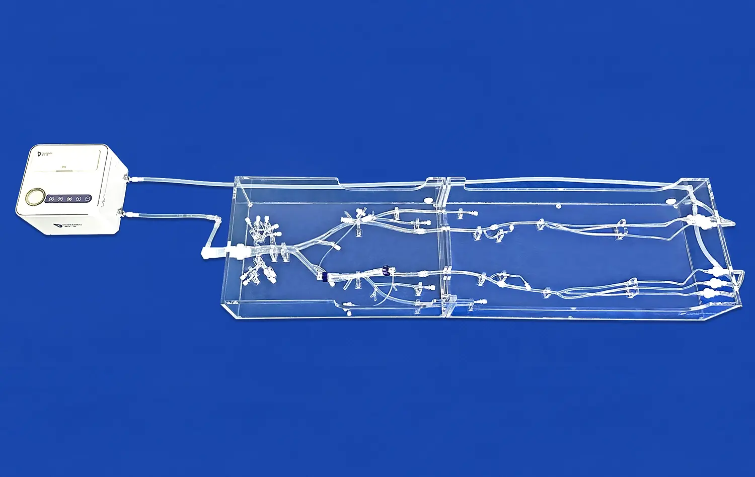
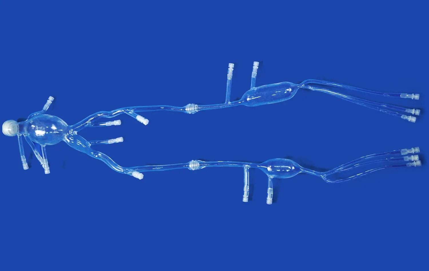
The complex network of arteries in the human cerebral system makes it hard to make new medical devices. The arteries have different lengths, curves, and branching angles. Models made from Silicone Shore 40A material, like the middle cerebral artery simulator (Product No.: SJX005), show the internal carotid artery and its branches in a way that is true to life. This makes them an important tool for medical professionals and device engineers to use for training and testing. This model goes beyond simple representations of the body by letting you test how well catheters and guidewires can be tracked, how well aneurysm tamponade procedures work, and how well device placement methods work.
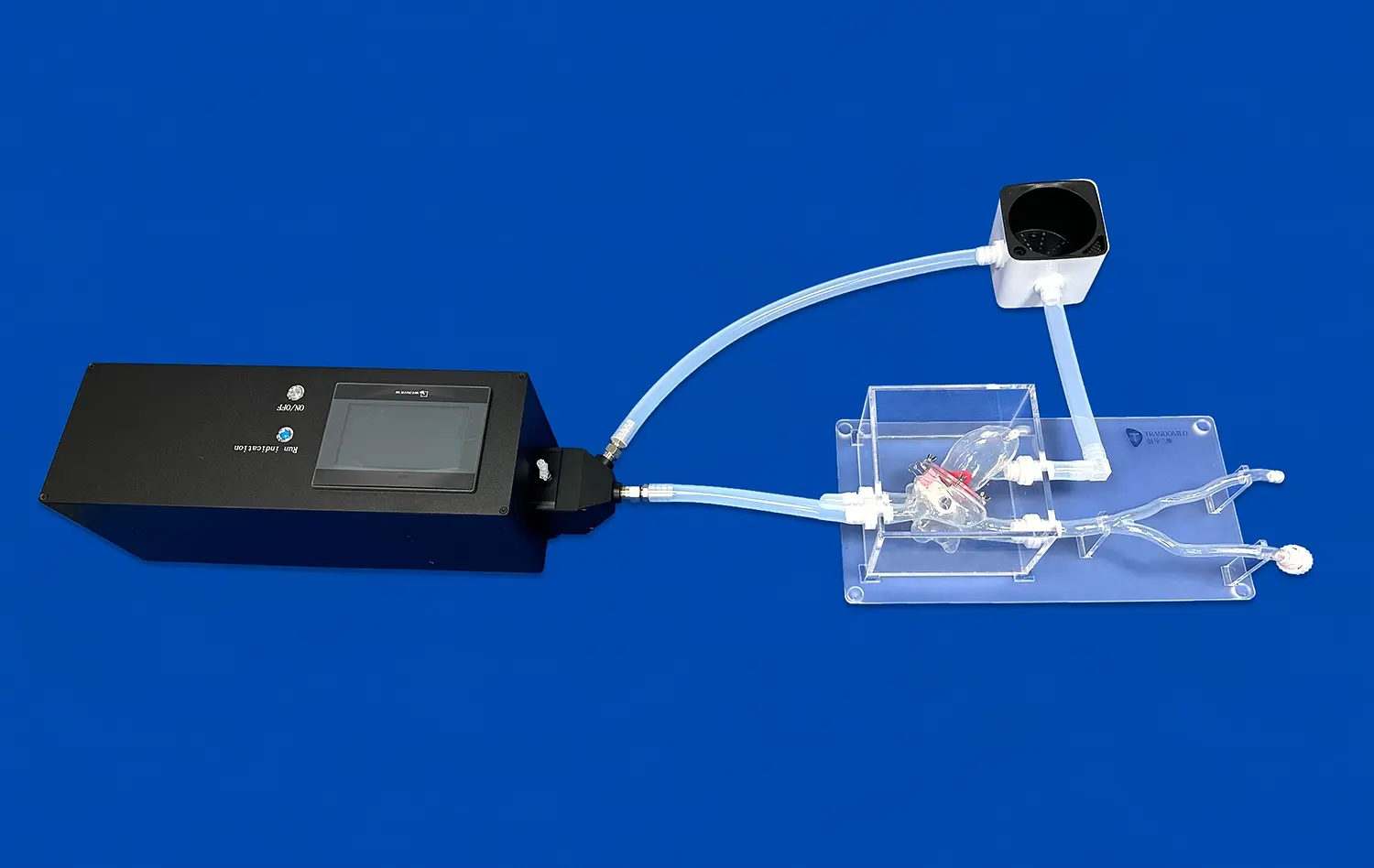
Because these plastic models feel like real tissue, users can get a true feel for how tissues work during virtual treatments. This feature is very helpful for testing how neurovascular devices move through complicated artery paths, adapt to curved vessels, and place themselves in treatment areas. The accuracy and flexibility offer big benefits, letting researchers and makers gain more knowledge and shorten the time it takes to make devices that fit each patient's unique anatomy. This sets a new bar for evaluating cardiovascular technology.
Applications Across Medical Sectors
There are many uses for these cerebral models in the medical field, going far beyond teaching basic anatomy. These models are used in medical school, nursing school, and clinical skills center training programs to teach difficult neurovascular anatomy and therapeutic methods. The realistic platform lets students get used to navigating catheters, learning how to deal with vessel tortuosity, and improving their hand-eye coordination needed for minimally invasive treatments before they work on real patients.
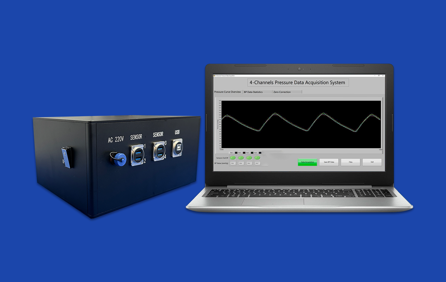
These physical tools can be used in a lot of different ways, including making, testing, and validating neuro-interventional devices. Medical device makers use these models to test catheters, guidewires, aneurysm coils, flow diverters, and stent retrievers in settings that are similar to real-life situations. This testing step is very important for finding mistakes in the design, making the gadget work better, and getting information needed for regulatory applications. Customizable models help research centers and labs with biomechanics analysis, trial studies, and prototype evaluation. This speeds up the process of turning new ideas into healthcare solutions.
Clinicians use models such as the 3D artery model to show complicated neurovascular conditions in clear and easy-to-understand ways when teaching patients, which is another useful use. Visual and physical examples using anatomically accurate models help patients understand and give informed consent when doctors talk about aneurysm sites, stenosis severity, or suggested treatment methods. These tools are useful for medical education, clinical practice, and gadget development because they show accurate shape and thorough anatomy.
Key Factors in Designing and Manufacturing 3D Artery Models
To make vascular models that are true to the human body, you need to use modern image technology, materials science, and precise manufacturing methods. From medical imaging data to a finished model, there are several important steps that must be taken. Each one affects the accuracy and usefulness of the end result.
Medical Imaging Integration and CAD Development
Anatomical data that is correct is the first step in building a good cerebrovascular model. To make sure the models are accurate, manufacturers combine complex CAD software with medical imaging data from CT angiography and MRI scans. Software like Mimics and 3D Slicer make accurate renderings of anatomy possible by turning DICOM images into three-dimensional digital models that show the shape of blood vessels, their branching patterns, and any abnormalities like aneurysms or stenoses.
Advanced modeling methods allow for small improvements to be made over and over again during the creation process. Based on the needs of the training or tests, engineering teams can change the shape of the vessel, the size and location of aneurysms, or the points at which branches meet. Manufacturers like Trandomed can accept client-specific data files in forms like CT, CAD, STL, STP, and STEP. This lets them turn patient-specific anatomy or standard anatomical versions into physical models without having to charge extra for design.
Material Selection and Biomechanical Properties
The choice of material is very important for how well a model reproduces the features of artery muscle. These days, flexible plastics and silicone blends are the best materials because they can behave mechanically like live blood vessels. The middle cerebral artery model uses Silicone Shore 40A, which is the best combination of toughness and tissue-like flexibility. This lets the model handle multiple catheter insertions while still providing realistic physical feedback.
Because of their physical features, these materials can be used to test devices in real-life situations. The silicone material reacts the same way that human arterial tissue does when guidewires move through bent parts of blood vessels or when balloon tubes grow against the walls of vessels. This accuracy is very important for testing how well a device works in settings that are very similar to clinical use. This helps makers find problems like the risk of vessel damage or not being able to track the device properly before they put it to the test on humans.
Quality Control and Validation Processes
Strict quality control procedures make sure that finished models meet the high standards of accuracy needed for neurovascular studies and device approval. Precision measurements check the accuracy of the dimensions against the source image data, and eye inspections make sure the surface finish is right and that the physical details are reproduced correctly. Following medical standards and best practices in the business makes sure that models work the same way in every production batch.
This all-around method to design and production for the 3D artery model guarantees the delivery of solid, working models that improve the evaluation processes for devices. With more than 20 years of experience in structural modeling, companies like Trandomed have improved their production processes to reduce variation and increase accuracy. This lets them give their clients simulation tools they can rely on for important training and development tasks.
Procurement Considerations for B2B Clients: How to Buy and Customize 3D Artery Models?
To successfully buy cerebral simulation models, it is important to make sure that the project's goals, funding, and technology requirements are all in line with those of the company. There are a lot of things that decision-makers need to think about to make sure that the goods they choose provide the best value over their entire lifecycle.
Aligning Model Complexity With Development Stages
The process of making a device goes through different stages, and each stage has its own modeling needs. In the early stages of idea proof, simple anatomy models that show how the gadget works in the most basic ways may be needed. As the project moves closer to developmental testing and regulatory filing, it needs more complex models that include the anatomy, pathology, and physical correctness of a real patient.
By comparing the complexity of the model to its current stage of growth, buying investments can be made more efficiently. When working on their first catheter designs, teams might start with standard models of the middle cerebral artery that show how the body normally looks. People who are getting ready for regulatory applications or clinical studies would benefit from custom models that replicate specific physical problems or patient groups. This would make sure that test results accurately predict how well the product will work in real life.
Volume Requirements and Customization Scope
Training centers that offer regular hands-on classes need models that are strong enough to be used by many students over and over again. Standardized structural configurations ordered by volume often help these uses because they make training uniform across groups. On the other hand, device makers might only need a few highly customized models that fit certain testing methods or variations in anatomy.
Trandomed can handle both situations because it can be manufactured in a variety of ways. Orders in bulk from educational institutions are given priority in the production schedule, while custom projects get focused engineering support to turn unique needs into real models. Since there are no design fees for customization, getting precisely set modeling tools is no longer financially difficult. This means that organizations can get models that exactly match their technology requirements without having to cut costs.
Logistics and Supply Chain Management
Lead times for a 3D artery model have a big effect on how projects are planned and how supplies are bought. Standard models, like the SJX005 middle cerebral artery simulator, usually ship 7–10 days after an order is confirmed. This makes them easy to use right away for training programs or testing plans. Custom projects need more time for design advice, reviewing prototypes, and final production. The amount of time needed depends on how complicated the project is and how much customization is needed.
When sending internationally, both cost and arrival dependability are affected by certain factors. Trandomed works with well-known shipping services like FedEx, DHL, EMS, UPS, and TNT to make sure that deliveries around the world are always on time. When planning project timelines, procurement workers should think about how to clear customs, possible import taxes, and shipping times in different regions. Talking to suppliers ahead of time about shipping needs helps avoid delays that come up out of the blue and affect training plans or device development goals.
After-Sales Support and Technical Assistance
Vascular computer models are valuable not only when they are first bought, but also when they get regular technical help and care. Organizations that are new to procedural modeling can benefit from advice on how to handle models properly, store them safely, and clean them in a way that makes the products last longer. When device makers try new ways to test their products, they might need help figuring out the best model setups or testing methods.
Comprehensive after-sales support adds reliability to procurement decisions. Customer service teams help people choose the right products, answer detailed questions about what each model can do, and offer solving advice when problems come up out of the blue. This support system is especially helpful for foreign clients who have to deal with different time zones and communication styles. It makes sure that everyone can get help quickly, no matter where they are in the world.
Cost Analysis and Value Optimization
To compare custom models to off-the-shelf models, you need to look at a lot of costs, not just the original buy amounts. Custom models made to fit specific study methods or device testing needs may cost more up front, but they are worth it because you don't have to settle on testing settings or physical arrangements. Standard models are easier to get and don't cost as much to start with, so they're good for general training where differences in anatomy aren't as important.
Total cost of ownership factors include how long the product lasts, how often it needs to be maintained, and how often it might need to be replaced. If you take good care of high-quality silicone models from well-known brands, they should last for hundreds of catheter insertions. This way, the cost of buying them is spread out over many training sessions or testing rounds. Companies should look at the cost per use instead of the total cost of the purchase, knowing that long-lasting models that are set up correctly are more valuable in the long run than cheaper models that need to be replaced often.
Conclusion
Neurovascular device creation, surgery training, and medical education have all been changed by advanced cerebrovascular simulations such as the 3D artery model. These simulators provide anatomically correct and biomechanically realistic environments for hands-on learning and thorough testing. Traditional solid models have been replaced by more complex silicone-based copies. This is a big improvement in the accuracy of simulations, which lets medical workers and gadget makers test new ideas in situations that are very close to real life. When procurement experts look at their choices in this changing market, they should think about things like physical correctness, the ability to customize, the manufacturer's knowledge, and the availability of a full support system. When companies buy high-quality vascular models from well-known brands, they get better training results, faster device development processes, and more confidence in how well the products will work before they are used in patients.
FAQ
In general, how long does it take to get normal cerebral models?
Most standard models, like the middle cerebral artery stimulator (SJX005), are shipped 7–10 days after the order is confirmed and payment is processed. This plan is for off-the-shelf setups that don't need to be changed to fit specific needs. Custom projects with specific anatomical variations, patient-specific anatomy reproduction, or special material needs may need more time for design consultation, prototype development, and final production. Based on the complexity of the project, timelines will be communicated during the quotation process.
How close are plastic models of arterial systems to real human anatomy?
When made from medical image data using modern CAD tools and precise production methods, high-quality cerebrovascular models have accuracy of less than one millimeter in all dimensions. The physical accuracy goes beyond shape and includes physiological qualities. For example, Shore 40A silicone materials can mimic the flexibility and feel of artery flesh. Because these models are accurate in both dimensions and mechanics, they can closely resemble the human body for both visual learning and testing functional devices.
Can these models be used to train surgeons and test medical devices at the same time?
Cerebrovascular models can be used for a lot of different things, from teaching surgeons to making new medical devices. These models are used in training programs to teach skills like catheter tracking, guidewire handling, and medical methods in settings that look and feel like the real body. To make sure their products work well in controlled situations that are like real life, device makers use the same models to test how well catheters can be tracked, how well release systems work, and how well their products work overall. Vascular models are useful for both neurovascular gadget research and teaching because they can be used for two different things.
What kinds of changes can be made to neurovascular models?
Customization options include changing the number, size, and location of aneurysms; the curve of the internal carotid artery; the tortuosity of the middle and anterior cerebral arteries; and creating a copy of the patient's structure from imaging data that has been given. Manufacturers like Trandomed can work with files in CT, CAD, STL, STP, and STEP forms. They can turn client requirements into actual models for no extra cost. This gives modeling tools the ability to perfectly match training goals or study methods.
Partner With Trandomed: Your Trusted 3D Artery Model Manufacturer
Trandomed's specialized knowledge will help organizations that need physically accurate cerebrovascular models for neurovascular device creation, surgery training, or study purposes. Our middle cerebral artery model and large vascular simulation library mix very accurate anatomy with high-level functionality. We also offer customization services to turn your unique needs into real training and testing tools. As a top 3D artery model supplier for over 20 years, we offer reliable products that meet international medical standards. You can make changes to our products without having to pay design fees, and we can deliver standard configurations quickly (7–10 days). We also offer dedicated technical support throughout the lifecycle of your project. You can email our expert team at jackson.chen@trandomed.com to talk about your specific application needs, get full product documentation, or set up sample evaluation units that show how committed we are to quality and accuracy in body modeling.
References
Anderson, J.R., Thompson, W.L., Alkattan, A.K., Diaz, O., Klucznik, R., Zhang, Y.J., Britz, G.W., Grossman, R.G., and Karmonik, C. (2016). Three-dimensional printing of anatomically accurate, patient specific intracranial aneurysm models. Journal of NeuroInterventional Surgery, 8(5), 517-520.
Mashiko, T., Otani, K., Kawano, R., Konno, T., Kaneko, N., Ito, Y., and Watanabe, E. (2015). Development of three-dimensional hollow elastic model for cerebral aneurysm clipping simulation enabling rapid and low cost prototyping. World Neurosurgery, 83(3), 351-361.
Russ, M., O'Hara, R., Setlur Nagesh, S.V., Mokin, M., Jimenez, C., Siddiqui, A., Ionita, C.N. (2015). Treatment planning for image-guided neuro-vascular interventions using patient-specific 3D printed phantoms. Proceedings of SPIE Medical Imaging, Volume 9417.
Ionita, C.N., Mokin, M., Varble, N., Bednarek, D.R., Xiang, J., Snyder, K.V., Siddiqui, A.H., Levy, E.I., Meng, H., and Rudin, S. (2014). Challenges and limitations of patient-specific vascular phantom fabrication using 3D Polyjet printing. Proceedings of SPIE Medical Imaging, Volume 9038.
Kimura, T., Morita, A., Nishimura, K., Aiyama, H., Itoh, H., Fukaya, S., Sora, S., and Ochiai, C. (2009). Simulation of and training for cerebral aneurysm clipping with 3-dimensional models. Neurosurgery, 65(4), 719-726.
Wurm, G., Lehner, M., Tomancok, B., Kleiser, R., and Nussbaumer, K. (2011). Cerebrovascular biomodeling for aneurysm surgery: simulation-based training by means of rapid prototyping technologies. Surgical Innovation, 18(3), 294-306.
_1734504221178.webp)
_1734507415405.webp)