The 3D artery model is a revolutionary learning tool that fills the gap between academic knowledge and real skill development. It is used by medical schools and research facilities to find effective neurovascular training solutions. Surgeons, interventional radiologists, and medical students can use these physically accurate models of cerebrovascular structures to practice complicated treatments without putting patients at risk. These models are made from medical-grade materials that feel like human flesh. They help doctors improve catheter tracking methods, try out different ways to treat aneurysms, and test new medical devices before they are used in real patients. The development of additive manufacturing technology has made these training tools easier to get and more flexible than ever. This has completely changed how doctors prepare for very important neurovascular treatments.
Understanding 3D Artery Models and Their Role in Neurovascular Training
The development of high-fidelity vascular models is a huge step forward in the field of medical modeling. These tools are more than just simple examples of anatomy; they're also full training platforms that mimic the dynamic properties and complicated structures of live flesh.
The Technology Behind Realistic Vascular Simulation
Modern neurovascular models are more realistic than ever before thanks to advances in materials science. Silicone compounds, especially those with a hardness of 40A Shore, are the best because they are both strong and flexible like flesh. This particular material makeup lets the catheter be put in more than once without losing its shape, which makes it perfect for long-term training programs in institutions.
Digital images from CT scans and angiography are combined with precise 3D printing methods to make the product. This combination makes sure that every change in anatomy, from the size of a blood vessel to the angle of a branch, correctly shows how the human body works. Medical engineers can now make aneurysms, stenoses, and arteriovenous defects that look like they are in real patients. This lets them create training models that are like real clinical problems.
Anatomical Precision in Middle Cerebral Artery Representation
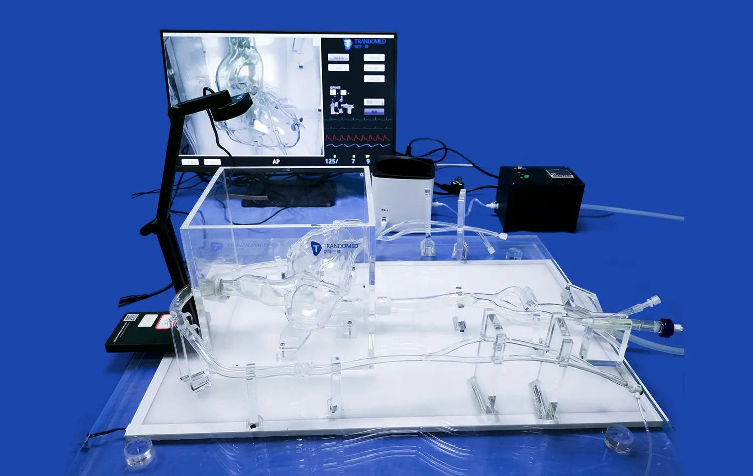
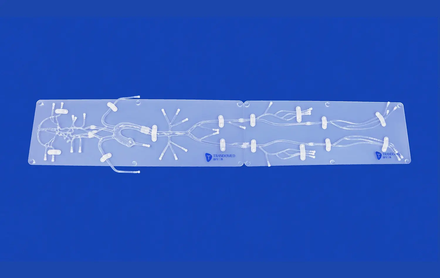
One of the most important and difficult areas to work on in neurovascular repair is the middle cerebral artery. Training models that correctly show this vessel's complicated growth patterns, tortuosity, and wall thickness are very helpful for practicing. The SJX005 model from Trandomed especially meets these needs by including the natural curve of the internal carotid artery and the complex bifurcations of the middle cerebral artery.
This amount of physical information is useful for many learning reasons. Residents learning diagnostic angiography can practice moving the tube through realistic vascular geometry. Experienced surgeons getting ready for difficult cases can practice specific steps of the procedure. When the guidewire is moved and the catheter is manipulated, the physical input closely resembles the body of a real patient. This helps build muscle memory that directly affects clinical performance.
Material Properties That Enhance Training Effectiveness
The choice of silicone as the main building material shows that training needs were carefully thought through. In contrast to hard plastic options, silicone has flexible qualities that are similar to artery walls. This means that it responds to catheter pressure in ways that are medically suitable. This trait comes in handy when doctors are practicing delicate moves near the heads of aneurysms or navigating complex segments of blood vessels, where too much force could harm real patients.
Model design choices for transparency in a 3D artery model let teachers see how the catheter is placed from outside, which makes it easier to give instant feedback during training classes. Visual access speeds up the learning process by making it easier for trainees to see how hand moves affect the reaction of the catheter tip in the three-dimensional venous space.
Comparison and Selection Criteria for 3D Artery Models in Neurovascular Applications
Before you can choose the right modeling tool, you need to know what the pros and cons of each training method are. Each method has its own benefits that depend on the goals of the organization, the available funds, and the teaching objectives.
Physical Models Versus Alternative Training Methods
Traditional training with cadavers has been thought of as the best way to learn surgery for a long time, but it has a lot of problems that make it hard to use in real life. Problems with preservation, social concerns, and a lack of access make cadaveric examples unsuitable for regular skill development. Also, changes in post-mortem tissue affect the mechanical qualities of arterial systems, which makes catheter handling less realistic.
Recently, virtual reality systems have become more popular because they let you experience realistic settings without being limited by your actual surroundings. When it comes to giving different situations and real-time success data, these digital tools really shine. But they don't have the sensory input that is needed to improve the fine motor skills needed for medical treatments. Computer systems can't fully recreate the feeling of the catheter rubbing against the walls of the vessel, the resistance that is felt when moving through a complicated body part, or the slight input that lets you know the device is in the right place.
Physical arterial models are one of a kind because they combine hands-on experience with uniform, repeated anatomy. They let you practice as much as you want without having to worry about time limits like in an operating room. This lets students build their confidence before moving on to controlled clinical procedures.
Key Selection Parameters for Institutional Procurement
When buying experts look at different providers and model setups, they should focus on a few key factors that have a direct effect on how well training works and how much it costs in the long run.
Material adaptability and longevity are the most important things to think about. For high-volume training programs, models must be able to handle hundreds of catheter insertions while still being anatomically correct. This toughness comes from the Shore 40A silicone used in high-end models. It will last for years, even with heavy use.
The ability to customize a model makes it much more useful for learning. Models that are specifically made for the type of patients an institution treats, like infant hospitals or centers that specialize in difficult aneurysm repair, are helpful. By letting trainees choose where aneurysms are located, change the tortuosity of the vessel, and include abnormal differences, training is made more like the problems doctors will face in real life.
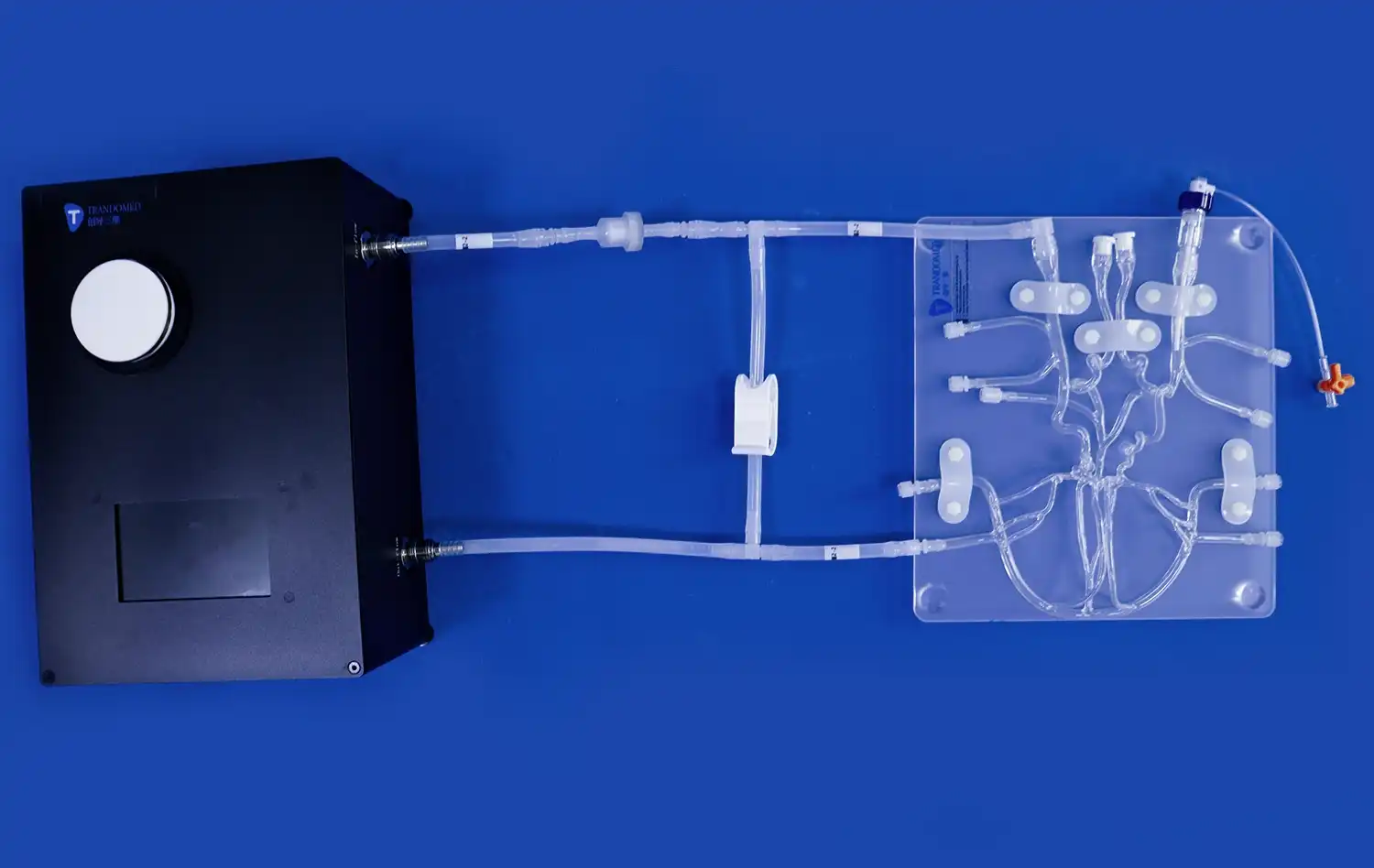
Pay close attention to how well a 3D artery model works with other tools you already have. Catheters, guidewires, and other medical devices that are used in real life should be able to fit in models so that skills can be transferred easily. Some advanced training programs mix physical models with fluoroscopy or angiography systems to practice full treatment processes. This means that the two can work together even when imaging is involved.
Cost-Effectiveness and Return on Investment
The original costs of acquisition are an important factor, but to find the real value, you need to look at the long-term benefits. A good neurovascular model helps many groups of trainees over a number of years, spreading the costs of the training over many rounds. When you add up the costs of getting a body, setting up a facility, and getting rid of it, durable plastic models often end up being cheaper.
In addition to directly affecting finances, these training tools help lower the number of problems that arise and make procedures run more smoothly for newly trained staff. The trust that comes from doing a lot of computer practice leads to better results for patients, which could lower the risk of lawsuits and the cost of correction procedures. For hospital managers and training heads, these secondary effects make the value argument a lot stronger.
How 3D Artery Models Can Be Used in Education and Medicine?
It is possible to use accurate circulatory models in many different areas of healthcare, and each area benefits from the tools in its own way.
Advancing Neurovascular Surgical Training
Neurovascular treatments require a high level of professional skill because the blood veins in the brain are very fragile and problems could be very bad. There are risks with traditional training models where trainees learn on real patients while being supervised. These risks can be eliminated with simulation-based options.
For complicated procedures like mechanical thrombectomy for treating an acute stroke, the tube needs to be moved quickly and precisely through the body's twisted structures. From groin entry sites, doctors have to make their way through the aortic arch, the carotid arteries, and into certain cerebral veins, all while keeping the treatment time as short as possible to protect brain tissue. Interventionalists can practice this difficult travel over and over with realistic models, which helps them improve their spatial awareness and hand-eye coordination.
Aneurysm coiling methods have extra technical challenges that can be greatly improved by practicing them in a computer. Moving microcatheters into aneurysm sacs, putting coils in place to get the best packing density, and dealing with possible problems like coil movement all take careful thought. Customizable aneurysm shapes in training models let practitioners experience a range of situations, giving them the hands-on experience they need for safe clinical practice.
Supporting Medical Device Development and Validation
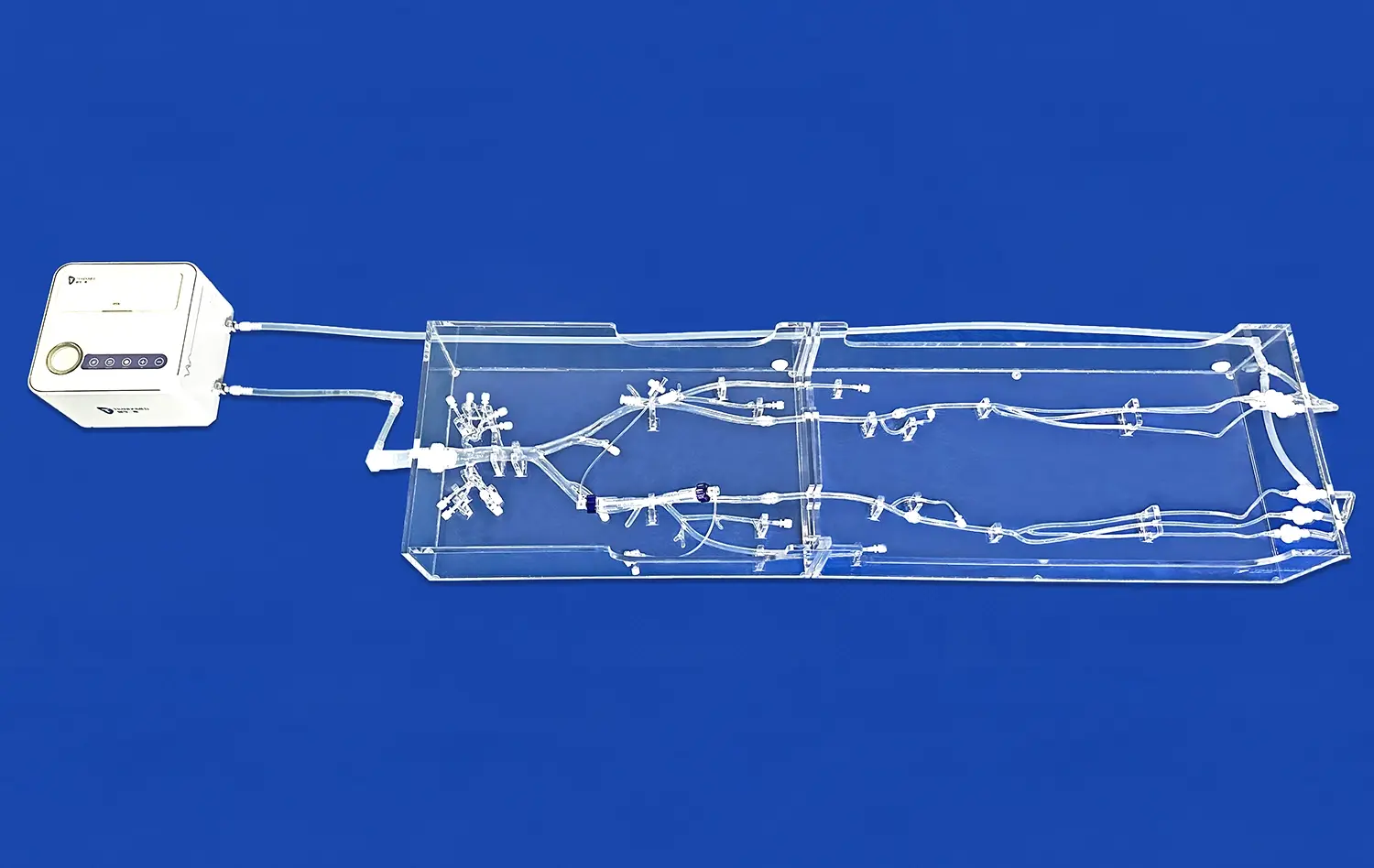
Before getting government approval and putting their products on the market, companies that make tubes, stents, guidewires, and other neurovascular devices have to put them through a lot of tests using a 3D artery model. Creating arterial models for each patient gives us a standard way to test how well devices work in a controlled environment.
Engineers can test trackability, which is how easily a tube follows a guidewire through a body part that is not straight, by using models that look like difficult venous shapes. Different types of vessels can be used to measure pushability, which is the amount of proximal force that is transmitted to the catheter tip. These mechanical tests find ways to improve designs before they are put through expensive clinical studies. This speeds up the development process and lowers costs.
Demonstration models are also very important for marketing because they let salespeople show doctors exactly how gadgets work in surroundings that look like real bodies. This hands-on experience is much more believable than academic explanations or two-dimensional pictures, which helps people make smart buying decisions.
Enhancing Patient Communication and Informed Consent
Patients often have trouble understanding suggested treatments because neurovascular procedures are so complicated. Verbal or graphic explanations that are too general may not be able to show the three-dimensional truth of their arterial structure and the suggested actions.
Making physical models from patients' own scan data gives us real-world tools for visualizing that change the way we talk about these things. The doctors can show the tube paths, where the devices will be put in place, and any possible problems in clear, specific language. This better understanding helps patients give truly informed consent while lowering their worry by making complicated treatments less mysterious.
Facilitating Research and Innovation
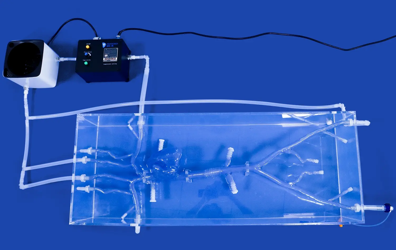
Anatomical models are used by academic medical centers and research institutions to look into new ways to treat patients and improve on methods that are already used. Standardized physical models that go along with computer simulations are helpful for biomechanical studies that look at vessel wall stress, flow dynamics, and the interactions between devices and tissues.
Before studying animals or doing clinical trials, researchers working on new embolization materials, flow-diverting stents, or thrombectomy devices can do basic testing in real-life body models. This middle step gives useful information while following moral rules that limit using animals and put human safety first.
Conclusion
The addition of high-fidelity vascular models such as the 3D artery model to neurovascular training classes is a big step forward in the way medical teaching is done. These tools get around some of the major problems with standard training methods while still giving people a safe, repeatable way to improve their skills. As patient safety and procedural skill become more important to healthcare systems, simulation-based training will continue to grow in its role in professional preparation. When institutions buy good anatomy models, they prepare their staff for clinical success and show that they are committed to teaching methods that are based on evidence. The technology can be used for direct procedural teaching, gadget development, study investigations, and communicating with patients. It is useful for many different organization offices and goals.
FAQ
In comparison to real human tissue, how exact are 3D artery models?
When made from thorough medical image data, high-quality neurovascular models are amazingly accurate in terms of anatomy. The level of accuracy is mostly determined by how clear the source images are and how precise the manufacturing methods are. High-quality plastic models accurately show the sizes of blood vessels, their branching angles, and the thickness of their walls, with enough accuracy to meet the needs of practical training. Even though there isn't a perfect synthetic model that exactly copies every property of living tissue, like temperature and pulsatility, the mechanical qualities and spatial layout of well-engineered models make them useful for training that works well in real life.
Can these models be changed to fit the bodies of different patients?
One of the best things about current 3D-printed anatomy models is that they can be changed to fit your needs. From each patient's CT scan, MRI data, or angiography series, manufacturers can make exact copies that show the patient's unique structural differences, diseases, and vessel configurations. This feature is especially useful for surgery practice before complicated surgeries, as it lets teams work on exact copies of the body parts they will be working on. Customized models that show specific disease conditions or structural variations that are important to the classroom focus are also useful for schools.
What kinds of materials work best for simulating neurovascular procedures?
Silicone materials with a Shore hardness between 30A and 50A are best for simulating blood vessels. These materials are flexible like artery walls and strong enough to withstand multiple catheter insertions. The Shore 40A silicone used in Trandomed's SJX005 model strikes a good mix between these qualities, giving accurate touch without breaking down too quickly. For some uses, clear materials are better because they let you see where the tube is placed, but for others, darkness that more closely matches clinical situations is more important. The training goals and expected level of use should guide the choice of materials.
How long do these training models usually last when they are used often?
How long neurovascular training models last relies on how often they are used, how they are handled, and the quality of the materials used. When used correctly, high-quality silicone models can handle hundreds of catheter insertions and last for years in busy training programs. Properly lubricating the model while it's being used, handling it carefully to avoid damage, and storing it away from high temperatures and direct sunlight are all things that can make it last longer. When institutions follow care standards, their equipment usually lasts a lot longer than when they don't and don't follow repair processes.
Partner With a Trusted 3D Artery Model Supplier for Enhanced Training Outcomes
Medical institutions, study centers, and device makers are welcome to contact Trandomed to learn more about how our specialized neurovascular models such as the 3D artery model can improve your training and development programs. Our team is ready to talk about your specific needs, give you thorough product specs, and come up with custom solutions that solve your specific problems. We provide quality, dependable, and quick service whether you need a single patient-specific model for practicing surgery or a lot of them for full training programs.You can email jackson.chen@trandomed.com to get more information about the product, talk about how it can be customized, or set up a sample review.
References
Anderson, J.R., Thompson, W.L., Alkattan, A.K., Diaz, O., Klucznik, R., Zhang, Y.J., Britz, G.W., Grossman, R.G., and Karmonik, C. (2016). "Three-Dimensional Printing of Anatomically Accurate, Patient-Specific Intracranial Aneurysm Models." Journal of Neurointerventional Surgery, Vol. 8, pp. 517-520.
Mashiko, T., Otani, K., Kawano, R., Konno, T., Kaneko, N., Ito, Y., and Watanabe, E. (2015). "Development of Three-Dimensional Hollow Elastic Model for Cerebral Aneurysm Clipping Simulation Enabling Rapid and Low Cost Prototyping." World Neurosurgery, Vol. 83, No. 3, pp. 351-361.
Ryan, J.R., Almefty, K.K., Nakaji, P., and Frakes, D.H. (2016). "Cerebral Aneurysm Clipping Surgery Simulation Using Patient-Specific 3D Printing and Silicone Casting." World Neurosurgery, Vol. 88, pp. 175-181.
Waran, V., Narayanan, V., Karuppiah, R., Owen, S.L., and Aziz, T. (2014). "Utility of Multimaterial 3D Printers in Creating Models with Pathological Entities to Enhance the Training Experience of Neurosurgeons." Journal of Neurosurgery, Vol. 120, No. 2, pp. 489-492.
Belykh, E., Zhao, X., Ngo, B., Farhadi, D.S., Byvaltsev, V.A., Eschbacher, J.M., Nakaji, P., and Preul, M.C. (2017). "Systematic Review of Patient-Specific Surgical Simulation: Toward Advancing Medical Education." Journal of Surgical Education, Vol. 74, No. 6, pp. 1028-1044.
Kono, K., Shintani, A., and Terada, T. (2013). "Hemodynamic Effects of Stent Struts versus Straightening of Vessels in Stent-Assisted Coil Embolization for Sidewall Cerebral Aneurysms." Journal of Biomechanical Engineering, Vol. 135, No. 6, Article 061011.