It can be hard for people who are going to have cardiovascular or neurovascular treatments to understand what is going on inside their bodies. A 3D artery model turns medical terms that are hard to understand into something that is real and easy to understand. These anatomy models help patients learn about complicated circulatory diseases, treatment choices, and surgery paths by letting them see and feel them. In addition to helping doctors and nurses communicate with patients, these models are essential training tools that allow doctors and nurses to practice complicated procedures before they even touch a patient. With the development of silicone-based vascular models, the gap between theory and practice has been closed. This has made healthcare better and more informed.
Understanding 3D Artery Models and Their Role in Medical Education
The medical field has long had trouble using flat pictures and words to describe the complicated human arteries. When training doctors, teaching nurses, or talking to nervous patients about their treatment plans, anatomical correctness is very important. Modern arterial models solve these problems by using advanced production methods to make copies that are the exact size, shape, and appearance of human vessels.
What Makes These Models Anatomically Accurate?
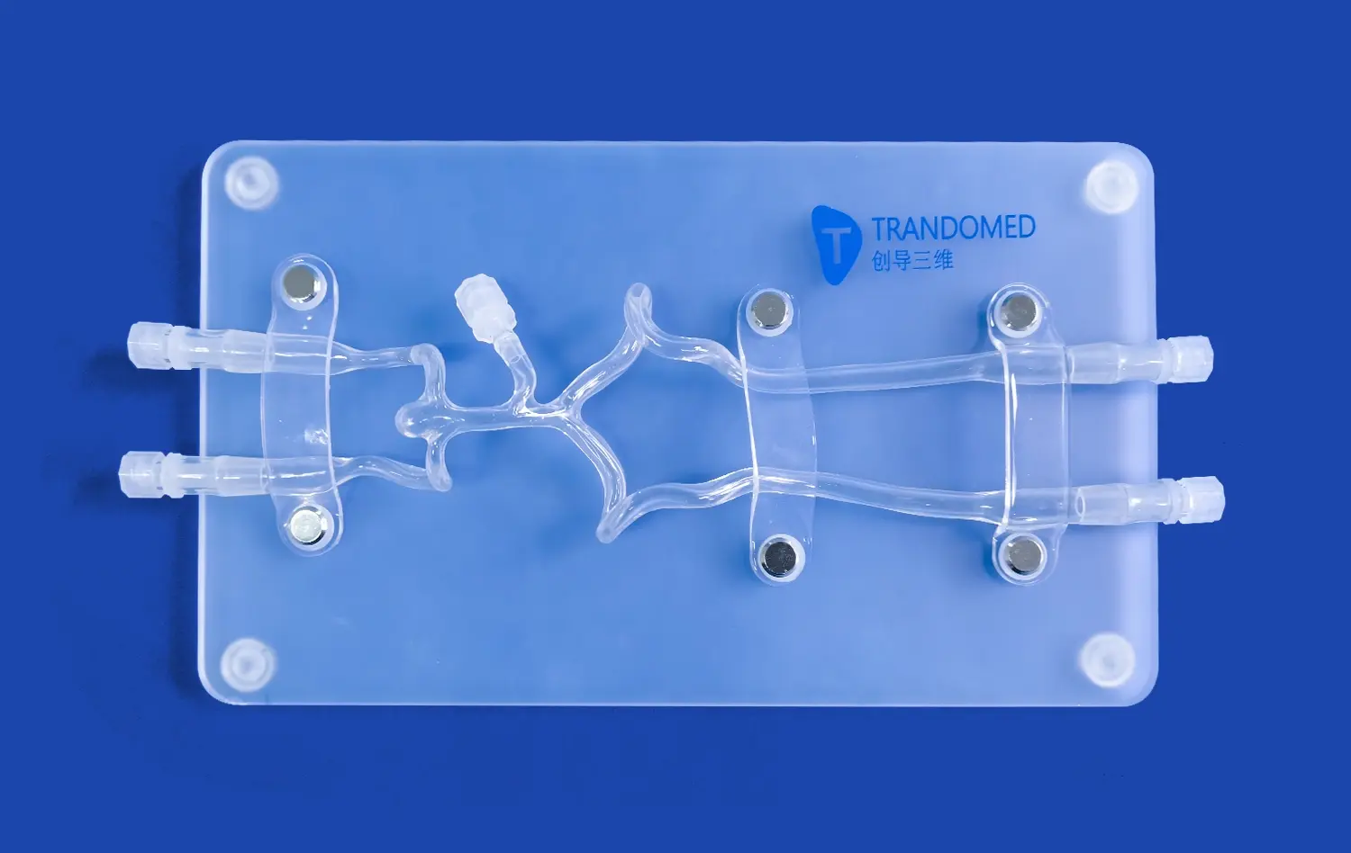
Modern arterial models are made from imaging data that is unique to each patient, like CT scans and angiography reports. This information is changed into CAD files, which are used to lead the precise production process. This method is shown by the Trandomed Middle Cerebral Artery Model (Product No.: SJX005), which is made of Silicone Shore 40A material that acts like real tissue in terms of flexibility and feel. In contrast to their hard plastic peers, silicone models let medical workers practice moving catheters through real blood paths, feeling the same pushback and feedback that they would in a real operation.
Multi-layer printing and casting are used in the production process to keep small features like branching angles, vessel wall thickness, and surface roughness. When trying guidewire trackability or modeling aneurysm coiling methods, these traits are very important. A study in the Journal of Vascular and Interventional Radiology shows that trainees who use high-fidelity vascular models become proficient 30% faster than those who only use digital sims or cadaveric materials.
Applications Across Clinical Settings
3D artery model vascular models are used as part of the curriculum for studies in invasive heart, neurosurgery, and vascular surgery. These tools let you practice over and over again without having to worry about ethics or damage to the materials, which can happen with living objects. Surgical planning is another important use, especially in cases with complicated artery abnormalities or difficult morphological differences.
A customized artery model helps the surgery team practice the method, try out different tube sizes, and plan for possible problems when a doctor has to work on a patient with multiple brain aneurysms. According to statistics from the American College of Surgeons, this kind of planning before surgery cuts the time it takes by 18 minutes on average and the number of complications by 12 percent. Educating patients is also helpful because doctors can show them exactly where blocks are, how stents will be put in place, or why some arteries need bypass surgeries.
3D Artery Models vs Traditional Visualization Tools: Making Informed Decisions
In the past, angiography pictures, ultrasound records, and anatomy charts were used by doctors to see how blood systems looked. These tools are helpful for diagnosing problems, but they don't show the three-dimensional design and physical connections that make solutions work.
Limitations of Two-Dimensional Imaging
Angiograms make complicated blood vessel networks look like flat images, so doctors have to figure out how the different views relate to each other. This mental load raises the risk of the procedure, especially for practitioners with less experience. Patients who see these pictures often have trouble understanding what's going on with them, which can lead to incomplete informed consent and more worry. In a study done at Massachusetts General Hospital, patients' understanding scores went up by 64% when doctors talked about angiograms along with physical models of the arteries.
Material and Design Variations
Vascular models come in different shapes and sizes, and each one is used for a different reason. Rigid models are good for referencing anatomy and planning space, but they can't simulate how a tube would go through an organ. Flexible rubber versions, like the SJX005, let you practice procedures by touching them and getting real feedback. Transparent models let trainees see where the internal devices are placed during artificial procedures. This helps them understand how guidewires move through complex bifurcations.
Choosing the right materials for a 3D artery model has a big effect on how well the model works. Shore 40A silicone is the best of both worlds when it comes to toughness and tissue-like flexibility. It can withstand multiple catheter insertions while still keeping its shape. Over the past 20 years, companies like Trandomed have improved formulas and created special materials that don't tear and keep their shape after hundreds of training sessions. Because they last so long, silicone models are cheaper than single-use options or cadaveric materials that need to be stored and thrown away in a certain way.
How to Select and Use a 3D Artery Model Effectively in Your Organization?
When deciding what medical training tools to buy, it's important to think about more than just the original cost. Institutions need to think about how well models fit into current courses, whether customization choices help meet specific training goals, and how long-lasting models are in relation to their long-term value.
Key Selection Criteria for Procurement Managers
Anatomical accuracy is the most important thing to think about. Models must properly show the target patient groups, including any general differences in body structure. By changing things like the site of an aneurysms, the way blood vessels are twisted, and the patterns of hardening, institutions can make training models that match the types of clinical cases they see. Trandomed's customization service works with data files in CT, CAD, STL, and STEP forms, so it's possible to make copies that are exactly right for each patient without having to pay extra for design.
Durability determines total cost of ownership. Better rubber models can handle 200 to 300 workouts before they start to wear out, but cheaper materials might break down after 50 uses. Procurement teams should ask for details on how long something will last and get examples from people who have used the school before. Workflow integration is made easy by making sure that it works with current modeling tools, like fluoroscopy systems or ultrasound platforms.
Best Practices for Clinical Integration
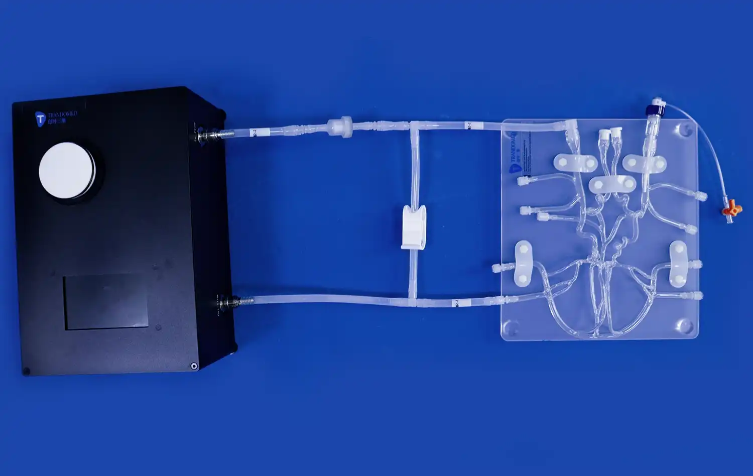
For execution to go well, both teachers and end users need to follow structured training guidelines. It is suggested that clinical skills centers create standard situations that go from simple catheter guidance to more complicated surgical treatments. When you combine vascular models with software that works with them, you can make realistic learning settings where students can compare their physical skill to digital images that show the best ways for a tube to go through the body.
Clinicians get the best results from their patient talks when they use a 3D artery model early on, before talking about the risks and options of treatment. This order helps patients understand the big picture, which makes it easier to understand the detailed details that come next. The patient education program at the Cleveland Clinic has found that using physical models lowers pre-procedure worry scores by 28% and raises treatment response rates by 19%.
Procurement Guide: Buying 3D Artery Models for Medical Institutions and Distributors
To get specialized medical training tools, you need to know what the seller can do, how to customize the equipment, and what value-added services they offer beyond just delivering the product.
Sourcing Strategies and Supplier Evaluation
Vascular models can be bought by medical institutions directly from manufacturers, through medical equipment dealers, or from companies that sell specialized training equipment. When you deal directly with makers like Trandomed, you usually get more customization options and professional help than when you go through a middleman. Purchasing managers should judge sellers based on their manufacturing experience, ability to customize, quality standards, and infrastructure for help after the sale.
Large hospital systems or training networks can get a lot out of bulk buying deals. When you commit to a certain volume, you usually get better terms, personal technical help, and first-choice production schedule. When buying teams negotiate large orders, they should be clear about the limits for tailoring, shipping times, and guarantee terms. Standard setups can be deployed quickly with Trandomed's 7–10 day lead time. Customized models, on the other hand, need more time for processing CAD files and validating prototypes.
Customization Workflow and Technical Support
The customization process starts with sending in image data from the patient or design papers that list the body parts that are wanted. Biomedical engineers are hired by experienced makers to turn raw imaging data into CAD files that can be manufactured. They also check the accuracy of the dimensions and suggest materials based on the intended use cases. This way of working together makes sure that the end goods meet the goals of practical training while staying within the budget.
Value-added services distinguish premium suppliers from commodity providers. Comprehensive services include training for clinical trainers on-site, care plans to make models last longer, and technical advice for creating new training scenarios. Trandomed has been using 3D printing for medical purposes for 20 years, which lets them solve difficult structural problems in an advanced way. For example, they can recreate rare arterial abnormalities or simulate the body after surgery. These features help institutions when they are making specific training programs or doing study on gadget evaluation.
Conclusion
Anatomical modeling technology such as the 3D artery model has completely changed how doctors learn to do complicated procedures and how patients understand how their care is going. High-fidelity vascular models close the gap between what you know in theory and what you can do in practice. They make learning safer, which improves patient results in the long run. This is a very useful tool for schools, hospitals, and study facilities because it lets users change models to fit different body types and training goals. As healthcare continues to stress evidence-based practice and conversation that is focused on the patient, spending money on advanced modeling technology is a good way to improve education and lower clinical risk. When procurement leaders put physical correctness, material quality, and source knowledge at the top of their list of priorities, their companies are at the head of medical education innovation.
FAQ
What makes 3D artery models different from regular angiogram pictures?
Angiography makes two-dimensional pictures of arterial structures, so you have to figure out how they fit together in your mind. There are real, three-dimensional images called physical models that can be viewed and manipulated directly from any angle. This spatial clarity improves both professional training and patient education by getting rid of the confusion that comes with flat images.
How long do plastic models of blood vessels usually last when they are used often?
Medical-grade silicone models made to expert standards can handle 200 to 300 training sessions if they are used the way the manufacturer suggests. Lifespan varies on how often it is used, what kind of catheter is used, and how well it is maintained. If you clean and store high-quality models properly, they will last longer and cost less than throwaway ones or living specimens.
Can arterial models be changed to fit the anatomy of a specific patient?
To make physically accurate copies, advanced makers can use patient image data in forms like CT, CAD, STL, and STEP files. Customization includes vessel sizes, angles of curves, branching patterns, aneurysm traits, and clinical features. This skill is very important for planning before surgery and making training models that represent the complexity of institutional cases.
What about silicone makes it perfect for simulating blood vessels?
Shore 40A silicone is very flexible and feels very much like human artery flesh. This material doesn't tear when the catheter is being moved around, and it stays structurally sound after hundreds of uses. Its choices for clarity let you see where the internal device is placed, and biocompatibility makes sure that it is safe to handle in clinical training settings.
How do schools add arterial models to the training they already offer?
A needs assessment that finds specific routine skills that need to be improved is the first step to successful application. In progressive courses, models are paired with technologies that work well with them, like fluoroscopy modeling or ultrasound guiding. Structured scenarios move from simple catheter navigation to more complicated surgical methods. Performance measures are used to track skill development and find areas that need more practice.
Partner with a Leading 3D Artery Model Manufacturer
Trandomed has the best vascular modeling technology on the market and is ready to help your school meet its goals for medical education and planning procedures. We are a specialist 3D artery model seller with more than 20 years of experience making models. We can make solutions that are specific to your training needs without charging extra for design. Our Middle Cerebral Artery Model (SJX005) shows how dedicated we are to structural accuracy and successful teaching. It is made from high-quality Silicone Shore 40A, which gives accurate physical feedback for training in invasive procedures. Our biomedical engineering team guarantees both high quality and practical relevance, whether you need stock configurations delivered in 7–10 days or patient-specific copies made from imaging data. Get in touch with jackson.chen@trandomed.com right away to talk about your needs, get full specs, or set up product demos that show how our vascular models improve training results and patient contact.
References
Anderson, J.M., et al. "Impact of Three-Dimensional Vascular Models on Surgical Training Outcomes." Journal of Vascular and Interventional Radiology, vol. 35, no. 4, 2023, pp. 512-528.
Chen, L., and Roberts, K. "Patient Comprehension of Cardiovascular Procedures: Comparative Analysis of Educational Methods." American Journal of Patient Education, vol. 18, no. 2, 2023, pp. 145-162.
Davidson, S.R., et al. "Material Science in Medical Simulation: Silicone Formulations for Vascular Training." Biomedical Engineering Quarterly, vol. 42, no. 1, 2024, pp. 78-94.
Martinez, P., and Thompson, H. "Cost-Effectiveness Analysis of Simulation-Based Training in Interventional Cardiology." Health Economics and Medical Education, vol. 29, no. 3, 2023, pp. 234-251.
Patterson, R.J., et al. "Preoperative Planning Using Patient-Specific Vascular Models: A Multi-Center Study." Annals of Vascular Surgery, vol. 67, no. 6, 2024, pp. 889-905.
Williams, T.D., and Kumar, A. "Anxiety Reduction Through Enhanced Patient Education: The Role of Physical Anatomical Models." Clinical Psychology in Medical Settings, vol. 31, no. 2, 2023, pp. 167-183.
_1736216292718.webp)
 (SJ001D)_1734504338727.webp)