How Does 3D Printing Technology Enhance Anatomical Fidelity?
Precision in Replication
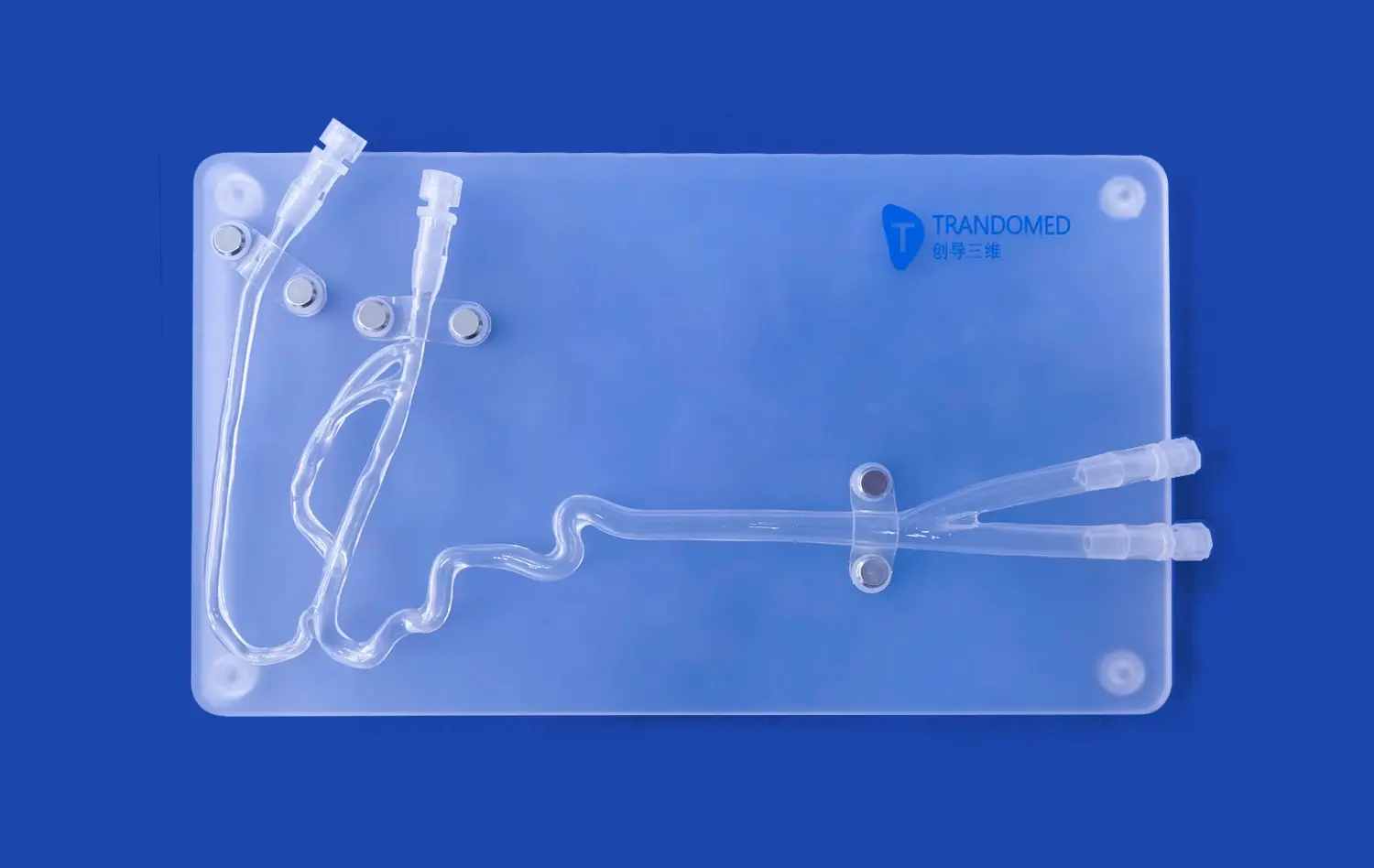
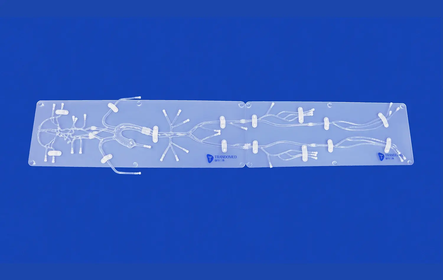
3D printing technology has revolutionized the creation of medical models, particularly in replicating the intricate anatomy of lower extremity arteries. By utilizing high-resolution CT and MRI scans, 3D printers can produce models with submillimeter accuracy. This level of precision allows for the faithful reproduction of complex arterial structures, including bifurcations, stenoses, and aneurysms. The ability to capture these fine details is crucial for creating realistic training environments and for planning complex endovascular procedures.
Customization Capabilities
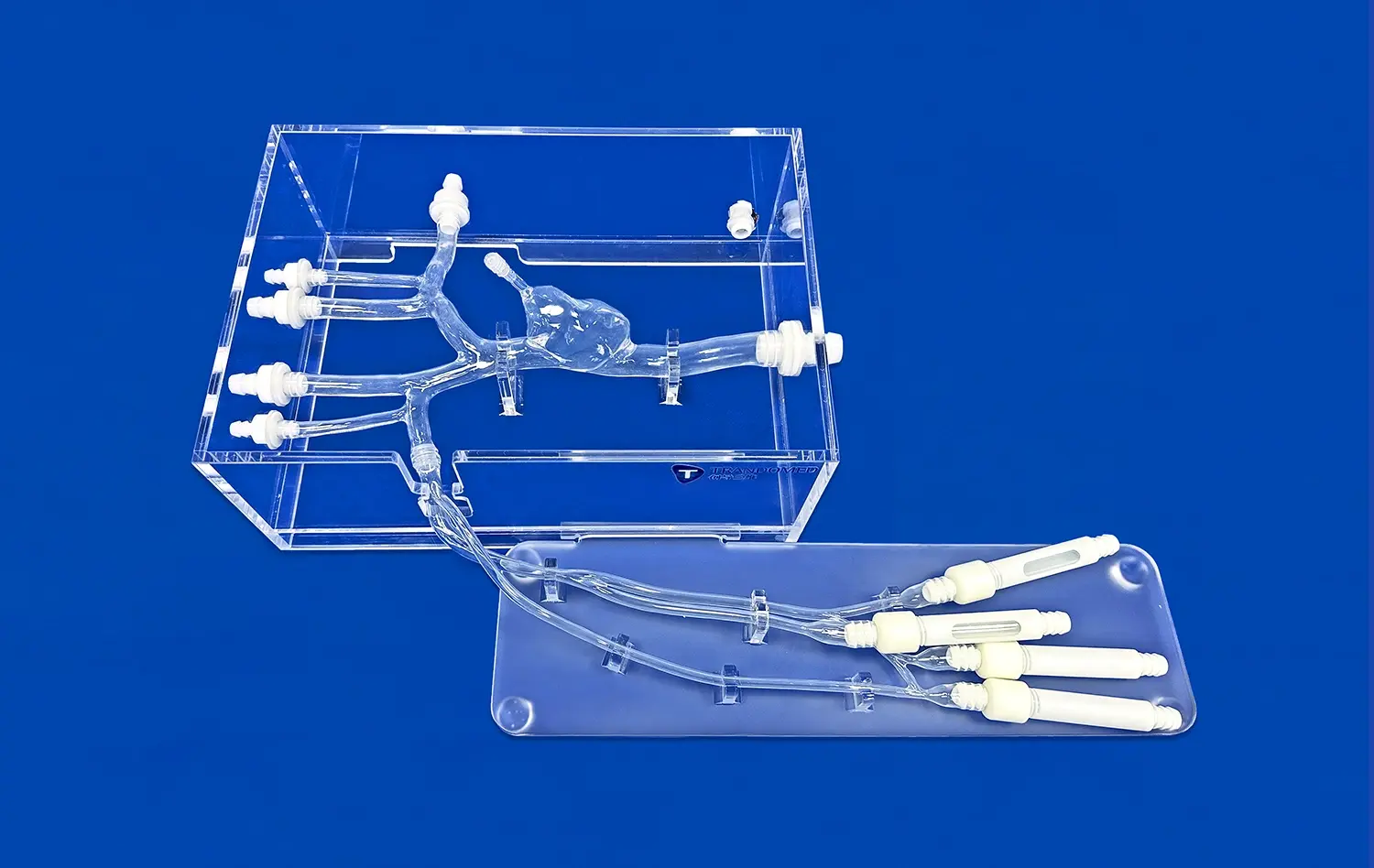
One of the most significant advantages of 3D printing in medical modeling is the ability to create patient-specific anatomies. This customization is particularly valuable in the context of lower extremity artery models. Surgeons and interventional radiologists can now practice on models that mirror the exact vascular anatomy of their patients, allowing for meticulous pre-procedural planning. This level of personalization not only enhances the training experience but also contributes to improved patient outcomes by allowing practitioners to anticipate and prepare for individual anatomical variations.
Integration of Pathological Features
3D printing technology excels in its ability to incorporate pathological features into lower extremity artery models. Complex conditions such as peripheral artery disease, with its characteristic plaque buildup and vessel narrowing, can be accurately represented. Additionally, rare vascular anomalies or post-surgical anatomies can be recreated with high fidelity. This capability allows medical professionals to gain experience with a wide range of clinical scenarios, from common presentations to rare and challenging cases, all within a controlled and repeatable environment.
Material Innovation for Realistic Tactile Feedback
Advancements in Silicone Technology
The development of advanced silicone materials has been a game-changer in the production of realistic lower extremity artery models. Modern silicone formulations, such as those with a Shore 40A hardness, closely mimic the elasticity and texture of human blood vessels. This similarity extends beyond mere visual resemblance; it provides tactile feedback that is crucial for interventional procedures. When guidewires and catheters are inserted into these silicone models, the sensation closely resembles that of navigating through actual arteries, allowing for a more immersive and authentic training experience.
Multi-Material Printing for Layered Structures
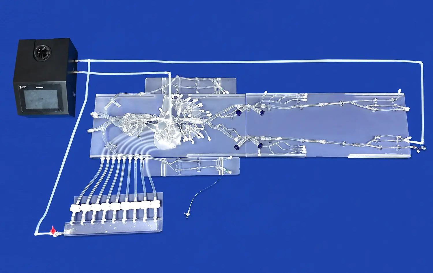
Recent advancements in 3D printing have enabled the use of multiple materials within a single print job. This capability is particularly valuable in creating lower extremity artery models that accurately represent the layered structure of blood vessels. By utilizing materials of varying densities and flexibilities, these models can replicate the distinct layers of the arterial wall - the intima, media, and adventitia. This layered approach not only enhances the visual and tactile realism of the model but also allows for more accurate simulation of procedures such as angioplasty and stenting, where the interaction between medical devices and different vessel layers is crucial.
Integration of Calcification and Plaque Simulation
One of the most challenging aspects of creating realistic lower extremity artery models is the accurate representation of calcifications and plaque buildup. Innovative materials and printing techniques now allow for the integration of these pathological features with remarkable accuracy. By incorporating materials of different densities and textures within the vessel walls, these models can simulate the resistance and tactile feedback associated with navigating through partially occluded or calcified arteries. This level of detail is invaluable for training in complex procedures such as chronic total occlusion (CTO) interventions, where the ability to feel and navigate through challenging lesions is paramount.
Why Realism Matters in Endovascular Simulation and Research?
Enhanced Procedural Proficiency
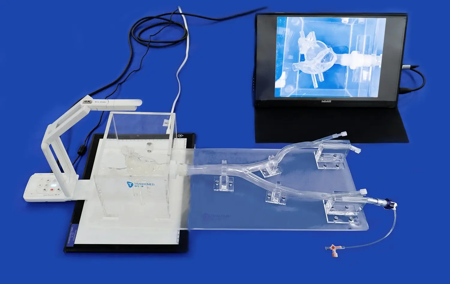
The high degree of realism in modern lower extremity artery models plays a crucial role in enhancing procedural proficiency among vascular specialists. These models allow practitioners to repeatedly practice complex endovascular techniques in a risk-free environment. The ability to perform procedures such as guidewire navigation, balloon angioplasty, and stent placement on anatomically accurate models helps in developing muscle memory and refining fine motor skills. This hands-on experience translates directly to improved performance in real clinical scenarios, potentially leading to reduced procedure times, decreased complication rates, and better patient outcomes.
Accelerated Learning Curve
Realistic lower extremity artery models significantly accelerate the learning curve for both novice and experienced practitioners. Traditional training methods often rely heavily on observation and limited hands-on experience during actual procedures. In contrast, these advanced models allow for unlimited practice sessions, enabling trainees to gain confidence and competence more rapidly. The ability to simulate a wide range of anatomical variations and pathological conditions exposes learners to diverse scenarios they might encounter in clinical practice. This comprehensive exposure helps in developing critical decision-making skills and adaptability, which are essential in the dynamic field of endovascular interventions.
Advancement of Medical Device Development
The realism of modern lower extremity artery models extends beyond training applications; it plays a pivotal role in the advancement of medical device development. These models serve as invaluable tools for testing and refining new endovascular devices and techniques. Manufacturers can use these highly accurate replicas to assess the performance of catheters, guidewires, stents, and other interventional tools in a setting that closely mimics real-world conditions. This capability not only accelerates the product development cycle but also enhances the safety and efficacy of new medical devices before they reach clinical trials. The ability to iterate and improve designs based on performance in realistic models ultimately contributes to the development of more effective and safer interventional tools for vascular procedures.
Conclusion
The integration of 3D printing technology in creating highly realistic lower extremity artery models marks a significant leap forward in medical training and research. These advanced simulators, with their unparalleled anatomical fidelity and tactile realism, are transforming the landscape of endovascular education and device development. By providing a platform for risk-free practice and experimentation, they not only enhance the skills of medical professionals but also contribute to the advancement of vascular interventions. As this technology continues to evolve, we can anticipate even more sophisticated models that will further bridge the gap between simulation and real-world clinical scenarios, ultimately leading to improved patient care and outcomes in vascular medicine.
Contact Us
Experience the cutting-edge realism of Trandomed's lower extremity artery models. As a leading manufacturer and supplier of 3D-printed medical simulators, we offer unparalleled quality and customization options. Our state-of-the-art factory combines innovative technology with expert craftsmanship to deliver products that meet the highest standards of anatomical accuracy and tactile feedback. Whether you're a medical institution, research facility, or device manufacturer, our team is ready to provide you with tailored solutions that elevate your training and development processes. Discover how our advanced simulators can transform your approach to endovascular education and research. Contact us today at jackson.chen@trandomed.com to explore our range of products and discuss your specific requirements.