What Imaging Modalities Benefit Most from Replaceable Models?
Computed Tomography Angiography (CTA) Advancements
CTA has emerged as a cornerstone in cardiovascular imaging, and the aortic arch replaceable model has significantly enhanced its capabilities. These models allow radiologists and technicians to optimize scanning parameters, reduce artifacts, and improve image quality. By simulating various pathological conditions, such as aneurysms or stenoses, these models enable the fine-tuning of contrast injection protocols and timing, resulting in more accurate and detailed visualizations of the aortic arch and its branches.
Moreover, the replaceable nature of these models facilitates the exploration of different anatomical variations, helping radiologists better understand and interpret complex cases. This adaptability is particularly valuable in planning interventional procedures, as it allows for precise measurements and assessment of potential challenges before the actual intervention.
Digital Subtraction Angiography (DSA) Enhancements
In the realm of DSA, aortic arch replaceable models have proven invaluable for both training and research purposes. These models provide a realistic environment for interventional radiologists to practice catheter navigation and device deployment without risking patient safety. The ability to replicate specific patient anatomies or create challenging scenarios helps in developing new techniques and refining existing ones.
Furthermore, these models serve as excellent tools for evaluating new contrast agents and injection techniques. By allowing for repeated imaging under controlled conditions, researchers can assess the efficacy of various contrast protocols and optimize image acquisition parameters, ultimately leading to improved diagnostic accuracy and reduced radiation exposure in clinical settings.
Magnetic Resonance Angiography (MRA) Innovations
MRA techniques have greatly benefited from the introduction of aortic arch replaceable models. These models enable researchers and clinicians to validate and improve MRA sequences, ensuring optimal visualization of the aortic arch and its branches without the need for ionizing radiation or iodinated contrast agents. By simulating different flow conditions and vessel geometries, these models help in developing more sensitive and specific MRA protocols for detecting vascular abnormalities.
Additionally, the use of these models in MRA research has led to advancements in 4D flow imaging techniques, allowing for better understanding of complex blood flow patterns in the aortic arch. This has significant implications for the assessment of hemodynamics in various pathological conditions, potentially leading to more accurate diagnoses and tailored treatment strategies.
Optimizing Model Compatibility with CTA, DSA, and MRA
Material Selection for Imaging Accuracy
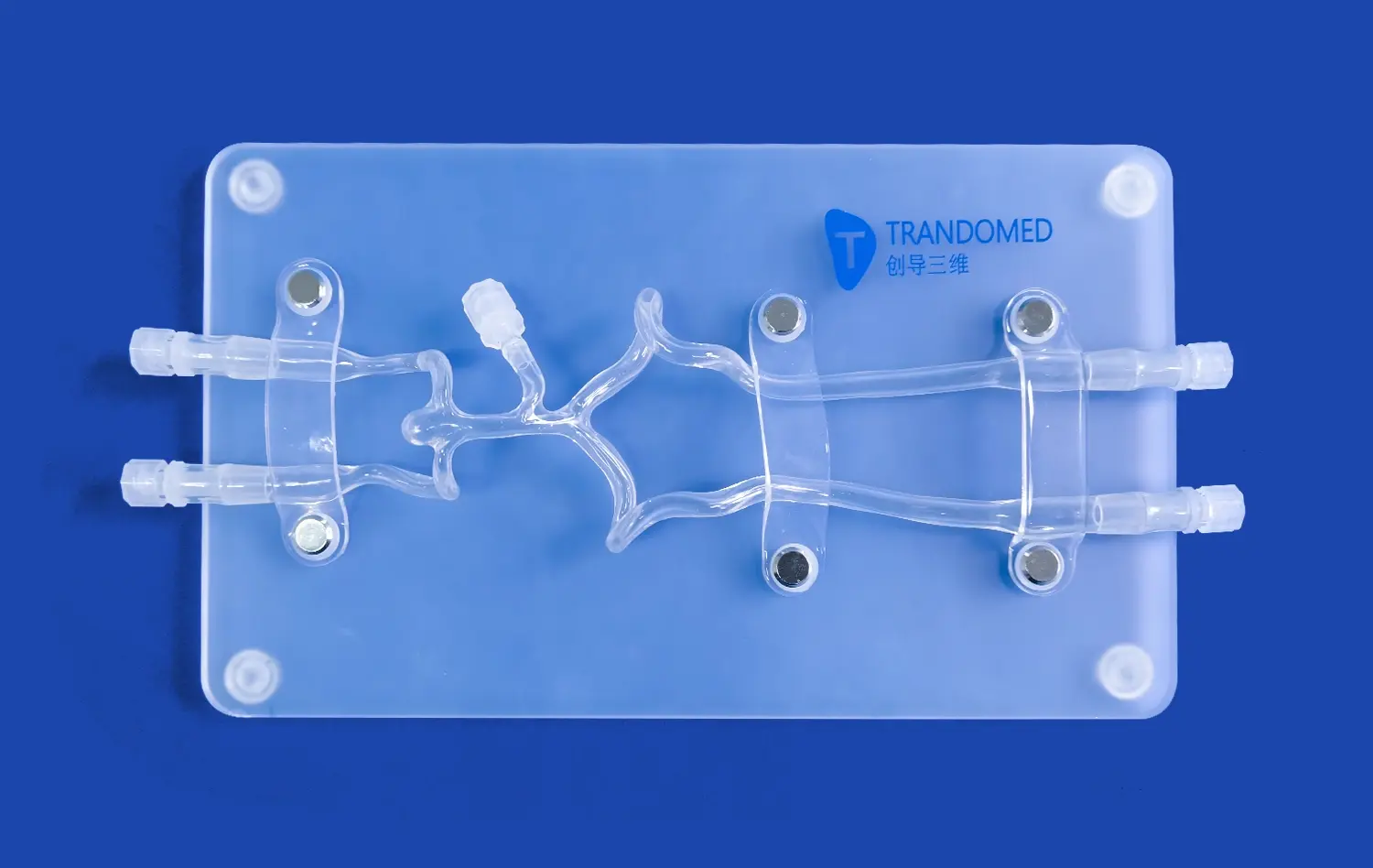
The choice of materials used in aortic arch replaceable models is crucial for ensuring compatibility with various imaging modalities. Trandomed's expertise in material science allows for the selection of silicone compounds that closely mimic the radiographic properties of human tissues. This careful material selection ensures that the models produce realistic images across CTA, DSA, and MRA platforms, minimizing artifacts and maximizing image quality.
For CTA applications, the models are designed with materials that have appropriate Hounsfield unit values, allowing for accurate representation of vessel walls and lumen. In DSA, the models incorporate radio-opaque markers strategically placed to aid in calibration and measurement. For MRA compatibility, materials with suitable magnetic properties are used to ensure proper signal generation and minimal distortion.
Structural Design for Multi-modality Use
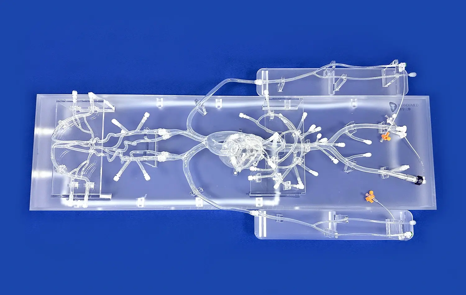
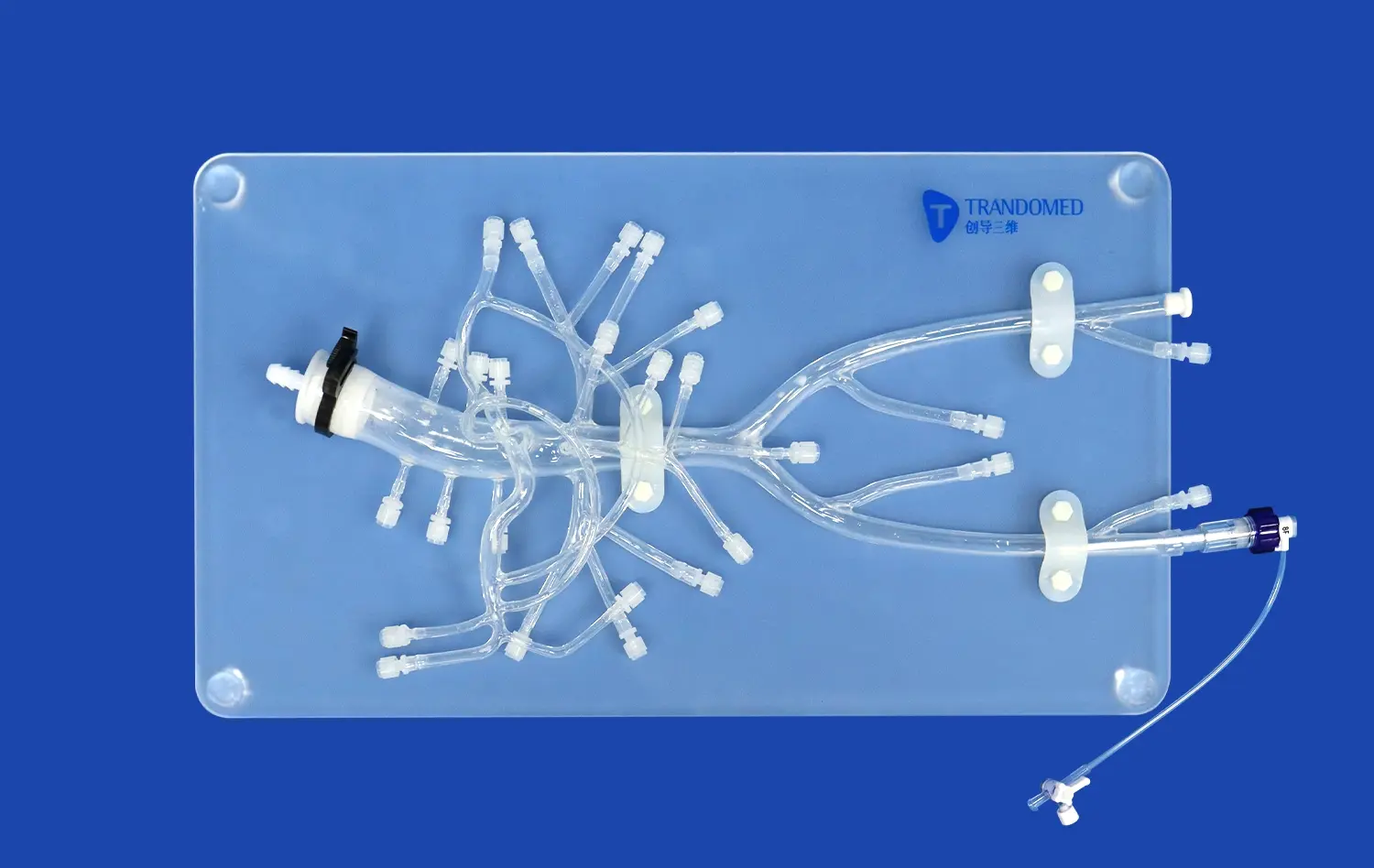
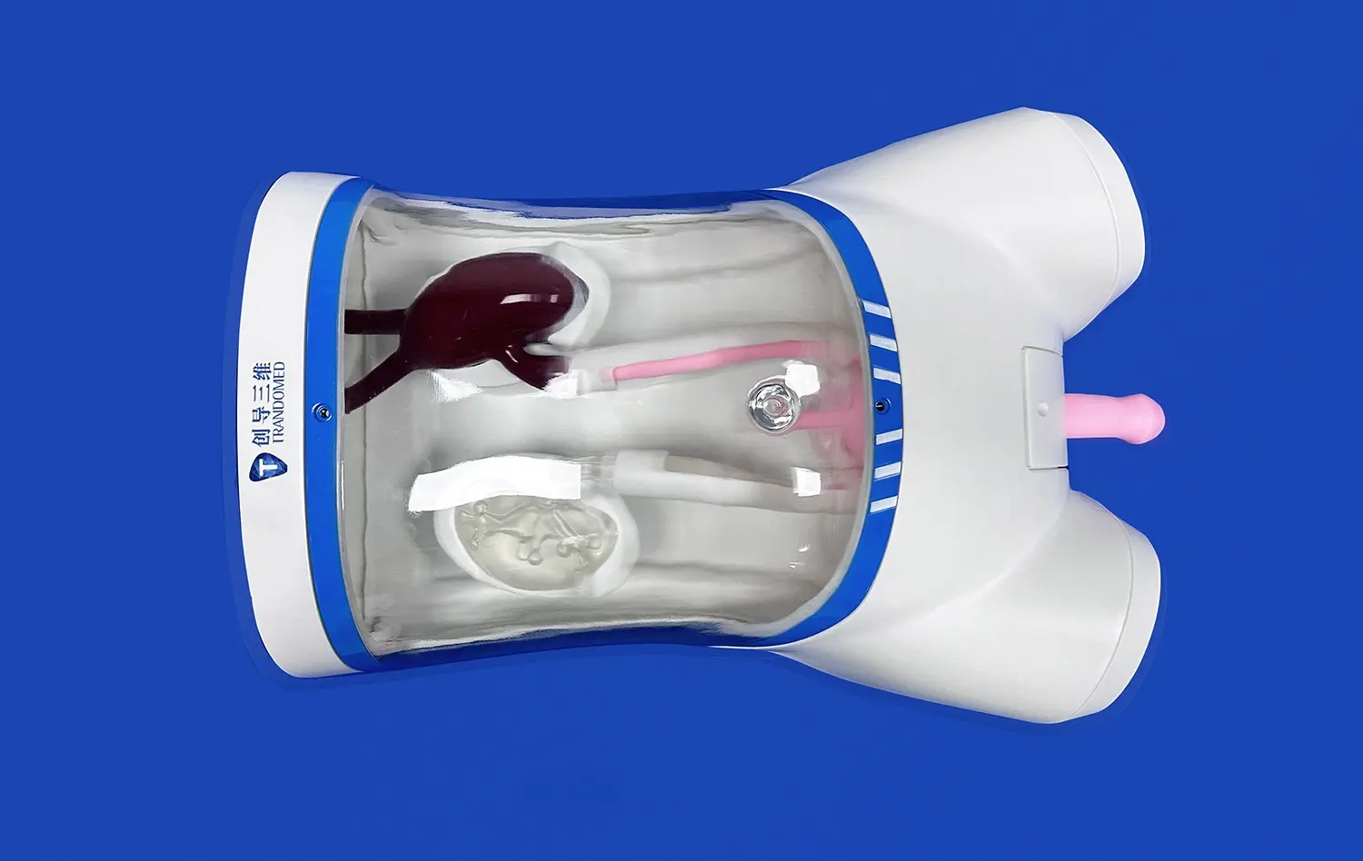
The structural design of aortic arch replaceable models plays a vital role in their compatibility across imaging modalities. Trandomed's models feature a modular design with easily detachable components, allowing for versatility in imaging setups. This design facilitates the integration of flow systems for dynamic imaging studies and enables the simulation of various pathological conditions.
The models incorporate special features such as reinforced access points for catheter insertion in DSA simulations and flow connectors for CTA and MRA studies. These design elements ensure that the models can withstand the rigors of repeated use while maintaining their structural integrity and imaging compatibility.
Customization for Specific Imaging Protocols
Recognizing that different imaging protocols may require specific model characteristics, Trandomed offers customization options to optimize compatibility. For CTA studies, models can be tailored with varying degrees of calcification or soft plaque to challenge image acquisition and post-processing algorithms. In DSA applications, customizable stenoses or aneurysms can be incorporated to simulate complex interventional scenarios.
For MRA studies, models can be designed with specific flow channels or turbulence-inducing geometries to test advanced imaging sequences. This level of customization ensures that researchers and clinicians can obtain the most relevant and accurate data for their specific imaging needs across all three modalities.
Enhancing Diagnostic Accuracy Through Realistic Vascular Simulation
Replicating Complex Anatomical Variations
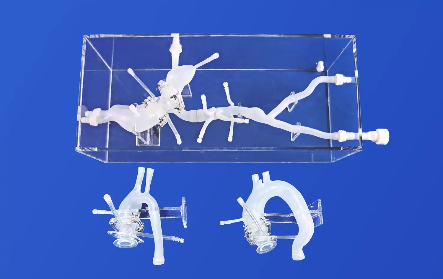
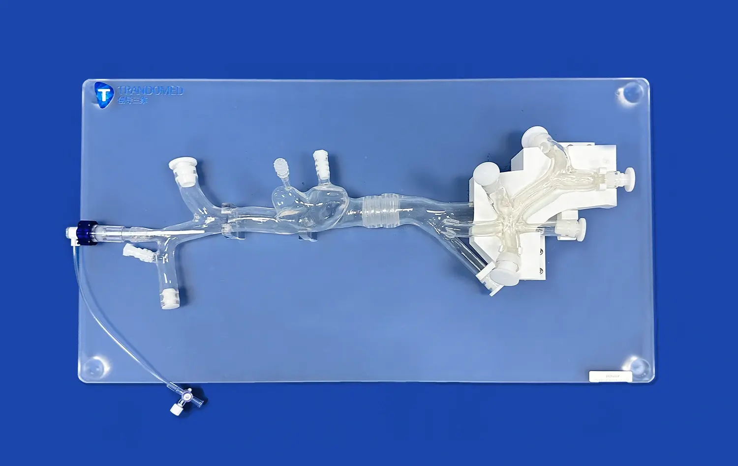
The aortic arch replaceable model excels in replicating complex anatomical variations, a crucial factor in enhancing diagnostic accuracy. By utilizing advanced 3D printing and molding techniques, Trandomed creates models that faithfully reproduce patient-specific anatomies derived from real CT and MRI data. This level of anatomical fidelity allows radiologists and clinicians to encounter and interpret a wide range of vascular configurations, from common variants to rare anomalies.
The ability to simulate various arch types (Type I, II, III, and abnormal arches) and incorporate realistic branch vessel anatomy enhances the diagnostic capabilities of imaging professionals. By practicing with these diverse anatomical models, radiologists can improve their ability to detect subtle abnormalities and differentiate between normal variants and pathological conditions in real patient cases.
Simulating Pathological Conditions
One of the most valuable aspects of the aortic arch replaceable model is its capacity to simulate various pathological conditions. Trandomed's customization options allow for the incorporation of stenoses, aneurysms, dissections, and other vascular lesions into the models. This feature is invaluable for training radiologists and interventionalists in identifying and characterizing different aortic pathologies across CTA, DSA, and MRA modalities.
By working with models that accurately represent different stages and severities of vascular diseases, healthcare professionals can refine their diagnostic skills and develop a more nuanced understanding of how various pathologies appear in different imaging modalities. This hands-on experience with realistic simulations translates directly to improved diagnostic accuracy in clinical practice.
Validating Imaging Protocols and Post-processing Techniques
The aortic arch replaceable model serves as an excellent tool for validating and optimizing imaging protocols and post-processing techniques. Researchers and clinicians can use these models to assess the effectiveness of various acquisition parameters, contrast injection protocols, and reconstruction algorithms across CTA, DSA, and MRA platforms. By providing a consistent and known anatomical reference, these models allow for objective evaluation of image quality and diagnostic accuracy.
Furthermore, the models facilitate the development and validation of advanced post-processing techniques, such as 3D volume rendering, centerline extraction, and automated vessel analysis. These validated techniques can then be applied with greater confidence in clinical settings, leading to more accurate diagnoses and improved patient care. The ability to repeatedly image the same model under different conditions also allows for rigorous comparison of different imaging approaches, contributing to the ongoing refinement of vascular imaging protocols.
Conclusion
The aortic arch replaceable model has emerged as an indispensable tool in advancing CTA, DSA, and MRA imaging techniques. By providing a realistic, customizable platform for simulating complex anatomies and pathologies, these models significantly enhance diagnostic accuracy, optimize imaging protocols, and improve interventional planning. The versatility and fidelity of these models make them invaluable assets in medical education, research, and clinical practice. As imaging technologies continue to evolve, the role of high-quality, adaptable simulation models will become increasingly crucial in pushing the boundaries of cardiovascular imaging and improving patient outcomes.
Contact Us
Trandomed is at the forefront of 3D printed medical simulation technology, offering state-of-the-art aortic arch replaceable models that can revolutionize your imaging practices. Our customizable, high-fidelity models are designed to meet the specific needs of your research, training, or clinical applications. Experience the benefits of enhanced diagnostic accuracy, optimized imaging protocols, and improved interventional planning. To learn more about how our aortic arch replaceable models can advance your work in CTA, DSA, and MRA imaging, contact us today at jackson.chen@trandomed.com. Let's collaborate to push the boundaries of cardiovascular imaging and improve patient care together.