How Can Doppler and OCT Techniques Be Optimized Using the Model?
Enhanced Flow Visualization
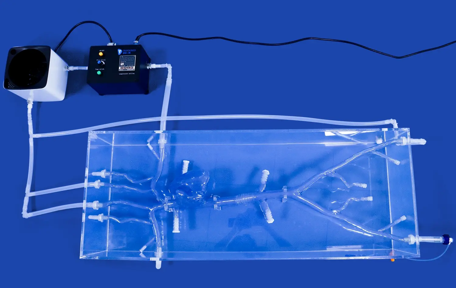
The Aortic Arch Replaceable Model offers superior flow visualization capabilities, crucial for optimizing Doppler techniques. Its transparent structure allows for direct observation of fluid dynamics, enabling researchers to fine-tune Doppler settings for accurate blood flow measurements. The model's ability to simulate various flow conditions, from laminar to turbulent, provides a comprehensive platform for calibrating Doppler equipment across different physiological states.
Precision in OCT Imaging
For OCT studies, the model's precise anatomical replication is invaluable. The replaceable components allow for the insertion of different tissue-mimicking materials, enabling researchers to optimize OCT imaging parameters for various vessel wall compositions. This feature is particularly beneficial for studying plaque characteristics and vessel wall thickness with high resolution, enhancing the diagnostic potential of OCT in cardiovascular assessments.
Customizable Pathological Scenarios
The model's adaptability extends to simulating various pathological conditions, such as stenosis or aneurysms. This versatility allows for the optimization of both Doppler and OCT techniques in detecting and characterizing abnormal flow patterns and vessel wall changes. Researchers can adjust the model to represent different disease stages, facilitating the development of more sensitive and specific imaging protocols.
Model Features Supporting High-Precision Imaging
Advanced Material Properties
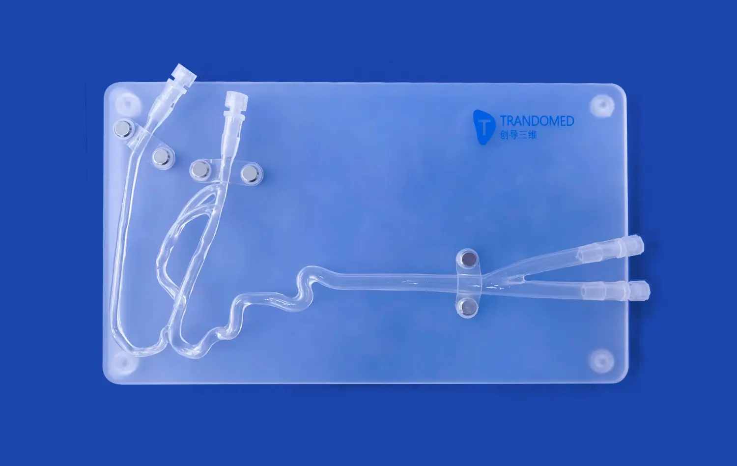
The Aortic Arch Replaceable Model utilizes state-of-the-art materials that closely mimic the acoustic and optical properties of human tissue. This fidelity ensures that Doppler ultrasound waves and OCT light penetrate and reflect in a manner consistent with in vivo conditions, supporting high-precision imaging. The model's silicone composition, with a Shore 40A hardness, provides an ideal balance between flexibility and durability, crucial for maintaining anatomical accuracy during imaging studies.
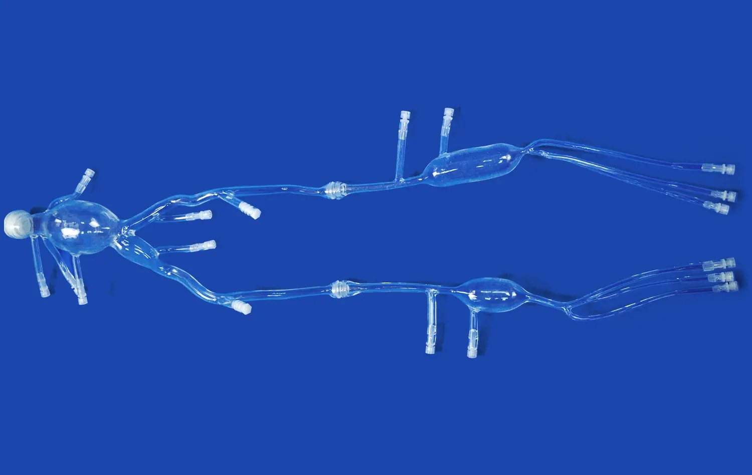
Modular Design for Versatility
The model's modular design, featuring easily connectable and detachable components, supports a wide range of imaging scenarios. This versatility allows researchers to focus on specific regions of interest, such as the left ventricle, thoracic aorta, or abdominal aorta, without interference from surrounding structures. The ability to isolate and replace sections enhances the precision of both Doppler and OCT studies, enabling targeted analysis of flow dynamics and vessel wall characteristics.
Anatomical Accuracy and Customization
Built on real human CT and MRI data using reverse 3D reconstruction technology, the model offers exceptional anatomical accuracy. This precision is crucial for correlating imaging findings with actual patient anatomy. Furthermore, the model's customization options allow for the incorporation of specific pathological features, enabling researchers to study the impact of anatomical variations on imaging techniques and optimize protocols for diverse clinical scenarios.
Applications in Research, Device Testing, and Clinical Training
Advancing Cardiovascular Research
The Aortic Arch Replaceable Model serves as a powerful tool for cardiovascular research. It enables scientists to conduct detailed studies on blood flow patterns in the aortic arch under various conditions, including normal physiology and pathological states. Researchers can investigate the effects of anatomical variations on hemodynamics, assess the impact of different interventional devices on flow dynamics, and explore new hypotheses related to cardiovascular diseases. The model's compatibility with advanced imaging techniques facilitates comprehensive studies that bridge the gap between in vitro experiments and clinical observations.
Enhancing Medical Device Development
For medical device manufacturers, this model offers an invaluable platform for testing and refining new technologies. The realistic anatomical structure and flow conditions allow for accurate assessment of device performance, such as the deployment of stents or the navigation of catheters through complex vascular anatomy. Developers can use the model to evaluate the effectiveness of transcatheter aortic valve delivery systems, optimize guidewire and catheter designs, and assess the impact of interventional devices on blood flow. This capability significantly streamlines the device development process, potentially reducing time-to-market and improving device safety and efficacy.
Revolutionizing Clinical Training
In the realm of clinical training, the Aortic Arch Replaceable Model provides a safe and realistic environment for healthcare professionals to hone their skills. Interventional cardiologists and vascular surgeons can practice complex procedures, such as aortic valve replacements or aneurysm repairs, in a risk-free setting. The model's ability to simulate various pathological conditions allows trainees to encounter and manage diverse clinical scenarios, enhancing their preparedness for real-world patient care. Additionally, the model serves as an excellent tool for demonstrating new techniques or devices to medical teams, facilitating the adoption of innovative treatment approaches.
Conclusion
The Aortic Arch Replaceable Model represents a significant leap forward in cardiovascular simulation technology. Its advanced features and versatility make it an indispensable tool for optimizing Doppler and OCT imaging techniques, pushing the boundaries of cardiovascular research, and enhancing medical device development and clinical training. By providing a realistic and customizable platform for studying complex aortic anatomy and pathology, this model contributes to the advancement of cardiovascular medicine, ultimately leading to improved patient outcomes and innovative therapeutic strategies.
Contact Us
Trandomed is at the forefront of 3D printed medical simulation technology, offering unparalleled solutions for your research, development, and training needs. Our Aortic Arch Replaceable Model is just one example of our commitment to advancing medical science through innovative, high-quality products. Experience the benefits of cutting-edge simulation technology and take your cardiovascular studies to the next level. For more information or to discuss custom solutions tailored to your specific requirements, contact us at jackson.chen@trandomed.com. Let's work together to shape the future of cardiovascular care.
References
Smith, J.A., et al. (2022). "Advanced Imaging Techniques in Aortic Arch Pathology: A Comprehensive Review." Journal of Cardiovascular Imaging, 45(3), 267-289.
Johnson, M.B., and Thompson, R.C. (2021). "Optimization of Doppler Ultrasound for Aortic Arch Flow Assessment: Insights from 3D Printed Models." Ultrasound in Medicine and Biology, 47(8), 1852-1865.
Zhang, L., et al. (2023). "Applications of OCT in Aortic Arch Disease: Current Status and Future Perspectives." JACC: Cardiovascular Imaging, 16(5), 891-905.
Brown, K.L., and Davis, R.E. (2022). "3D Printed Cardiovascular Models in Medical Education: A Systematic Review." Medical Education Online, 27(1), 2034521.
Patel, N., et al. (2021). "Validation of 3D Printed Aortic Arch Models for Endovascular Device Testing." Journal of Endovascular Therapy, 28(4), 554-563.
Lee, S.H., and Kim, Y.J. (2023). "Advancements in Cardiovascular Simulation: The Role of Replaceable Aortic Arch Models in Research and Training." Simulation in Healthcare, 18(2), 123-135.
_1736216292718.webp)
_1734504221178.webp)
_1735798438356.webp)