What Makes These Models Suitable for Experimental Validation?
Anatomical Accuracy and Realism
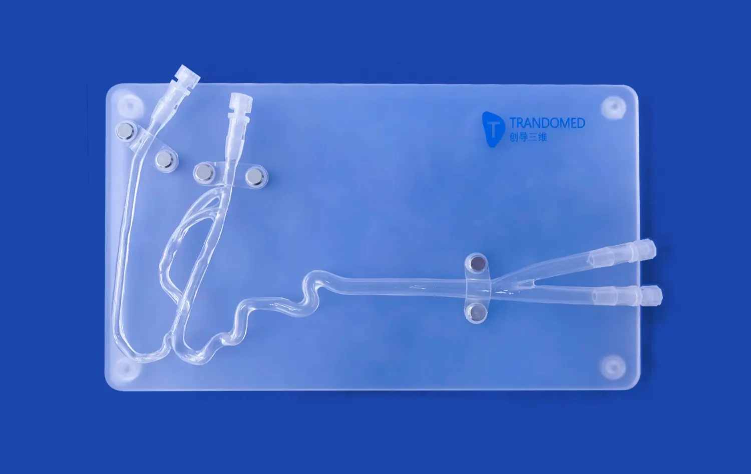
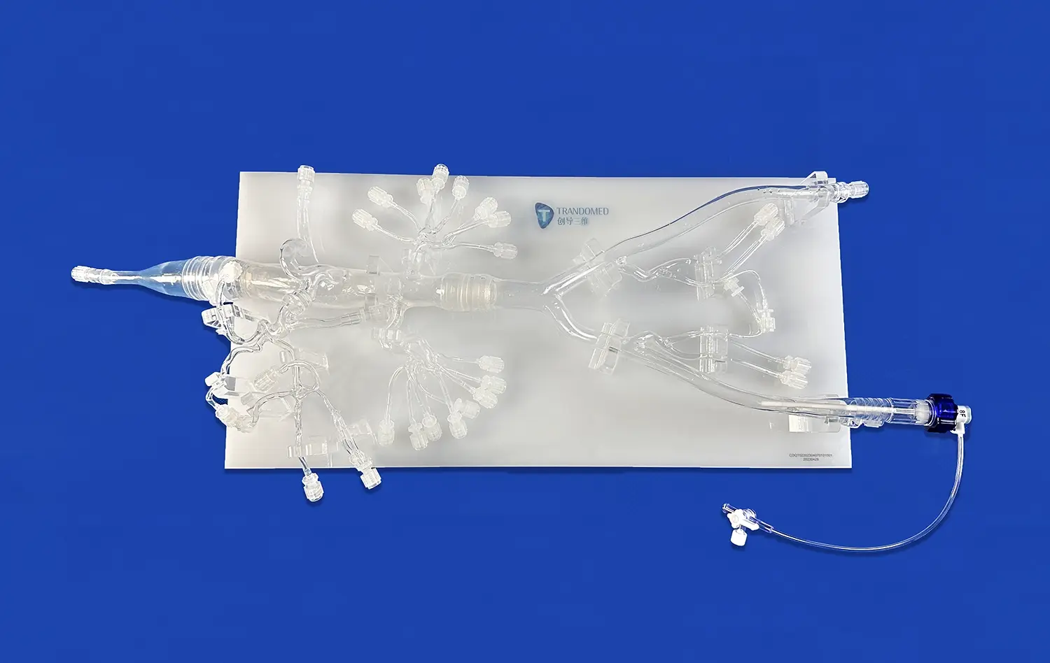
The effectiveness of aortic dissection models in experimental validation stems from their exceptional anatomical accuracy. These models, such as the Aortic Dissection Model (XXK004D), are meticulously crafted to replicate the intricate structure of the aorta and its major branches. By incorporating details like the femoral artery, iliac artery, abdominal aorta, renal arteries, celiac trunk, thoracic aorta, aortic arch, ascending aorta, and subclavian artery, these models provide a comprehensive representation of the vascular system involved in aortic dissections.
The centerpiece of these models is the realistic depiction of the aortic dissection lesion, typically located in the thoracic aorta segment. This level of detail allows researchers to study the initiation, propagation, and consequences of aortic dissections with unprecedented clarity. The ability to visualize and manipulate these anatomical features makes these models indispensable for validating hypotheses, testing treatment strategies, and assessing the effectiveness of surgical interventions.
Physiological Fidelity
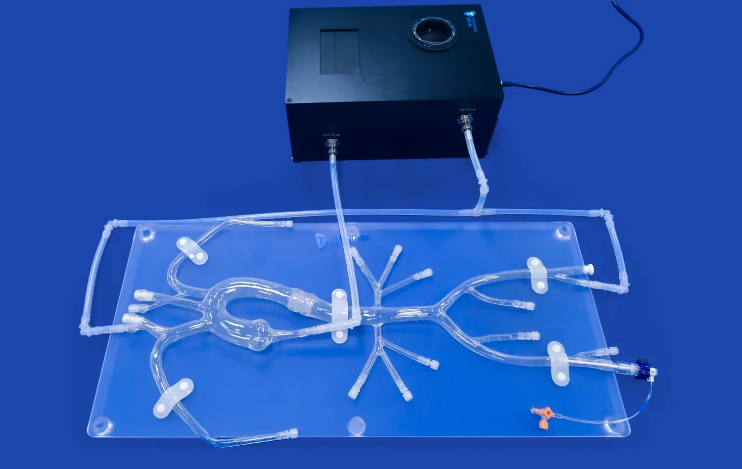
Beyond anatomical accuracy, aortic dissection models excel in replicating the physiological aspects of this condition. Advanced models incorporate features that mimic the hemodynamics of blood flow through the dissected aorta, including the true and false lumens characteristic of aortic dissections. This physiological fidelity allows researchers to study how blood flow patterns change in the presence of a dissection, how these changes affect organ perfusion, and how various interventions might alter these dynamics.
Some models even incorporate pulsatile flow systems, enabling the simulation of cardiac cycles and their effects on the dissected aorta. This level of physiological realism is crucial for validating computational fluid dynamics models, testing the performance of endovascular devices, and assessing the impact of surgical repairs on blood flow patterns.
Versatility and Customization
The versatility of aortic dissection models significantly enhances their suitability for experimental validation. Many models, like those offered by Trando 3D Medical Technology Co., Ltd, can be customized to represent different types of aortic arch anatomies (Type I, II, or III) or to include additional features such as thoracic or abdominal aortic aneurysms. This customization capability allows researchers to tailor the models to specific research questions or patient scenarios, increasing the relevance and applicability of their experimental findings.
Furthermore, the ability to create models based on diverse data file formats, including CT, CAD, STL, STP, and STEP, enables researchers to develop patient-specific models. This personalization is particularly valuable for validating treatment strategies in complex cases or for conducting pre-surgical planning simulations.
Simulated Environments for Evaluating Device Safety and Performance
Replicating In Vivo Conditions
Aortic dissection models provide an unparalleled simulated environment for evaluating the safety and performance of medical devices. These models create a controlled setting that closely mimics the in vivo conditions encountered during actual aortic dissection cases. By replicating the physical and mechanical properties of the aortic wall, including its elasticity and strength, these models allow researchers to assess how devices interact with the vessel tissue under various conditions.
The ability to simulate different stages of aortic dissection, from acute to chronic, enables comprehensive testing of devices across various clinical scenarios. This versatility is crucial for evaluating the adaptability and efficacy of new technologies in managing diverse presentations of aortic dissections.
Testing Endovascular Devices
For endovascular devices such as stent grafts and dissection-specific stents, aortic dissection models serve as an ideal platform for performance evaluation. These models allow researchers to assess crucial factors such as device deliverability, deployment accuracy, and seal integrity. The realistic anatomical features of the models, including the curvature of the aortic arch and the presence of branch vessels, provide a challenging yet accurate environment for testing device navigation and positioning.
Moreover, the incorporation of flow dynamics in some models enables the evaluation of how these devices affect blood flow patterns and pressures within the true and false lumens. This capability is particularly valuable for assessing the potential of endovascular interventions to promote false lumen thrombosis or to ensure adequate perfusion to vital organs.
Surgical Technique Validation
Beyond device testing, aortic dissection models play a crucial role in validating surgical techniques. These models provide a risk-free environment for surgeons to practice and refine complex procedures such as aortic root replacements, arch reconstructions, or hybrid repair techniques. The ability to replicate various anatomical variations and dissection patterns allows for the development and validation of tailored surgical approaches for challenging cases.
Additionally, these models serve as excellent tools for assessing the effectiveness of novel surgical materials, such as synthetic grafts or biological patches, in repairing aortic dissections. The realistic tissue properties of advanced models enable researchers to evaluate factors like suture retention strength, graft integration, and overall repair durability.
Advancing Clinical Research Through Realistic Vascular Models
Enhancing Understanding of Disease Progression
Realistic vascular models, particularly those replicating aortic dissections, are instrumental in advancing clinical research by providing unprecedented insights into disease progression. These models allow researchers to study the complex interplay between hemodynamic forces and vessel wall mechanics that contribute to the initiation and propagation of aortic dissections. By simulating various physiological conditions, such as hypertension or connective tissue disorders, researchers can investigate how these factors influence the risk and progression of aortic dissections.
The ability to create longitudinal studies using these models offers a unique opportunity to observe and analyze the natural history of aortic dissections over time. This capability is particularly valuable for understanding the factors that contribute to aneurysmal degeneration, false lumen expansion, or spontaneous healing in some cases. Such insights are crucial for developing more effective risk stratification strategies and tailored treatment approaches.
Facilitating Comparative Studies
Aortic dissection models excel in facilitating comparative studies of different treatment modalities. Researchers can use identical model setups to evaluate and compare the outcomes of various interventional approaches, such as medical management, endovascular repair, or open surgical techniques. This standardized approach allows for more objective assessments of treatment efficacy, potentially leading to the development of more evidence-based clinical guidelines.
Furthermore, these models enable the comparison of different device designs or surgical techniques under controlled conditions. For instance, researchers can assess how various stent graft configurations affect false lumen thrombosis or how different surgical repair techniques impact long-term aortic remodeling. Such comparative studies are invaluable for refining existing therapies and guiding the development of novel treatment strategies.
Bridging the Gap Between Bench and Bedside
Perhaps one of the most significant contributions of realistic vascular models to clinical research is their role in bridging the gap between bench-side experiments and bedside applications. These models serve as an essential intermediate step in the translational research pipeline, allowing for the validation of concepts and technologies before progressing to animal studies or human trials.
By providing a platform that closely mimics human anatomy and physiology, aortic dissection models reduce the reliance on animal testing and potentially accelerate the development of new therapies. They offer a cost-effective and ethically sound approach to early-stage research, enabling iterative refinement of ideas and technologies before committing to more resource-intensive clinical studies.
Moreover, these models play a crucial role in medical education and training. They provide a hands-on learning experience for medical students, residents, and practicing clinicians, enhancing their understanding of aortic dissection pathophysiology and treatment approaches. This educational aspect contributes to the overall advancement of clinical practice by ensuring that future generations of healthcare providers are well-equipped to manage this complex condition.
Conclusion
Aortic dissection models have emerged as indispensable tools in preclinical testing and research, offering unparalleled opportunities for advancing our understanding and treatment of this critical cardiovascular condition. Their anatomical accuracy, physiological fidelity, and versatility make them ideal for experimental validation, device testing, and clinical research. As technology continues to evolve, these models will undoubtedly play an increasingly crucial role in driving innovation, improving patient outcomes, and shaping the future of cardiovascular medicine.
Contact Us
To explore how Trandomed's advanced aortic dissection models can enhance your research or training programs, contact us at jackson.chen@trandomed.com. Our team is dedicated to providing customized solutions that meet your specific needs, ensuring you have the most realistic and effective tools for your cardiovascular studies.
_1736215128474.webp)
_1736216292718.webp)
_1732863962417.webp)