How Do Models Drive Innovation in Cardiovascular Devices?
Anatomically accurate aortic dissection models serve as a catalyst for innovation in cardiovascular devices by providing a platform for testing, refinement, and validation. These models bridge the gap between conceptual design and clinical application, allowing researchers and engineers to iterate and optimize their inventions in a controlled environment.
Enhancing Device Design and Performance
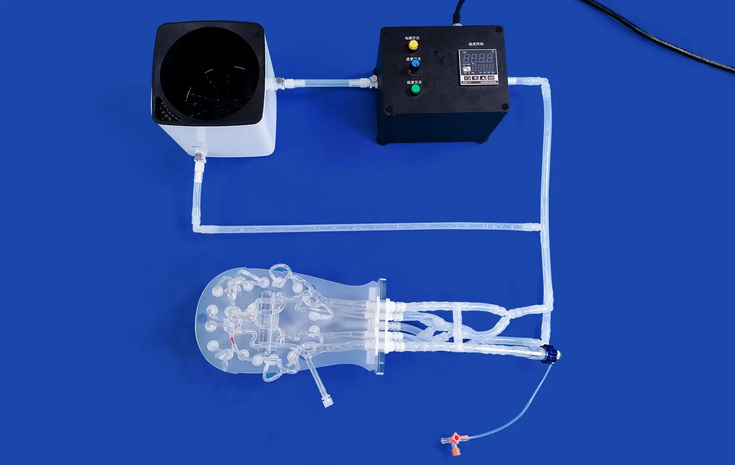
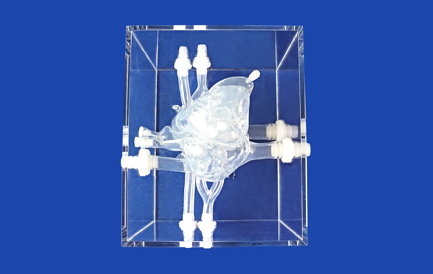
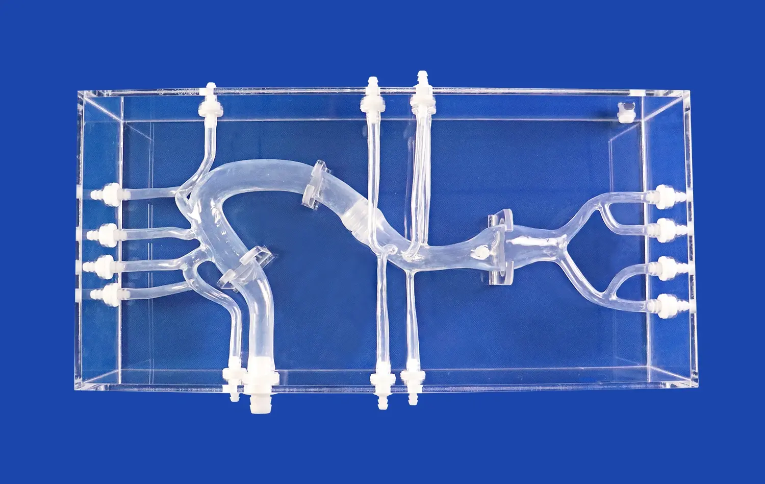
The intricate details captured in high-fidelity aortic models, such as the aortic dissection model (XXK004D) from Trandomed, enable developers to fine-tune the design and functionality of cardiovascular devices. By replicating the complex anatomy of the aorta, including the femoral artery, iliac artery, and aortic arch, these models allow for precise evaluation of device performance under various physiological conditions.
Simulating Challenging Scenarios
Advanced aortic dissection models can simulate a wide range of pathological conditions, from simple type A dissections to more complex scenarios involving multiple entry tears or aneurysmal dilatation. This versatility allows device manufacturers to test their products in diverse clinical situations, ensuring robust performance across a spectrum of patient cases.
Accelerating the Innovation Cycle
By providing a reliable testbed for new ideas, aortic models significantly reduce the time and resources required to bring innovative cardiovascular devices from concept to market. Developers can rapidly prototype, test, and refine their designs without the need for extensive animal studies or high-risk human trials in the early stages of development.
Supporting the Development of Novel Stent-Graft Technologies
The evolution of stent-graft technologies for treating aortic dissections has been greatly facilitated by the use of sophisticated aortic models. These models provide an ideal platform for evaluating new designs, materials, and deployment techniques, ultimately leading to more effective and less invasive treatment options for patients.
Optimizing Stent-Graft Design
Realistic aortic dissection models allow engineers to assess the performance of various stent-graft designs in a controlled setting. Factors such as radial force, flexibility, and conformability can be evaluated and optimized to ensure optimal sealing and long-term durability in the challenging environment of a dissected aorta.
Improving Deployment Techniques
The complex anatomy of aortic dissections, particularly in the aortic arch and descending thoracic aorta, presents significant challenges for stent-graft deployment. Using anatomically accurate models, clinicians and device manufacturers can refine deployment techniques, develop new delivery systems, and address common complications such as endoleaks or malpositioning.
Evaluating Novel Materials and Coatings
Aortic models provide an excellent platform for testing innovative materials and surface treatments designed to enhance the biocompatibility and long-term performance of stent-grafts. Researchers can assess the interaction between these novel materials and simulated aortic tissue, paving the way for next-generation devices with improved healing characteristics and reduced risk of complications.
Bridging R&D and Clinical Practice Through Anatomical Validation
Aortic dissection models serve as a crucial link between research and development efforts and clinical practice, offering a means of validating new techniques and technologies before their introduction into patient care. This bridge ensures that innovations are thoroughly tested and refined before reaching the operating room, ultimately leading to safer and more effective treatments for aortic dissections.
Enhancing Preoperative Planning
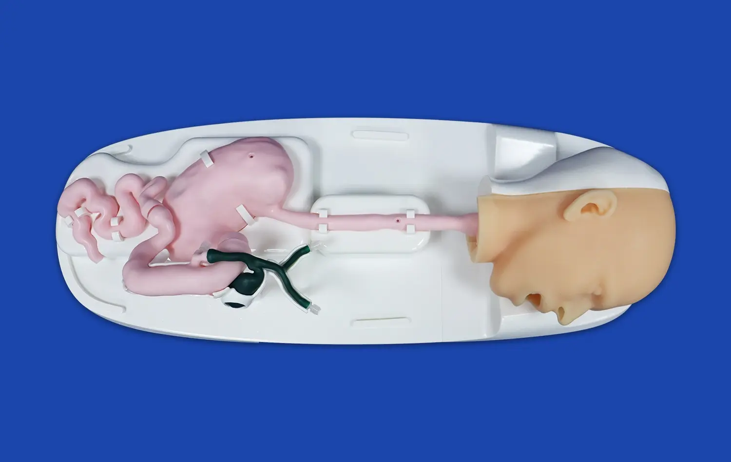
Patient-specific aortic dissection models - produced through high-resolution imaging and 3D printing - provide an accurate and tactile representation of an individual's vascular anatomy. These models allow surgical teams to examine key anatomical challenges in advance, such as tortuous pathways, variable tear locations, or branch vessel involvement. Surgeons can simulate the complete procedure beforehand, refining access strategies and device placement. This proactive planning enhances procedural safety, reduces intraoperative decision-making pressure, and contributes to more efficient, personalized interventions in the operating room.
Facilitating Hands-On Training
Aortic dissection models like the XXK004D provide a high-fidelity simulation environment for skill development without patient risk. These anatomically correct models replicate realistic flow dynamics and tissue behavior, offering valuable opportunities for practicing catheter navigation, endograft deployment, and complex repair techniques. Trainees and seasoned professionals alike benefit from repeated, hands-on exposure to diverse clinical scenarios. The ability to make and learn from mistakes in a risk-free setting not only improves procedural competency but also builds confidence and enhances team readiness in high-pressure clinical environments.
Validating Computational Models
Physical aortic dissection models offer a critical benchmark for validating computational simulations used in cardiovascular device development and surgical planning. Researchers can compare simulated flow patterns, pressure gradients, and wall stress predictions against in vitro data obtained from physical models. This iterative validation process helps improve the accuracy, realism, and clinical relevance of virtual models. As a result, digital simulations become more reliable tools for predicting treatment outcomes, optimizing device design, and guiding personalized care strategies in patients with complex aortic pathologies.
Conclusion
The use of advanced aortic dissection models in cardiovascular innovation represents a significant leap forward in our ability to develop, refine, and validate new technologies for treating this complex and life-threatening condition. From enhancing device design and performance to supporting the development of novel stent-graft technologies and bridging the gap between R&D and clinical practice, these models are instrumental in driving progress in the field of cardiovascular medicine.As we continue to push the boundaries of what's possible in treating aortic dissections, the role of high-fidelity anatomical models will only grow in importance. By providing a realistic and versatile platform for innovation, these models are helping to shape the future of cardiovascular care, promising improved outcomes and new hope for patients facing this challenging condition.
Contact Us
For those at the forefront of cardiovascular innovation, partnering with a leading provider of advanced medical simulation models is crucial. Trandomed offers state-of-the-art aortic dissection models that combine anatomical accuracy with versatility, supporting your research and development efforts every step of the way. To learn more about how our models can accelerate your innovation pipeline and improve patient outcomes, contact us at jackson.chen@trandomed.com.
1_1732869849284.webp)