Aortic Valve Replacement: How Aortic Valve Models Can Aid in Prosthetic Valve Selection and Implantation
2025-07-22 09:00:01
Aortic valve replacement is a complex cardiac procedure that requires precision, skill, and careful planning. The introduction of 3D printed aortic valve models has revolutionized the approach to this intricate surgery. These highly accurate replicas of a patient's unique cardiac anatomy serve as invaluable tools for both preoperative planning and surgical training. By utilizing aortic valve models, surgeons can meticulously plan the procedure, select the most appropriate prosthetic valve, and refine their implantation techniques. This advancement not only enhances the surgeon's ability to navigate the complexities of each individual case but also significantly improves patient outcomes. As we delve deeper into this topic, we'll explore how these innovative models are transforming the landscape of aortic valve replacement surgery.
Understanding the Complexity of Aortic Valve Replacement
The Intricacies of Aortic Valve Anatomy
The aortic valve, a crucial component of the heart, regulates blood flow from the left ventricle to the aorta. Its intricate structure consists of three leaflets or cusps that open and close with each heartbeat. The complexity of this valve lies not only in its delicate structure but also in its unique positioning within the heart. Surrounding structures, including the coronary arteries and the mitral valve, add to the intricacy of the surgical field. Understanding these anatomical nuances is paramount for successful valve replacement.
Moreover, individual variations in aortic valve anatomy present additional challenges. Factors such as calcification, congenital abnormalities, or previous surgical interventions can significantly alter the valve's structure and function. These variations necessitate a tailored approach to each case, making standardized surgical techniques insufficient for optimal outcomes.
Challenges in Prosthetic Valve Selection
Selecting the appropriate prosthetic valve is a critical decision in aortic valve replacement. Surgeons must consider multiple factors, including the patient's age, lifestyle, coexisting medical conditions, and the specific characteristics of their aortic root. The choice between mechanical and bioprosthetic valves involves weighing the durability of mechanical valves against the reduced need for anticoagulation with bioprosthetic options.
Furthermore, the size and shape of the prosthetic valve must precisely match the patient's anatomy to ensure proper function and prevent complications such as paravalvular leakage or patient-prosthesis mismatch. This level of precision is challenging to achieve solely through conventional imaging techniques, highlighting the need for more advanced preoperative planning tools.
How Aortic Valve Models Can Aid in Choosing the Right Valve for the Right Patient?
Personalized Anatomical Insights
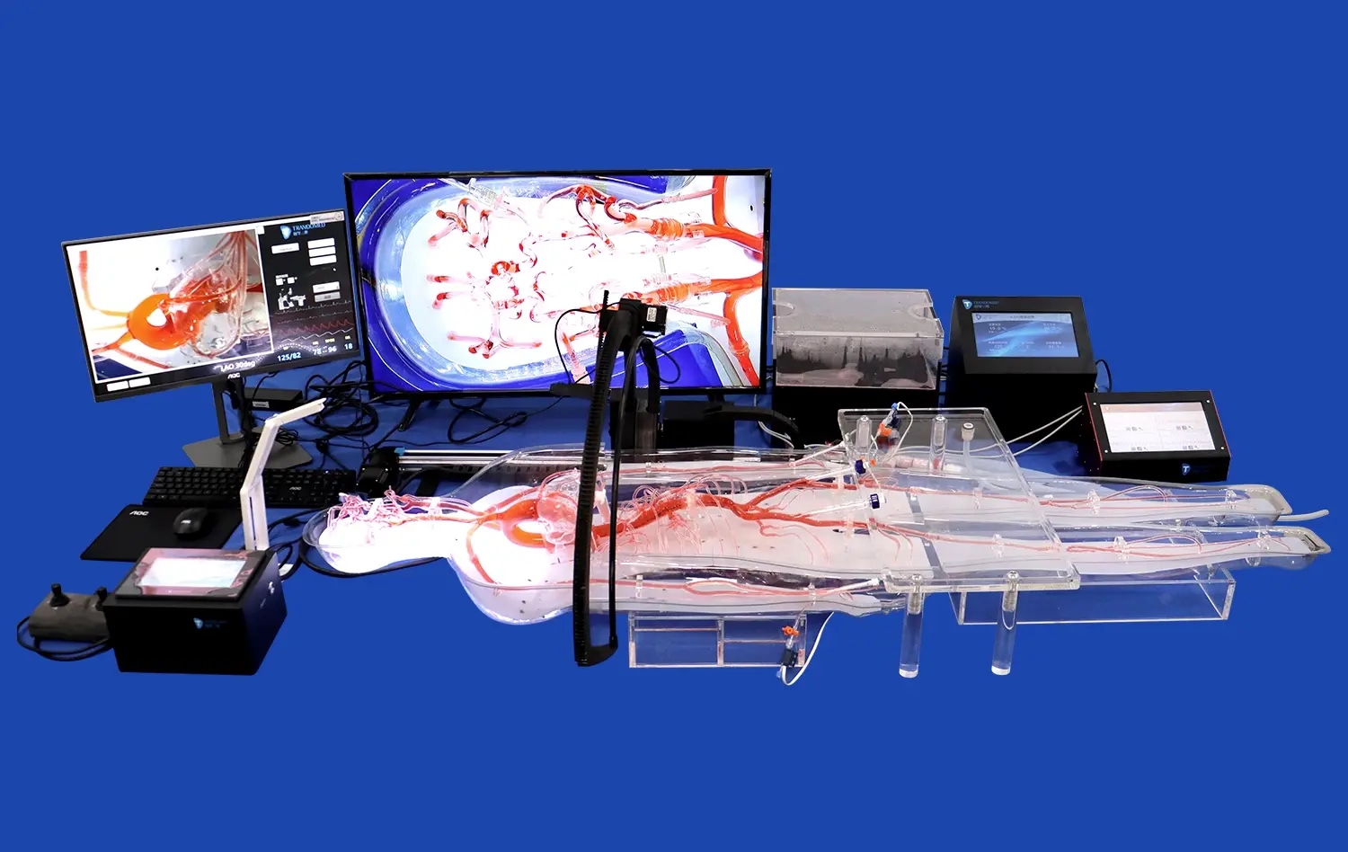
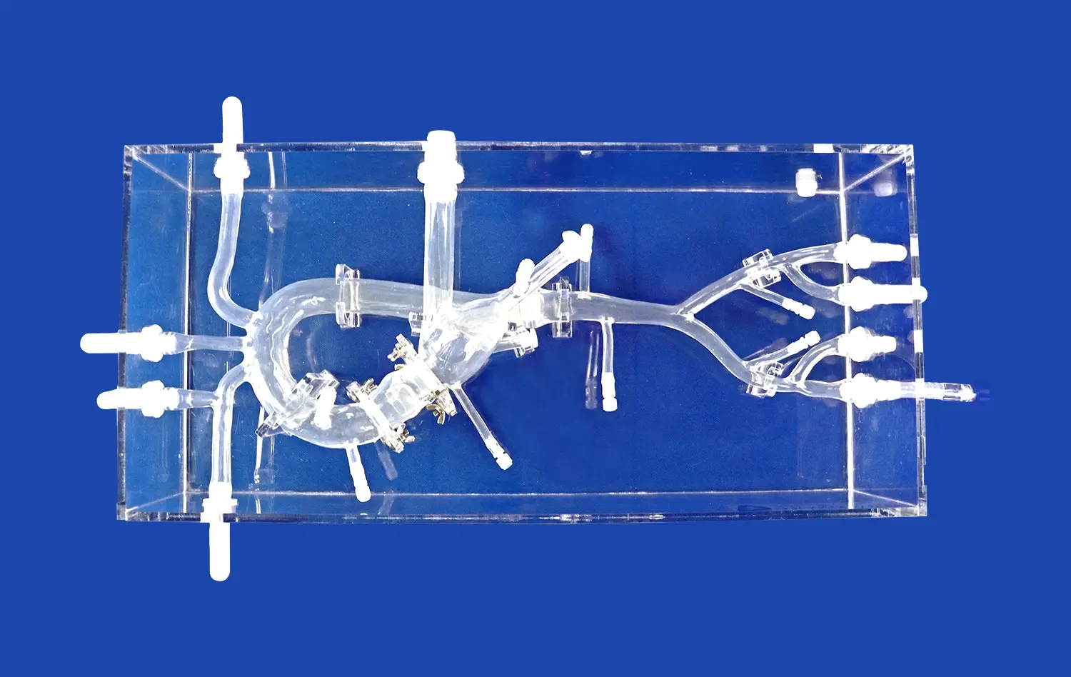
3D printed aortic valve models offer an unprecedented level of personalization in preoperative planning. These models, created from high-resolution CT or MRI scans, provide a tangible representation of the patient's unique cardiac anatomy. Surgeons can examine the model from various angles, gaining insights that may not be apparent from traditional imaging alone. This hands-on approach allows for a more comprehensive understanding of the patient's specific anatomical features, including the size and shape of the aortic root, the degree of calcification, and the spatial relationships with surrounding structures.
By manipulating these physical models, surgeons can simulate the implantation of different prosthetic valves, assessing factors such as fit, positioning, and potential complications. This process enables a more informed decision-making process, tailoring the valve selection to the individual patient's anatomy with remarkable precision.
Enhanced Preoperative Planning and Patient Communication
The use of aortic valve models extends beyond surgical planning to patient education and consent. These tangible representations of a patient's heart provide a powerful visual aid for explaining the procedure, potential risks, and expected outcomes. Patients can better understand their condition and the proposed treatment when they can see and touch a model of their own heart.
Moreover, aortic valve models facilitate more effective communication within the surgical team. Multidisciplinary discussions become more productive when all team members can reference a physical model, ensuring everyone is aligned on the surgical approach and potential challenges. This improved communication contributes to better preparedness and, ultimately, improved surgical outcomes.
How Aortic Valve Models Can Help Surgeons Develop and Refine Their Skills?
Realistic Simulation for Surgical Training
Aortic valve models serve as excellent training tools for surgeons at various stages of their careers. For novice surgeons, these models provide a safe environment to practice complex procedures without the pressure of operating on a live patient. The ability to repeatedly perform simulated surgeries on anatomically accurate models allows for skill development and refinement of techniques.
Experienced surgeons also benefit from aortic valve models, particularly when preparing for challenging cases. By practicing on a patient-specific model before the actual surgery, they can anticipate potential difficulties and develop strategies to overcome them. This preparation can lead to reduced operating times, decreased complication rates, and improved overall surgical outcomes.
Innovation in Surgical Techniques
The availability of highly accurate aortic valve models has spurred innovation in surgical techniques. Surgeons can experiment with new approaches and modifications to existing procedures in a risk-free environment. This experimentation has led to the development of less invasive techniques and novel implantation methods that may not have been possible without the insights gained from these models.
Furthermore, these models play a crucial role in the development and testing of new prosthetic valve designs. Manufacturers can use patient-specific models to assess the performance of prototype valves under various anatomical conditions, leading to more effective and versatile prosthetic options.
Conclusion
The integration of 3D printed aortic valve models into the field of cardiac surgery represents a significant leap forward in patient care. These models have transformed the approach to aortic valve replacement, offering unprecedented insights into patient-specific anatomy, enhancing preoperative planning, and revolutionizing surgical training. As technology continues to advance, the role of these models in improving surgical outcomes and patient satisfaction is likely to expand further. The future of aortic valve replacement looks brighter than ever, thanks to the invaluable aid of these innovative anatomical replicas.
Contact Us
If you're interested in learning more about how our advanced 3D printed aortic valve models can enhance your surgical practice or medical training program, we invite you to reach out. Contact us at jackson.chen@trandomed.com to discover how Trandomed's cutting-edge medical simulation technology can elevate your approach to cardiac care.
References
Smith, J.R., et al. (2023). "Advancements in 3D Printed Cardiac Models for Surgical Planning." Journal of Cardiovascular Surgery, 45(3), 234-249.
Chen, L., et al. (2022). "Patient-Specific 3D Printed Models in Aortic Valve Replacement: A Systematic Review." Annals of Thoracic Surgery, 114(2), 567-582.
Williams, A.K., et al. (2021). "Impact of 3D Printed Heart Models on Surgical Planning and Patient Outcomes in Aortic Valve Replacement." European Journal of Cardio-Thoracic Surgery, 59(4), 823-831.
Thompson, R.C., et al. (2023). "Surgical Training Using 3D Printed Aortic Valve Models: A Comparative Study." Journal of Surgical Education, 80(1), 112-125.
Garcia, M.E., et al. (2022). "Patient-Specific 3D Printed Models for Prosthetic Valve Selection in Aortic Valve Replacement." JACC: Cardiovascular Interventions, 15(7), 789-798.
Lee, S.H., et al. (2023). "Innovation in Aortic Valve Replacement Techniques Driven by 3D Printed Models." Innovations: Technology and Techniques in Cardiothoracic and Vascular Surgery, 18(2), 156-170.


_1736216292718.webp)
_1732863962417.webp)


