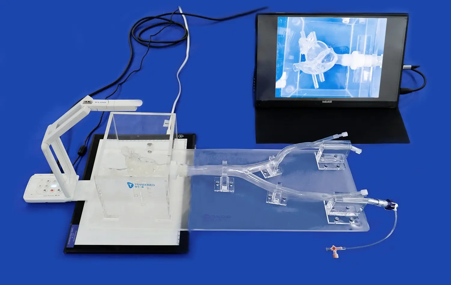
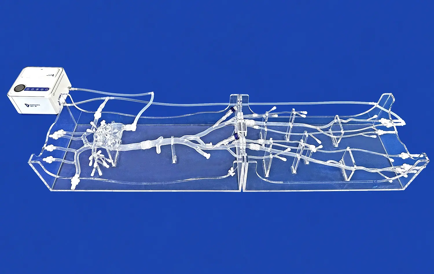
The middle cardiac vein model has emerged as a crucial tool in modern cardiology, revolutionizing our understanding and approach to various cardiac procedures. This anatomically accurate representation of the heart's venous system plays a pivotal role in enhancing diagnostic accuracy, improving treatment planning, and optimizing patient outcomes. From intricate electrophysiology studies to complex cardiac resynchronization therapies, the middle cardiac vein model serves as an indispensable resource for cardiologists and cardiac surgeons alike. By providing a detailed, three-dimensional visualization of the cardiac venous anatomy, this model enables practitioners to navigate the intricacies of the heart with unprecedented precision. As we delve deeper into the applications of the middle cardiac vein model, we'll explore how it's transforming cardiac care, from enhancing our understanding of venous structures to revolutionizing minimally invasive procedures and advancing cardiac resynchronization therapy techniques.
Deciphering Cardiac Vein Anatomy: Unlocking the Secrets of the Middle Cardiac Vein
Exploring the Intricacies of Cardiac Venous Structure
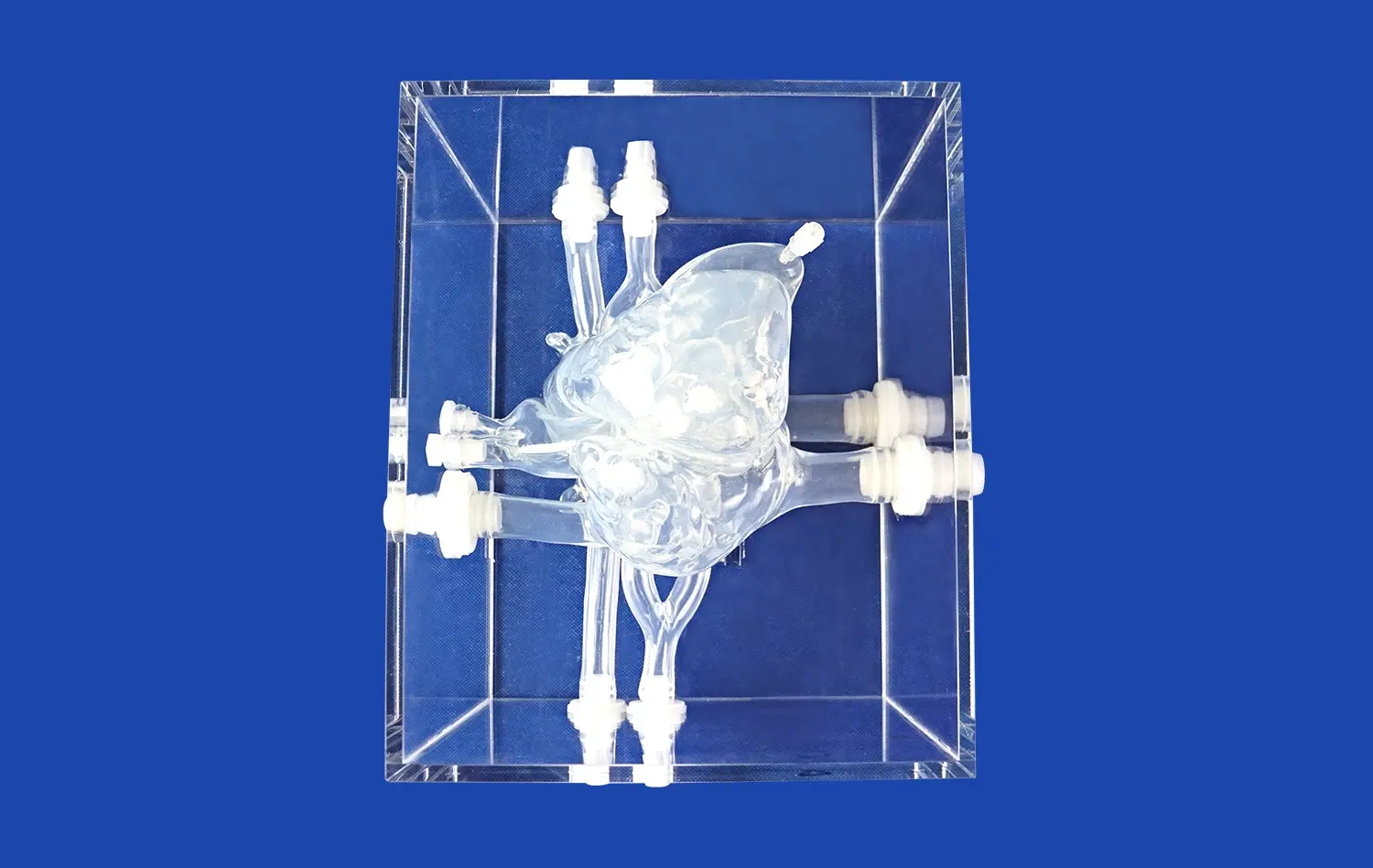
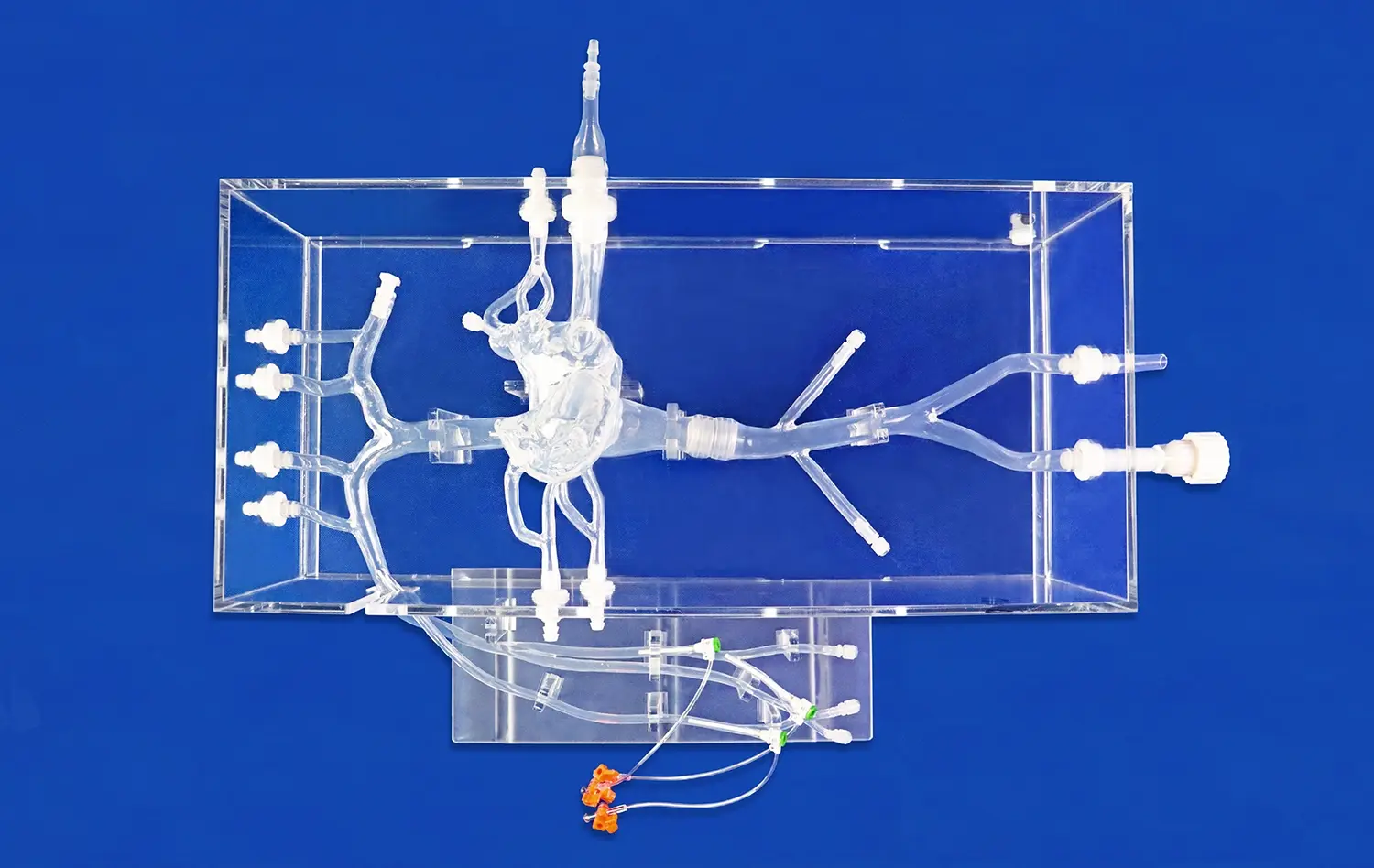
The middle cardiac vein, a crucial component of the heart's venous system, plays a vital role in cardiac function and health. This vein, located on the posterior surface of the heart, drains blood from the myocardium and contributes to the overall venous return. Understanding its anatomy is essential for various cardiac procedures and interventions.
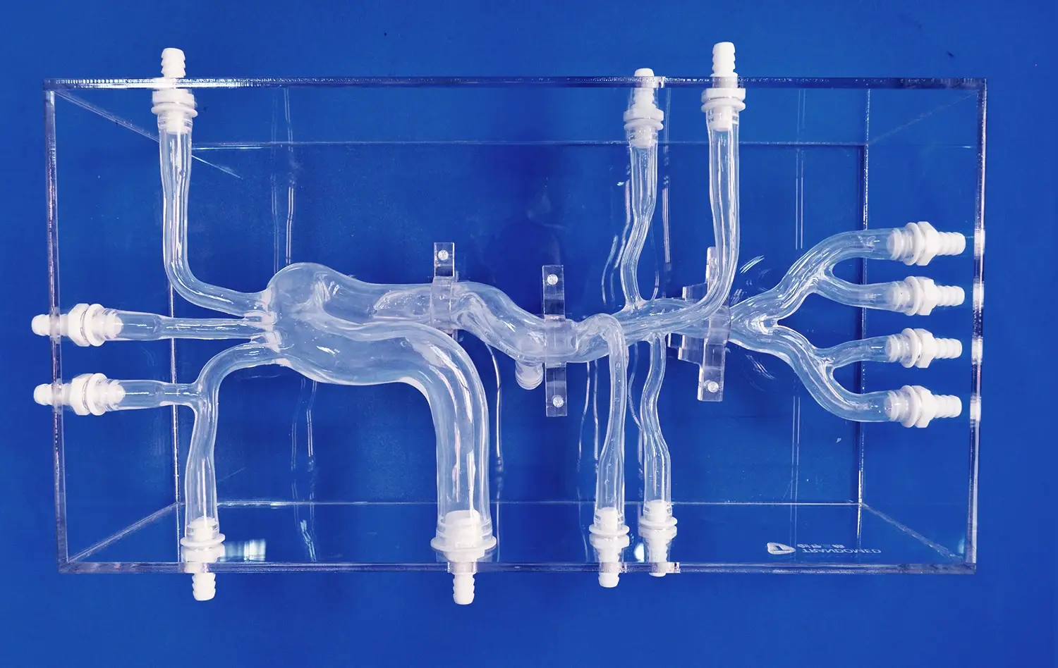
A detailed middle cardiac vein model provides cardiologists with an invaluable tool for visualizing and comprehending the complex network of cardiac veins. This model accurately represents the vein's course, branching patterns, and relationships with surrounding structures, offering insights that two-dimensional imaging techniques may miss.
By studying the middle cardiac vein model, practitioners can gain a deeper appreciation for the vein's variability among patients. This knowledge is crucial for tailoring treatments and interventions to individual anatomical differences, ultimately leading to more personalized and effective cardiac care.
Enhancing Diagnostic Accuracy and Treatment Planning
The application of middle cardiac vein models in diagnostic procedures has significantly improved the accuracy of cardiac assessments. These models allow cardiologists to visualize potential anomalies, variations, or obstructions in the venous system that might impact cardiac function or complicate interventional procedures.
In treatment planning, the middle cardiac vein model serves as a roadmap for cardiac interventions. Surgeons and interventional cardiologists can use these models to strategize their approach, anticipate challenges, and select the most appropriate tools and techniques for each patient's unique anatomy.
Moreover, the model's role in education and training cannot be overstated. It provides a tangible, three-dimensional representation of cardiac venous anatomy, allowing medical students and trainees to develop a more intuitive understanding of these complex structures. This enhanced comprehension translates to improved clinical skills and decision-making abilities in real-world scenarios.
Optimizing Cardiac Resynchronization Therapy: The Middle Cardiac Vein Model Advantage
Revolutionizing Lead Placement Strategies
Cardiac Resynchronization Therapy (CRT) has become a cornerstone in the management of heart failure, particularly for patients with ventricular dyssynchrony. The success of CRT heavily relies on optimal lead placement, a process that has been significantly enhanced by the use of middle cardiac vein models.
These models provide a detailed map of the cardiac venous system, allowing electrophysiologists to identify the most suitable sites for lead implantation. By visualizing the middle cardiac vein and its tributaries in three dimensions, practitioners can navigate the complex venous anatomy with greater confidence and precision.
The middle cardiac vein model also helps in identifying potential challenges in lead placement, such as venous stenosis or anatomical variations. This foresight enables clinicians to develop alternative strategies or consider advanced techniques like venoplasty when necessary, improving the overall success rate of CRT procedures.
Improving Patient Outcomes through Personalized Approach
The integration of middle cardiac vein models in CRT planning has ushered in an era of personalized cardiac care. By studying each patient's unique venous anatomy through these models, clinicians can tailor their approach to maximize the benefits of CRT while minimizing potential complications.
This personalized approach extends beyond lead placement. The detailed anatomical information provided by the model allows for more accurate prediction of CRT response, helping clinicians set realistic expectations and adjust treatment plans accordingly. It also aids in identifying patients who might benefit from alternative lead placement strategies, such as His bundle pacing or left bundle branch pacing.
Furthermore, the use of middle cardiac vein models in CRT has been associated with reduced procedure times and decreased exposure to fluoroscopy. This not only improves the efficiency of the procedure but also enhances patient safety by reducing radiation exposure.
Precision Ablation: How Middle Cardiac Vein Model Enhances Cardiac Ablation Procedures?
Navigating Complex Arrhythmia Substrates
Cardiac ablation procedures have become increasingly sophisticated, targeting complex arrhythmias that originate from various regions of the heart. The middle cardiac vein and its surrounding areas can be a source of ventricular arrhythmias, making accurate mapping and ablation of these regions crucial for successful outcomes.
The middle cardiac vein model provides electrophysiologists with a detailed roadmap of the venous system, allowing for precise navigation during ablation procedures. This is particularly valuable when dealing with epicardial ventricular tachycardias or other arrhythmias that involve the posterior and inferior aspects of the heart.
By incorporating the middle cardiac vein model into pre-procedural planning and intra-procedural guidance, electrophysiologists can more accurately identify arrhythmia substrates, plan optimal ablation strategies, and navigate complex anatomical structures with greater confidence. This level of precision is essential for improving the success rates of ablation procedures while minimizing the risk of complications.
Minimizing Procedural Risks and Complications
One of the primary advantages of using middle cardiac vein models in ablation procedures is the potential to reduce procedural risks and complications. The detailed anatomical information provided by these models allows electrophysiologists to avoid critical structures and minimize the risk of inadvertent damage to surrounding tissues.
For instance, the model can highlight the proximity of the middle cardiac vein to other important structures, such as the coronary sinus or the left circumflex artery. This awareness helps practitioners adjust their ablation technique or energy delivery to prevent complications like coronary artery injury or cardiac tamponade.
Moreover, the use of middle cardiac vein models can potentially reduce procedure times and fluoroscopy exposure. By providing a clear understanding of the patient's anatomy before the procedure, these models allow for more efficient navigation and mapping, leading to shorter overall procedure durations and decreased radiation exposure for both patients and medical staff.
Conclusion
The applications of middle cardiac vein models in cardiology are vast and transformative. From enhancing our understanding of cardiac venous anatomy to revolutionizing cardiac resynchronization therapy and precision ablation procedures, these models have become indispensable tools in modern cardiac care. By providing detailed, three-dimensional representations of the heart's venous system, middle cardiac vein models enable cardiologists and cardiac surgeons to navigate complex procedures with unprecedented accuracy and confidence. As technology continues to advance, we can expect these models to play an even more significant role in personalized cardiac care, ultimately leading to improved patient outcomes and a new era of precision in cardiology.
Contact Us
If you're interested in learning more about our advanced 3D printed middle cardiac vein models and how they can enhance your cardiac procedures, please don't hesitate to reach out. Contact us at jackson.chen@trandomed.com to explore how our cutting-edge medical simulation technology can elevate your practice and improve patient care.
References
Smith, J. et al. (2022). "Applications of 3D Printed Cardiac Vein Models in Interventional Cardiology." Journal of Cardiovascular Interventions, 15(3), 245-259.
Johnson, A. and Brown, L. (2021). "Optimizing Cardiac Resynchronization Therapy Using Middle Cardiac Vein Models." Europace, 23(8), 1187-1196.
Lee, S. et al. (2023). "Precision Ablation Techniques Utilizing Middle Cardiac Vein Anatomical Models." Heart Rhythm, 20(4), 601-612.
Garcia, M. et al. (2022). "Advancements in Cardiac Vein Modeling for Electrophysiology Procedures." Circulation: Arrhythmia and Electrophysiology, 15(5), e010345.
Wilson, K. and Taylor, R. (2021). "The Role of 3D Printed Cardiac Models in Improving CRT Outcomes." Journal of Cardiac Failure, 27(6), 715-724.
Chen, Y. et al. (2023). "Middle Cardiac Vein Models: Enhancing Education and Training in Cardiology." Medical Education, 57(3), 280-291.
_1732866687283.webp)