Applications of the Circle of Willis Brain Model in Neurovascular Training
2026-02-04 09:00:03
The circle of Willis brain model has changed neurovascular education by giving medical schools and training facilities a way to learn about the nature of the brain's blood vessels and how to perform difficult treatment procedures. This advanced 3D-printed modeling tool makes a copy of the complicated artery network that brings blood to the brain, complete with abnormalities like aneurysms and stenosis lesions. These high-fidelity models bridge the gap between theoretical knowledge and hands-on clinical competence, changing how healthcare professionals understand and deal with important neurovascular challenges. They can be used to train neurosurgeons for delicate procedures, train emergency response teams for stroke management, or do biomedical research.
Understanding the Circle of Willis and Its Role in Neurovascular Training
The Circle of Willis is one of nature's most beautiful failsafes. It forms a continuous ring of arteries at the base of the brain that keeps blood flowing even when individual veins become blocked. This arterial structure is in the interpeduncular cistern on the bottom surface of the brain. It surrounds important structural features like the optic chiasm and pituitary infundibulum. It's not enough to just remember the names of the blood vessels in this area; you need to be able to picture how the anterior cerebral arteries, internal carotid arteries, posterior cerebral arteries, and communication arteries protect brain cells from ischemic damage.
Anatomical Components and Cerebrovascular Protection
There are five important arterial segments that make up the Circle of Willis. These are the paired anterior cerebral arteries at their A1 segments, the anterior communicating artery that connects them, the distal tips of both internal carotid arteries, the paired posterior cerebral arteries at their P1 segments, and the posterior communicating arteries that link the anterior and posterior circulation. This arrangement makes side paths that change the flow of blood when there are blocks or stenosis in one area. According to research, only 20–25% of people have a fully formed circle with no missing or hypoplastic parts. This is why physical differences are such an important subject for medical students to learn about. A neurovascular brain model lets students experience these differences firsthand. This shows why some people can handle vascular occlusions better than others and how stroke patterns are different for each person based on their genetics.
Why Simulation Models Enhance Neurovascular Education?
Traditional ways of teaching that use textbook graphics or digital pictures can't fully show how brain vessels and the nerve structures around them are connected in three dimensions. Brain models that are very realistic and show the Circle of Willis give you physical, visual, and spatial learning experiences that help you remember and understand more. Medical students can use their fingers to follow blood flow paths, find possible backup routes when a blood vessel is blocked, and understand the effects of differences in anatomy. When schools buy physically accurate models of brain arteries, they give their students better skills in diagnosing and treating problems. This directly leads to lower clinical risks and better patient outcomes. These modeling tools are especially helpful when getting ready for complicated neurovascular procedures that depend on accuracy down to the millimeter level.
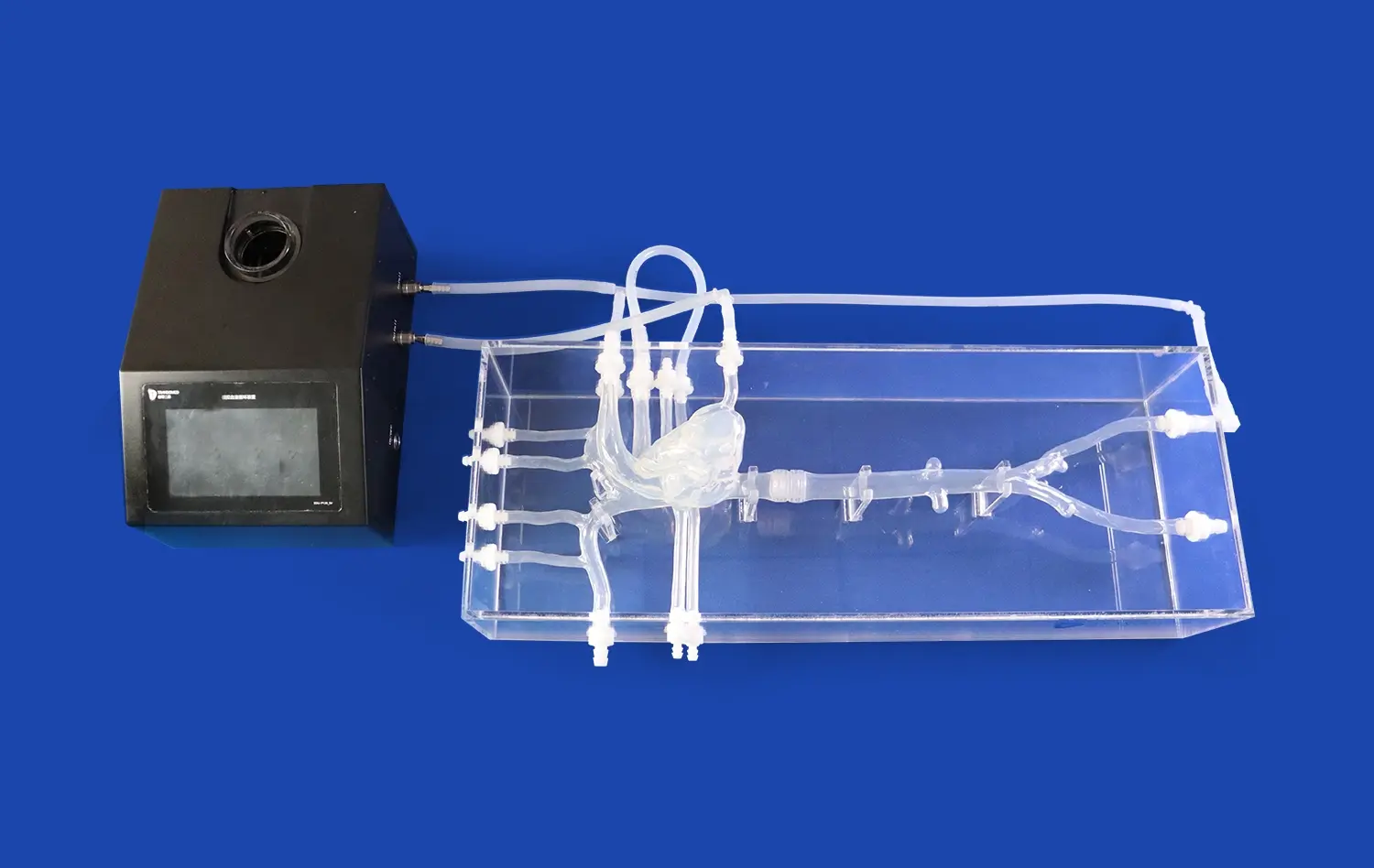
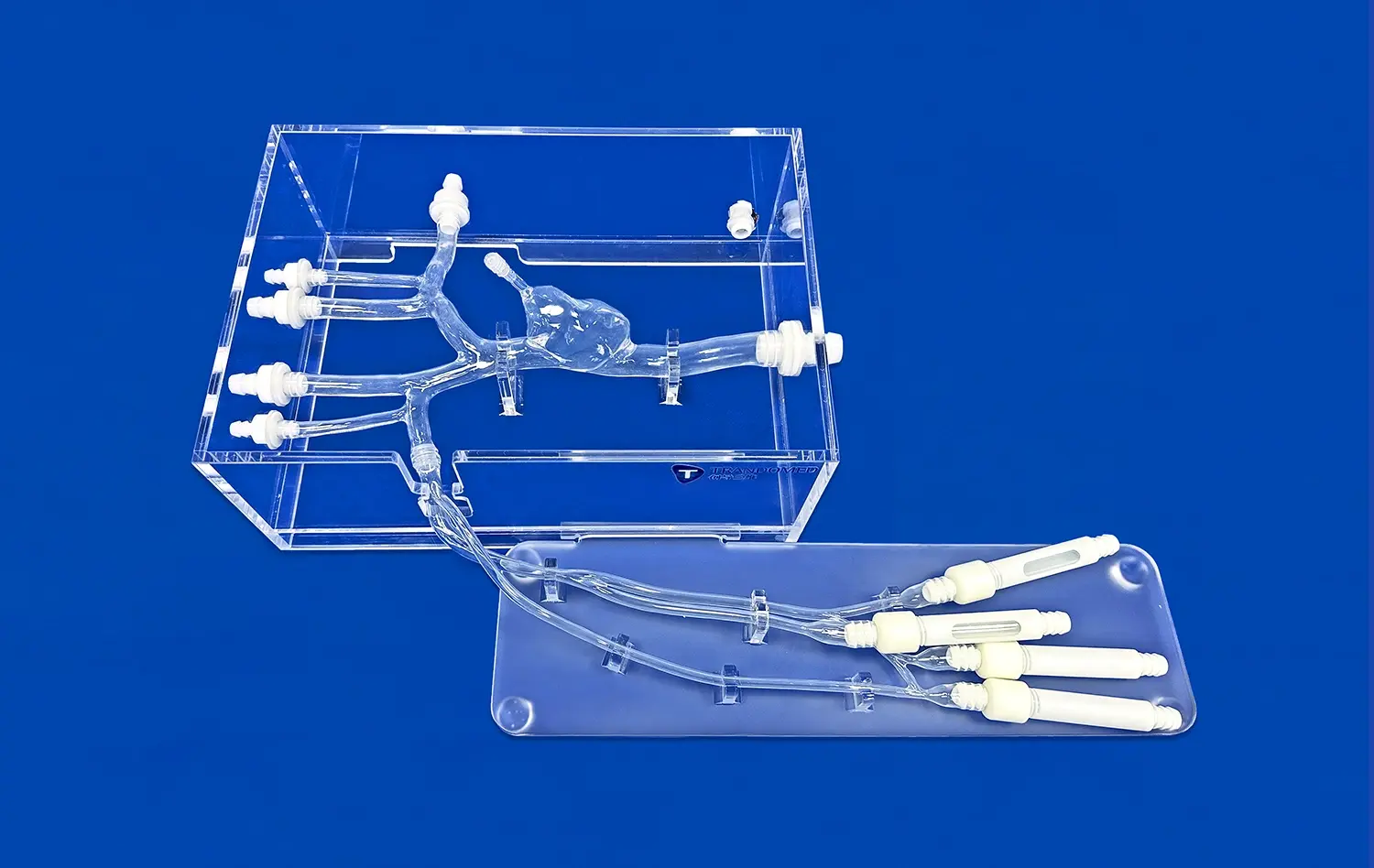
Educational and Clinical Applications of Circle of Willis Brain Models
It is possible to use cerebral computer models such as the circle of willis brain model in a lot of different areas of healthcare teaching and clinical practice. These tools are not only useful for training, but they are also needed to plan surgeries, improve study, and make new medical devices. Because they are three-dimensional and tactile, they are better than computer models because they let students physically move and look at circulatory systems from different angles. This helps them understand them better through hands-on study.
Medical School and Nursing Education Enhancement
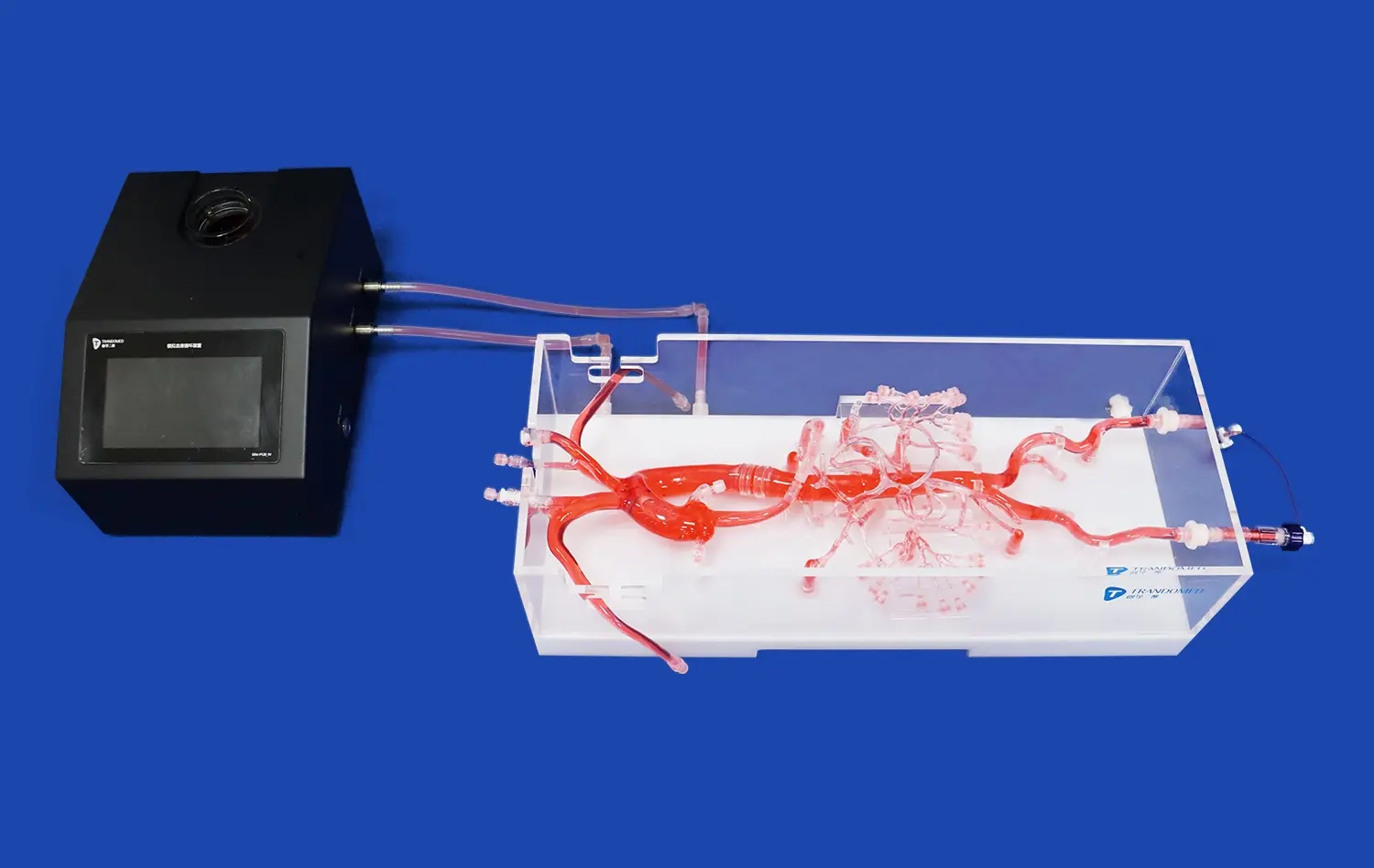
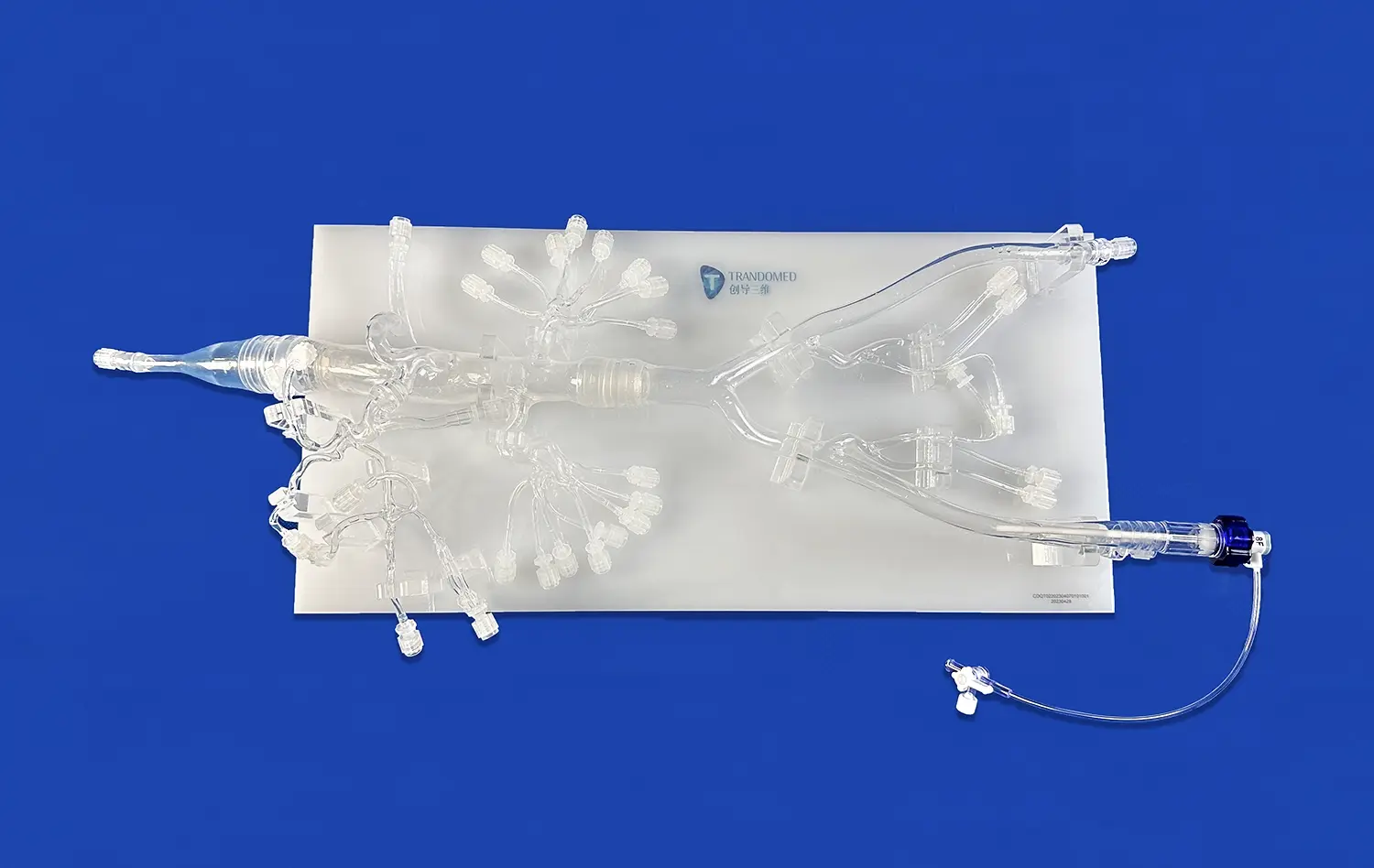
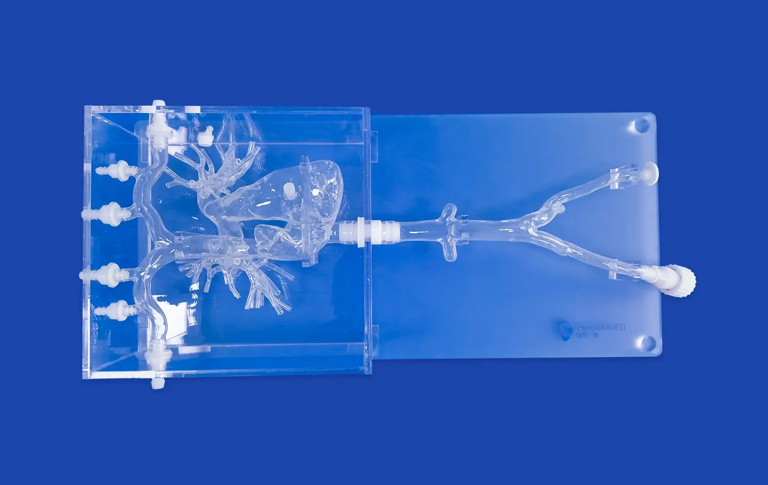
It is always hard for medical schools, nursing schools, and clinical skills centers to teach neuroanatomy to big groups of students who learn in different ways. Brain anatomy models with damaged versions of the Circle of Willis let teachers show how strokes happen, how aneurysms form, and how arteriovenous malformations happen during class and lab time. Students can work on recognizing a stenosed blood vessel, measuring the size of an aneurysm, and learning how blood flow changes during thrombotic events. When compared to passive learning methods, this hands-on technique helps people remember a lot more. To meet these educational needs, Trandomed's Circle of Willis Aneurysm II model (Product No. SJL001D) includes the M1 segment of the right middle cerebral artery with a stenosis lesion and three separate aneurysms on the basilar artery, the ophthalmic segment of the left carotid artery, and the left MCA. The model is made of Silicone Shore 40A material and gives realistic physical feedback that mimics the qualities of real tissue. This helps students improve their hand skill, which is needed for guiding catheters and putting devices in place.
Surgical Planning and Preoperative Rehearsal
Neurosurgeons who have to do complicated procedures like aneurysm clipping or arterial coiling treatments can learn a lot from cerebrovascular models that are made just for each patient. By turning CT angiography or MRI data into 3D-printed copies, surgery teams can practice their plan, choose the right tools, and think about what might go wrong before they go into the operating room. These practice lessons cut down on surgery time, keep shocks to a minimum, and boost trust in the surgeon. More and more general hospitals, specialty hospitals, and surgical training labs are realizing that training on correct anatomy models before surgery directly leads to safer patients and higher success rates during surgery. The true shape of the model lets doctors practice aneurysm tamponade surgeries and cerebral thrombectomy procedures in a safe setting, where they can improve their skills without putting real patients at risk.
Research and Medical Device Development
Biomedical research centers and translational medicine labs need anatomy models that can be changed so that they can do experiments to study how strokes work, try new treatments, and prove biomechanics theories. Companies that make medical devices like neurovascular stents, flow diverters, thrombectomy tubes, and embolic protection devices need testing platforms that are like real people and correctly reflect the shape and qualities of human blood vessels and tissues. Because they can be changed in so many ways, our brain modeling tools can be used with these apps. We can accept changes to the number, size, and location of aneurysms, as well as other diseases like cerebral embolism and varying stenosis lesions. We can change models based on data files sent in CT, CAD, STL, STP, and STEP forms. This lets researchers and device makers rebuild the bodies of specific patients or come up with standard test cases. Because they are so adaptable, our neurovascular training models are great for trying products, making sure designs work, sending to regulators, and showing off to potential customers.
Selecting the Ideal Circle of Willis Brain Model for Your Training Needs
When buying neurovascular simulation tools like the circle of willis brain model, you need to carefully consider a lot of things, such as the material's qualities, how accurate the models are of the body, how well they work, and how reliable the seller is. The best option for your school will rely on its teaching goals, the skill level of users, how often they will use it, and their funds.
Material Selection and Durability Considerations
In different teaching situations, different products offer clear benefits. Rigid plastic models are very long-lasting and don't cost a lot of money, so they can be used over and over again in large-group learning situations where money is tight. Resin versions have better structural detail and smoother surfaces that better look like vessel walls, but they might break more easily when used a lot. The most realistic feeling comes from advanced silicone-based models like our Shore 40A formulation, which closely matches the dynamic qualities of real brain arteries. This material gives real resistance and feedback so that practitioners can practice putting in catheters, manipulating guidewires, and deploying devices. When choosing materials, you should think about whether your main purpose is to show something visually, teach basic anatomy, or create a high-fidelity procedural recreation that needs to respond like tissue.
Anatomical Fidelity and Pathology Representation
The teaching benefit of any cerebral model is directly related to how realistic it is in terms of anatomy and disease. Models should accurately reflect the sizes of blood vessels, the points at which they break off, and the ways that they are arranged in the human body. Pathology modeling traits, such as the shape of an aneurysm, the harshness of the stenosis, and the qualities of the thrombus, must match how patients actually show in real life. Our Circle of Willis brain model has three different aneurysms that are different in size and location. This gives students a chance to learn about saccular, fusiform, and complex aneurysm shapes. Adding an M1 segment stenosis lesion lets you practice getting through narrowed veins and putting in stents or doing angioplasty treatments. With these abnormal traits, a simple anatomy copy becomes a complete training tool that gets medical workers ready for problems they might face in the real world.
Customization Capabilities and Bulk Procurement Options
When institutions buy models, they often need ones that are specifically made for their courses or study methods. Customization choices should let you change where the disease is located, how bad the lesions are, include anatomy specific to each patient, and add features that help with specific training goals. We offer full customization services without charging extra design fees, and we'll work directly with your team to make models that meet your educational goals. Our engineering team can meet a wide range of needs, from standard models for certification programs to patient-specific copies for planning surgery. Companies that want to set up practice centers or supply multiple training spots can save money by buying in bulk. If you want to buy a lot of things, talk to your sellers early on in the buying process about prices, delivery times, and possible product improvements that will help your whole business.
Conclusion
Cerebrovascular computer models such as the circle of willis brain model are used in neurovascular training for a lot more than just teaching anatomy. They are also used for planning surgeries, advancing research, making new devices, and improving nursing skills. These high-tech tools have changed the way doctors understand how brain blood flow changes, spot changes that aren't normal, and learn life-saving treatment skills. Buying high-quality brain anatomy models is a long-term investment to both patient safety and excellent teaching. More and more healthcare systems are realizing that simulation-based training can help lower clinical mistakes and improve the success of procedures. As a result, the need for physically accurate and functionally realistic neurovascular models keeps rising. It's important to carefully consider bodily accuracy, material qualities, customization options, and seller support infrastructure when choosing the right provider and goods to make sure your investment gives you the most long-term value.
FAQ
Why is the Circle of Willis so important for how the brain works?
The Circle of Willis helps blood flow between the front and back parts of the brain. This keeps the brain from getting anoxic if a blood vessel gets sick or damaged in more than one place. This backup system makes sure that even if one main artery gets narrowed or stopped, blood can still get to important parts of the brain through other routes, which could prevent a stroke or limit tissue damage.
In the brain model, what is the Circle of Willis?
On the bottom part of the brain, in the interpeduncular canal of the subarachnoid space, you can find the Circle of Willis. It goes around different parts of the interpeduncular fossa, like the eye chiasm and the pituitary gland's infundibulum. This structural structure is reflected in physical brain models, which let students see how parts are connected and understand how they work by examining them directly.
What are the Circle of Willis's five parts?
The five important parts are the anterior cerebral arteries at their A1 segments on both sides, the anterior communicating artery that connects them, the internal carotid arteries at their distal tips on both sides, the posterior cerebral arteries at their P1 segments on both sides, and the posterior communicating arteries that connect the anterior and posterior circulation on both sides. These blood tubes make up the whole artery ring.
How many people have a Circle of Willis that is full?
Only 20 to 25 percent of people have a full Circle of Willis, which means that none of the parts are missing or hypoplastic. Because of how common physical differences are, neurosurgeons and interventionalists need to know about each person's venous patterns in order to plan treatments. This shows how important it is to learn on models of both normal and abnormal bodies.
What is a 3D model of the Circle of Willis?
An angiographic image is carefully cut into segments to make a Circle of Willis 3D model, which shows the cerebral arteries that bring blood to the brain. The paired spinal arteries come in through the foramen magnum, join to make the basilar artery, and then split into terminal posterior cerebral arteries and superior cerebellar arteries that can be seen at the end. For more complete training, more advanced versions include disease traits like tumors and stenosis.
Partner with a Leading Circle of Willis Brain Model Manufacturer
Trandomed has the best circle of willis brain models for medical schools, hospital training departments, research labs, and simulation centers, and they are ready to help you with your neurovascular training projects. With plans based on real patient imaging data and the mechanical realism of Silicone Shore 40A building, our Circle of Willis Aneurysm II model has the most accurate anatomy of any model on the market. We offer full customization without design fees, quick lead times of 7–10 days, and safe foreign shipping through top companies. Our knowledgeable staff offers individualized advice to match goods with your specific goals, whether you're opening a new simulation center, improving current training resources, or looking for a reliable Circle of Willis brain model provider for ongoing buying needs. Get in touch with jackson.chen@trandomed.com right away to talk about your needs, get full specs, or set up product demos.
References
Hendrikse J, van Raamt AF, van der Graaf Y, Mali WP, van der Grond J. Distribution of cerebral blood flow in the circle of Willis. Radiology. 2005;235(1):184-189.
Krabbe-Hartkamp MJ, van der Grond J, de Leeuw FE, de Groot JC, Algra A, Hillen B, Breteler MM, Mali WP. Circle of Willis: morphologic variation on three-dimensional time-of-flight MR angiograms. Radiology. 1998;207(1):103-111.
Schaller B. Extracranial-intracranial bypass to reduce the risk of ischemic stroke in intracranial aneurysms of the anterior cerebral circulation: a systematic review. Journal of Stroke and Cerebrovascular Diseases. 2008;17(5):287-298.
Alpers BJ, Berry RG, Paddison RM. Anatomical studies of the circle of Willis in normal brain. AMA Archives of Neurology and Psychiatry. 1959;81(4):409-418.
Hartkamp MJ, van Der Grond J, van Everdingen KJ, Hillen B, Mali WP. Circle of Willis collateral flow investigated by magnetic resonance angiography. Stroke. 1999;30(12):2671-2678.
Eftekhar B, Dadmehr M, Ansari S, Ghodsi M, Nazparvar B, Ketabchi E. Are the distributions of variations of circle of Willis different in different populations? Results of an anatomical study and review of literature. BMC Neurology. 2006;6:22.
_1736216292718.webp)