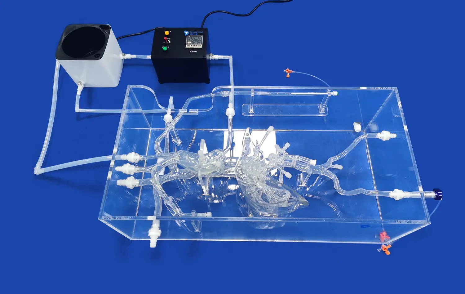
An advanced venous cardiac electrophysiology model is a big step forward in cardiac simulation technology. It gives doctors accurate 3D models of the heart's veins for training and study. The inferior vena cava, superior vena cava, right atrium, right ventricle, and subclavian vein are all very accurately modeled by these high-tech simulators. This lets doctors practice catheter navigation, electrophysiological mapping, and ablation methods without any risk. These models change how healthcare institutions train their staff for complex heart procedures by bridging the gap between theory knowledge and hands-on experience. They also speed up the development of new medical devices.
Understanding Venous Cardiac Electrophysiology Models
What Makes These Models Unique?
These models of venous cardiac electrophysiology are specialized training aids that are made to look and work like the heart's venous structures. Unlike flat anatomical charts or computer models, these three-dimensional copies let you feel how real tissue reacts when you insert and move a catheter through it. Shore 40A silicone, which closely mimics the consistency of human tissue, is often used in the models. This lets trainees build muscle memory and procedural confidence before they treat real patients.
The main difference between venous and arterial heart models is what they focus on. Venous models focus on parts that are important for electrophysiology procedures, especially the routes that are used for arrhythmia treatment during catheter ablation. Because of this, cardiologists, cardiac nurses, and medical device makers can focus on the venous access points and navigation problems that are unique to EP studies.
Key Anatomical Features Replicated
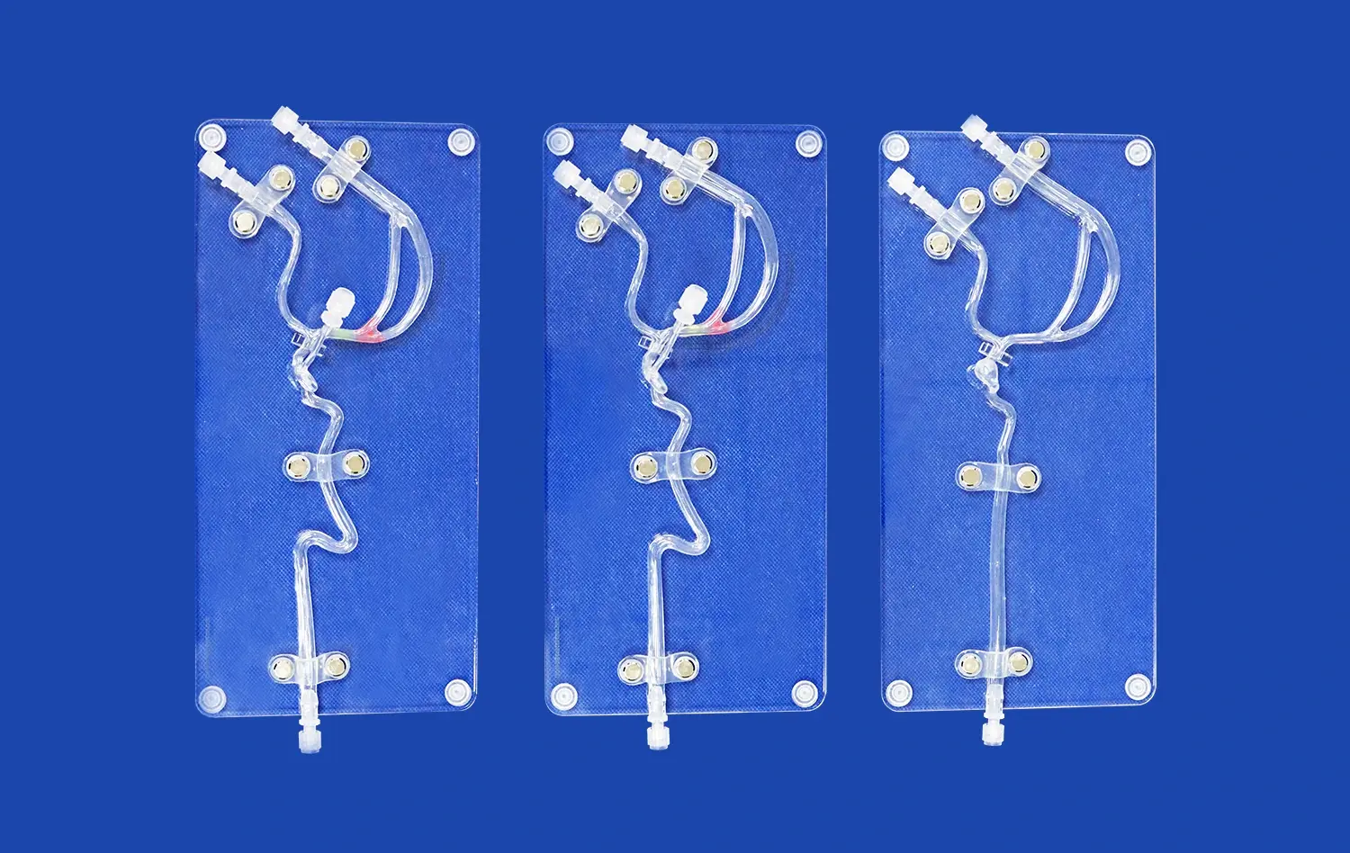
High-quality venous cardiac models have a number of important anatomical parts built in. In order to insert a catheter, the inferior vena cava and superior vena cava are the main entry points. The right atrium offers the space for placing electrodes during electrophysiological mapping. The right ventricle and subclavian vein finish off the anatomy, giving students a wide range of training settings that are like real-life medical situations.
Modern ways of making things, especially 3D printing from real CT and MRI scans, make sure that these models are more accurate than ever in terms of anatomy. With reverse 3D reconstruction technology, exact measures can be taken from medical images, so replicas can be made that take into account differences in each person's heart anatomy. With this new technology, training can now take into account different body types, which better prepares doctors for the wide range of patient situations they will see.
Major Benefits of Using Venous Cardiac Electrophysiology Models
Enhanced Training Outcomes for Medical Professionals
Medical schools know that hands-on practice is a great way to keep skills fresh and boost confidence during procedures. A venous cardiac electrophysiology model lets residents and students do treatments over and over again without having to worry about time or patient safety. This practice over and over again helps you remember how to do things, lowers your nervousness about performing, and speeds up your learning curve when you move on to real patient care.
These simulators are especially helpful for teaching cardiac catheterization methods in nursing schools and clinical skills centers. Before taking part in real procedures, staff can learn how to handle tools, find the right positions, and understand how different vein structures are connected in space. This preparation directly leads to safer patients and fewer complications in the first cases that are watched.
Cost-Effective Alternative to Animal Models and Cadavers
Animal models or cadaveric specimens were used a lot in traditional heart training, which had a lot of problems. Animal models come with ethical concerns, rules that must be followed, and high ongoing costs for care and purchase. Cadaveric specimens have realistic tissue qualities, but they are hard to get, hard to keep safe, and can't be used again and again.
Silicone-based cardiac models get rid of these worries and give you a lot of chances to practice. One model can be used for hundreds of catheter placements without breaking down, so the original cost can be spread out over many training sessions. Medical schools and hospitals can plan training at convenient times without having to coordinate with outside suppliers or manage biological materials. This makes training programs much more flexible and efficient.
Accelerated Medical Device Development and Testing
Before they can sell new cardiac equipment, companies that make medical devices have to go through strict approval processes. A lot of testing needs to be done on prototype tubes, ablation tools, and imaging systems to make sure they work, are safe, and can be used. Anatomically correct cardiac models create safe testing settings where engineers can check how well a device works, find problems with the design, and improve features without involving the patient.
Advanced simulators are especially useful for product creation because they can be changed to fit specific needs. Companies that make medical devices can ask for models with specific changes to the body's structure, diseases, or sizes that fit the patients they want to treat. This customization makes it possible to try specific scenarios that would be hard or impossible to repeat consistently using other methods. This leads to better-designed devices that meet real clinical needs.
Support for Preoperative Planning and Procedure Rehearsal
Procedure-specific planning and practice are very helpful for complicated heart cases. Surgical teams can order unique models based on CT or MRI scans of the real patient when they have to deal with cases with complicated anatomy. Before going into the operating room, these patient-specific models help surgeons picture the exact anatomy they will be working with, plan catheter approaches, think about possible problems, and talk about their plans with their teams.
This preoperative simulation cuts down on the time needed for the operation, the amount of radiation exposure from fluoroscopy, and the chance of complications during the procedure. When you practice on an exact copy, you feel more confident, which leads to steadier hands and better decisions in critical situations. This improves patient outcomes and institutional quality measures in the long run.
How to Choose the Best Venous Cardiac Electrophysiology Model for Your Needs?
Evaluating Material Quality and Durability
Material choice has a big effect on both how realistic something is and how long it lasts. Shore 40A silicone strikes the perfect mix between being flexible like tissue and staying structurally sound even after many uses. Lower-durometer materials may feel more real at first, but they tear easily when the catheter is put under stress. Harder materials don't tear easily, but they don't give practitioners the physical feedback they need to improve their skills.
Procurement managers should ask what the materials are made of, how long they are expected to last with normal use, and what the replacement plans are. Models meant for intense training programs need to be built with more strength than models meant for occasional shows. Knowing how things are used in your school can help you choose the right quality level that balances cost with durability needs.
Assessing Anatomical Accuracy and Customization Options
It's not always possible for cardiac models to accurately show how the heart works. Models that are based on real human imaging data are more accurate than models that are based on generic templates or artistic opinion. When vendors use CT and MRI data in their design process, they can show that their models accurately reflect differences in the human body, such as differences in vessel diameters, branching angles, and chamber sizes.
The ability to customize a model makes it useful in situations other than normal training scenarios. The ability to change the size of the foramen ovale, the shape of the atrial chamber, or to include pathological variations lets institutions meet certain study or educational goals. Customization service providers usually accept data files in a number of different formats, such as STL, STP, and STEP. This makes it easy to connect their services to your current imaging infrastructure.
Considering Vendor Support and Technical Expertise
Your relationship with the company that sells you simulators goes far beyond the initial buy. Reliable sellers offer full technical support that helps institutions get the most out of their venous cardiac electrophysiology models and fix any problems that come up during implementation. Training programs for teachers and staff make sure that everyone knows how to properly clean, care for, and store models so that they last as long as possible.
Companies that have been around for a long time have decades of experience making medical training technology. Their research and development (R&D) teams are always improving how things are made, using user feedback to make designs better, and keeping up with changes in professional practices. Because this is an ongoing project, your school has access to state-of-the-art modeling tools that keep up with changes in cardiac electrophysiology techniques and tools.
Understanding Lead Times and Logistics
Project timelines often depend on simulator availability. Standard venous cardiac electrophysiology models typically ship within seven to ten days, allowing rapid program initiation. Custom models require additional production time for data processing, design verification, and specialized manufacturing, potentially extending lead times to several weeks depending on complexity.
International logistics considerations affect both cost and delivery schedules. Reputable suppliers maintain relationships with major carriers including FedEx, DHL, EMS, UPS, and TNT, ensuring reliable shipping with tracking capabilities. Understanding customs requirements, import duties, and delivery coordination helps prevent unexpected delays when acquiring simulators from international manufacturers.
Practical Insights: Implementation and Use Cases
Integrating Models into Medical School Curricula
Medical schools incorporating venous cardiac electrophysiology models into their anatomy and clinical skills courses report improved student confidence and competency in cardiac procedures. The models work particularly well in small-group laboratory sessions where instructors can supervise multiple students simultaneously, providing individualized feedback on catheter handling techniques and anatomical recognition skills.
Progressive skill-building approaches maximize learning outcomes. Students might begin with basic catheter insertion and navigation exercises, advance to electrode positioning challenges, and culminate in simulated ablation procedures that integrate multiple skills. This scaffolded approach ensures mastery at each level before introducing additional complexity, reducing frustration and building sustainable competence.
Hospital Training Programs and Competency Assessment
Hospitals implementing simulation-based training for cardiac catheterization teams observe measurable improvements in procedural efficiency and complication rates. Structured training programs using realistic models allow new staff members to achieve competency benchmarks before participating in patient care, while experienced practitioners can maintain skills or learn new techniques without patient risk.
Competency assessment becomes more objective when using standardized simulators. Evaluators can observe catheter navigation accuracy, procedure completion times, and complication avoidance across multiple candidates under identical conditions. This standardization eliminates the variability inherent in patient-based training, producing fairer assessments that accurately reflect individual capabilities.
Research Applications and Data Generation
Research institutions utilize cardiac venous cardiac electrophysiology models to investigate catheter design innovations, evaluate imaging modality performance, and study electrophysiological phenomena under controlled conditions. The ability to repeat experiments with identical anatomical substrates eliminates a major confounding variable, improving data quality and statistical power.
Biomechanical studies particularly benefit from model-based research. Investigators can measure forces applied during catheter manipulation, assess tissue deformation under various conditions, and correlate mechanical parameters with clinical outcomes. These insights inform best-practice guidelines and equipment specifications that enhance procedural safety across the cardiology community.
Future Trends and Innovations in Venous Cardiac Electrophysiology Modeling
Integration with Digital Simulation Platforms
When physical models, virtual reality, and augmented reality come together, they could completely change the way people train their hearts. When you put together hybrid systems that use real-time imaging displays, performance analytics, and tactile feedback from physical models, you get more complicated learning experiences that are more immersive.
These platforms work together to keep track of where the catheter is in three dimensions, show mistakes in technique right away, and automatically come up with objective competency measures. Trainees get quick feedback on their performance, which helps them improve their skills faster, and program directors learn more about how effective the curriculum is and how each student learns.
Personalized Medicine and Patient-Specific Models
Rapid prototyping has come a long way, making it possible to make patient-specific heart models cheaply and in time for clinical use. As personalized treatment becomes more common in healthcare, being able to make custom models for difficult cases becomes more useful. Surgical teams can practice difficult procedures on models that exactly match the anatomy of their patients. This improves results for people with unusual anatomy or complicated pathology.
This personalized approach also includes the creation of medical devices. Companies can test their products on a range of body types that match the real-life variation in the population. Knowing how devices work across a wide range of body types makes designs more reliable and increases the number of patients who can benefit from new technologies.
Artificial Intelligence and Adaptive Learning Systems
By looking at how well trainees do on cardiac simulators, machine learning algorithms can find specific skill gaps and suggest specific tasks for practice. These adaptive learning systems make sure that each person gets teaching that is right for their current level of skill and speed of learning by customizing their training pathways.
AI also improves model design by looking at huge amounts of cardiac imaging data to find anatomical patterns, common variants, and features that are pathologically important. With this data-driven method, simulators are made that are more like real clinical situations. This helps doctors get ready for all the different types of cases they will see in their careers.
Conclusion
Venous cardiac electrophysiology models are important for teaching medicine, training doctors in the field, and making new medical devices. These simulators give healthcare professionals realistic, repeatable practice chances that help them get better at their jobs and boost their confidence while also speeding up the development of new cardiac care technologies. Advanced 3D printing and CT-based design make physical accuracy possible, which makes training useful and easy to apply to real patient care. When organizations buy high-quality simulators, they put themselves at the cutting edge of cardiac education and study. This leads to better patient outcomes and advances in the field of electrophysiology. As simulation technology keeps getting better, it will be able to do even more. This makes strategic investments in tried-and-true methods more valuable.
FAQ
What advantages do physical cardiac models offer compared to virtual simulations?
Physical models provide tactile feedback essential for developing catheter manipulation skills and understanding tissue response during electrophysiology procedures. While virtual simulations excel at visualizing electrical activity and anatomical relationships, they cannot replicate the haptic sensations practitioners experience when navigating catheters through actual venous structures. The combination of visual and tactile learning engages multiple sensory pathways, improving skill retention and procedural confidence.
How does model customization work for specialized training scenarios?
Customization begins with submitting patient imaging data in formats such as CT, CAD, STL, STP, or STEP files. Manufacturers utilize reverse engineering technology to reconstruct anatomical features, then modify specific dimensions according to your specifications. Common customizations include adjusting foramen ovale size, altering chamber proportions, or incorporating pathological variations relevant to your training objectives. Design modifications typically involve no additional fees beyond the base model cost.
Can these models accommodate different catheter types and sizes?
Quality venous cardiac simulators accommodate the full range of catheters used in clinical electrophysiology procedures, from diagnostic mapping catheters to ablation equipment. The Shore 40A silicone material maintains structural integrity while allowing catheter passage, and the anatomically accurate lumen dimensions ensure realistic navigation challenges. Models withstand repeated insertions without significant wear, supporting extensive training programs across diverse equipment platforms.
Partner with Trandomed for Advanced Cardiac Simulation Solutions
Trandomed stands as a premier manufacturer specializing in high-fidelity venous cardiac electrophysiology models that meet the demanding requirements of medical education, hospital training, and device development applications. Our XXS004 model incorporates anatomically precise replicas of the inferior vena cava, superior vena cava, right atrium, right ventricle, and subclavian vein, manufactured from medical-grade Shore 40A silicone for optimal realism and durability. With over two decades of experience in medical 3D printing innovation, our R&D team leverages extensive CT and MRI datasets to create simulators that accurately represent human cardiac anatomy. We offer comprehensive customization services at no additional design cost, accepting data files in multiple formats and tailoring models to your exact specifications. Contact jackson.chen@trandomed.com today to discuss how our cardiac simulation solutions can enhance your training programs or accelerate your product development initiatives. Our team provides rapid turnaround with seven to ten day lead times and reliable international shipping through major carriers, ensuring your institution receives the tools needed to advance cardiac care excellence.
References
Josephson, M.E. (2016). Clinical Cardiac Electrophysiology: Techniques and Interpretations. Fifth Edition. Wolters Kluwer Health.
Zipes, D.P. & Jalife, J. (2018). Cardiac Electrophysiology: From Cell to Bedside. Seventh Edition. Elsevier.
Issa, Z.F., Miller, J.M. & Zipes, D.P. (2019). Clinical Arrhythmology and Electrophysiology: A Companion to Braunwald's Heart Disease. Third Edition. Elsevier.
Huang, S.K.S. & Wood, M.A. (2015). Catheter Ablation of Cardiac Arrhythmias. Third Edition. Saunders.
Nattel, S., Harada, M. & Nattel, S. (2020). Electrophysiology and Mechanisms of Cardiac Arrhythmias. Journal of Cardiovascular Electrophysiology, 31(4), 1045-1063.
Stevenson, W.G. & Tedrow, U.B. (2017). Ventricular Arrhythmias: From Mechanisms to Therapy. Circulation Research, 120(2), 331-345.
_1736215128474.webp)
_1736214519364.webp)
_1734507815464.webp)
_1732863713705.webp)