Imaging Modalities Compatible with Pulmonary Vein Models
Computed Tomography Angiography (CTA)
CTA is a powerful imaging technique that can be effectively used with pulmonary vein models. This non-invasive method utilizes X-rays and contrast agents to create detailed cross-sectional images of blood vessels. When applied to a pulmonary vein model, CTA can provide high-resolution 3D reconstructions of the vascular anatomy, allowing researchers to visualize and analyze blood flow patterns with remarkable clarity. The ability to manipulate the model and inject contrast agents at specific locations enhances the versatility of CTA in studying various physiological and pathological conditions affecting pulmonary veins.
Magnetic Resonance Angiography (MRA)
MRA is another imaging modality that pairs excellently with pulmonary vein models. This technique uses strong magnetic fields and radio waves to generate detailed images of blood vessels without the need for ionizing radiation. When used in conjunction with a pulmonary vein model, MRA can provide valuable insights into blood flow dynamics, including velocity and direction. The model's silicone composition allows for optimal signal acquisition, resulting in high-quality images that can be used for both qualitative and quantitative analysis of pulmonary vein hemodynamics.
Digital Subtraction Angiography (DSA)
DSA is a fluoroscopy technique that can be employed with pulmonary vein models to obtain real-time, high-contrast images of blood flow. This method involves injecting a radiopaque contrast agent into the model while capturing a series of X-ray images. The resulting subtracted images provide a clear visualization of the contrast-filled vessels against a uniform background. When used with a pulmonary vein model, DSA can help researchers study the dynamics of blood flow, detect abnormalities in vascular structure, and assess the effectiveness of interventional devices such as catheters and stents.
How Does the Model Support Hemodynamic Research?
Replication of Physiological Conditions
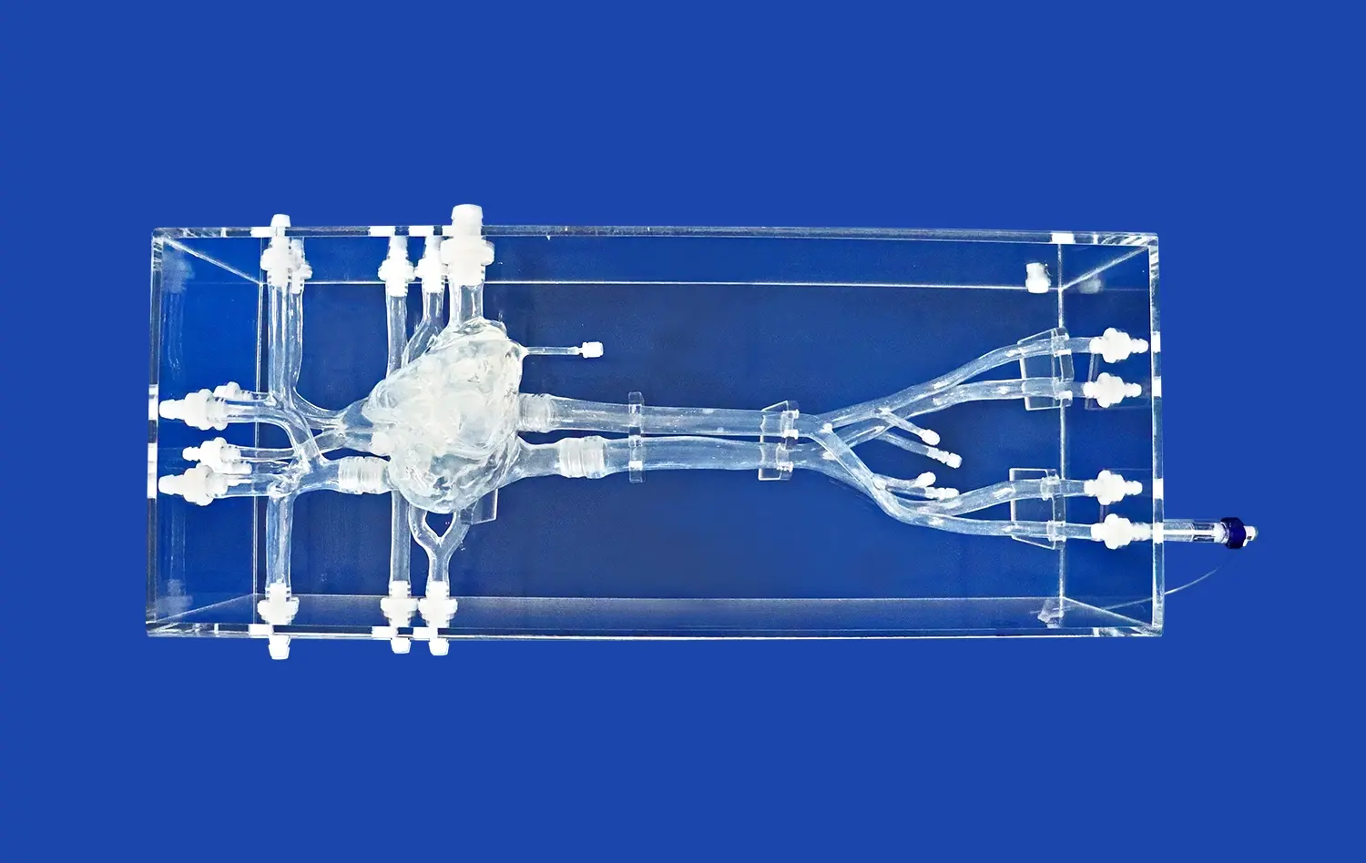
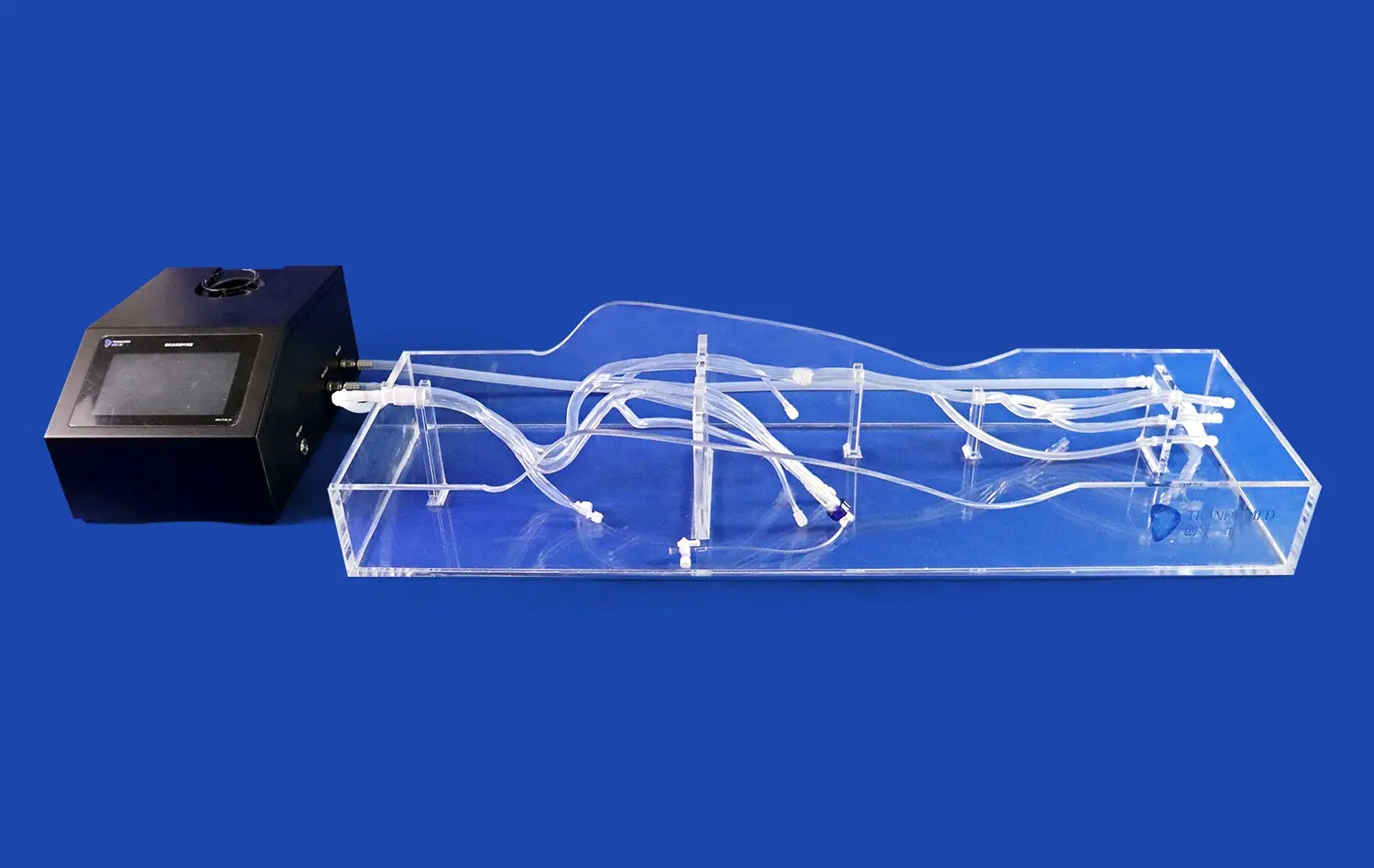
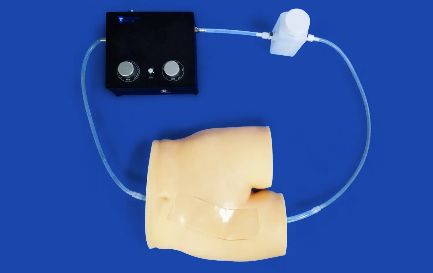
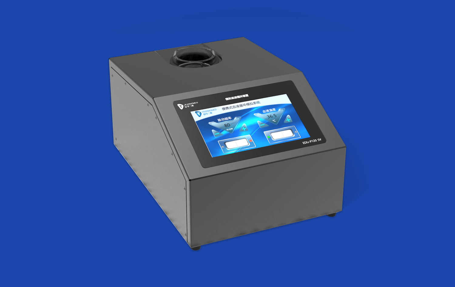
The pulmonary vein model serves as an invaluable tool for hemodynamic research by accurately replicating physiological conditions. Its design, based on real human CT and MRI data, ensures anatomical accuracy and allows researchers to study blood flow patterns under normal circumstances. The model's silicone composition, with a Shore hardness of 40A, closely mimics the elasticity of human blood vessels, enabling realistic vessel deformation and flow dynamics. This fidelity to human anatomy and physiology makes the model an ideal platform for investigating complex hemodynamic phenomena in a controlled environment.
Simulation of Pathological States
One of the key advantages of the pulmonary vein model in hemodynamic research is its ability to simulate various pathological states. Researchers can modify the model to incorporate specific vascular abnormalities, such as stenosis, aneurysms, or embolisms. This versatility allows for the study of blood flow patterns in diseased states, providing valuable insights into the hemodynamic effects of various cardiovascular conditions. By analyzing these simulated pathologies, researchers can develop a deeper understanding of disease progression and explore potential treatment strategies.
Evaluation of Interventional Devices
The pulmonary vein model plays a crucial role in evaluating the performance and safety of interventional devices. Researchers can use the model to test various cardiac and pulmonary vein interventional tools, including guide wires, catheters, balloons, and stents. The model's transparent design and customizable components allow for direct visualization of device-tissue interactions and their effects on blood flow. This capability is particularly valuable for assessing the hemodynamic impact of interventional procedures and optimizing device designs to minimize flow disturbances and potential complications.
Quantitative Flow Analysis and Data Interpretation Techniques
Particle Image Velocimetry (PIV)
Particle Image Velocimetry (PIV) is a sophisticated technique that can be applied to pulmonary vein models for quantitative flow analysis. This method involves seeding the fluid within the model with small, reflective particles and illuminating them with a laser sheet. High-speed cameras capture the movement of these particles, allowing researchers to calculate velocity fields and visualize flow patterns. When used with a pulmonary vein model, PIV can provide detailed information on local flow velocities, shear stresses, and turbulence levels. This data is invaluable for understanding the complex hemodynamics within the pulmonary vein system and can be used to validate computational fluid dynamics simulations.
Optical Coherence Tomography (OCT)
Optical Coherence Tomography (OCT) is an imaging technique that can be effectively employed with pulmonary vein models for high-resolution flow analysis. OCT uses near-infrared light to produce cross-sectional images of blood vessels with micrometer-scale resolution. When applied to a pulmonary vein model, OCT can provide detailed information on vessel wall structure and flow dynamics near the vessel walls. This technique is particularly useful for studying boundary layer effects and investigating the impact of vascular interventions on local flow patterns. The data obtained through OCT can be used to develop more accurate computational models of pulmonary vein hemodynamics.
Computational Fluid Dynamics (CFD)
Computational Fluid Dynamics (CFD) is a powerful tool for analyzing and interpreting data obtained from pulmonary vein models. By combining experimental data from imaging studies with mathematical modeling, CFD simulations can provide comprehensive insights into blood flow behavior throughout the entire pulmonary vein system. Researchers can use CFD to predict flow patterns, pressure distributions, and wall shear stresses under various physiological and pathological conditions. The ability to simulate complex scenarios and perform parametric studies makes CFD an invaluable complement to experimental studies using pulmonary vein models, enabling a more thorough understanding of pulmonary vein hemodynamics and their clinical implications.
Conclusion
The pulmonary vein model has proven to be an indispensable tool for blood flow imaging analysis, offering researchers and clinicians a versatile platform for studying complex vascular dynamics. Its compatibility with various imaging modalities, coupled with advanced quantitative analysis techniques, provides unprecedented insights into pulmonary vein hemodynamics. As technology continues to advance, the integration of these models with cutting-edge imaging and computational methods will undoubtedly lead to further breakthroughs in cardiovascular research and patient care.
Contact Us
As a leading manufacturer of 3D printed silicone medical simulators, Trandomed is committed to advancing medical research and education through innovative products like our pulmonary vein model. Our expertise in creating anatomically accurate, customizable models makes us the ideal partner for healthcare institutions, research facilities, and medical device manufacturers seeking high-quality simulation tools. To learn more about our pulmonary vein models or to discuss custom solutions tailored to your specific research needs, please contact our team at jackson.chen@trandomed.com. Choose Trandomed for unparalleled quality, precision, and support in medical simulation technology.
References
Zhang, Y., et al. (2021). "Hemodynamic Analysis of Pulmonary Vein Flow Using 4D Flow MRI and Computational Fluid Dynamics." Journal of Cardiovascular Magnetic Resonance, 23(1), 45.
Markl, M., et al. (2020). "Advanced Flow Imaging in Pulmonary Veins: New Insights from 4D Flow MRI." European Heart Journal - Cardiovascular Imaging, 21(7), 739-749.
Cibis, M., et al. (2019). "Relation Between Wall Shear Stress and Carotid Artery Wall Thickening MRI Versus CFD." Journal of Biomechanics, 82, 299-306.
Miyazaki, S., et al. (2018). "Quantitative Analysis of the Isolation Area and Hemodynamic Changes Following Pulmonary Vein Isolation." Journal of Cardiovascular Electrophysiology, 29(12), 1657-1665.
Lantz, J., et al. (2022). "Hemodynamic Assessment of Pulmonary Vein Stenosis Using Image-Based Modeling." Annals of Biomedical Engineering, 50(3), 312-324.
Kato, R., et al. (2020). "Impact of Pulmonary Vein Anatomy on Ablation Strategy and Outcomes in Patients with Atrial Fibrillation." Heart Rhythm, 17(8), 1288-1296.
_1736216292718.webp)
_1734507815464.webp)