Cardiac Electrophysiology Training With Realistic Venous Models
2026-05-21 09:00:04
With the addition of anatomically realistic simulation tools, cardiac electrophysiology training has changed a lot. The venous cardiac electrophysiology model is a unique way to train that copies the heart's venous system, which includes the inferior vena cava, the superior vena cava, the right atrium, the right ventricle, and the subclavian vein. Clinicians, medical students, and people who make medical devices can use these realistic models to practice ablation, catheter guidance, and electrophysiological mapping without any risk. These realistic venous models are now required in all medical education and procedural training programs in the United States. They bridge the gap between theory knowledge and real-life clinical use.
Understanding Venous Cardiac Electrophysiology Models
Physiological Basis and Anatomical Accuracy
The shape and function of venous cardiac models are very different from those of arterial models. The venous pathways make it easier for deoxygenated blood to return to the heart and are very important for getting to the heart during electrophysiology treatments. When choosing training tools that truly reflect clinical situations, it is important to know the anatomical differences between the venous and arterial systems.
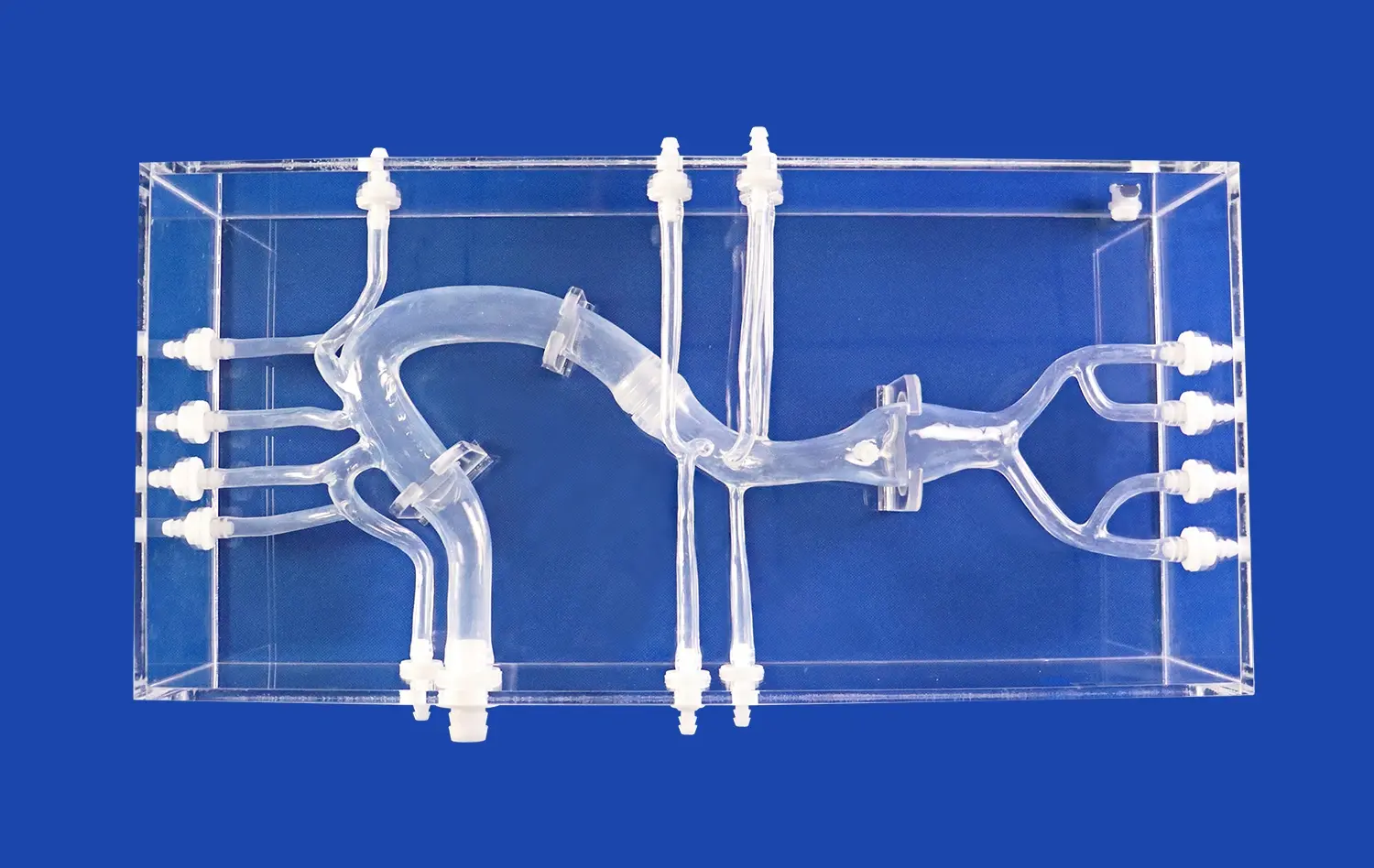
When we look at the heart's veins, the superior and inferior vena cava are the main ways that blood gets to the right ventricle. The right atrium is an important region where electrical signals gather before they cause the ventricles to contract. For these structures to be simulated in a realistic way, the dimensions must be exact, the tissue must be flexible enough, and the physical feedback must be the same as how human tissue reacts when a catheter is manipulated.
Electrical Conduction Principles in Venous Structures
Heartbeats are coordinated in large part by the electrical activity in the heart's veins. As part of an electrophysiology study, doctors move catheter electrodes through veins to record electrical signals and find the cause of an arrhythmia. The best way to understand how electrical impulses move through cardiac muscle is to use the bidomain model of cardiac tissue modeling. This way of computing takes into account both the electrical properties inside and outside of cells. This lets training models show conduction patterns more accurately.
Through built-in sensor systems, modern simulation methods add these electrical qualities to physical models. Traditional electrocardiograms only show electrical data at the surface level. EP studies, on the other hand, give very thorough maps of the inside of the heart. When making training models that look like venous anatomy, they need to be able to put catheters at certain anatomical landmarks where electrical measurements are most useful for diagnosis.
Key Parameters for Realistic Simulation
Several important factors decide how well venous cardiac training models work. The choice of material has a direct effect on how realistic the touch feels. Silicone compounds with Shore hardness ratings around 40A are very close to the properties of human flesh. Anatomical accuracy makes sure that all the important parts are shown correctly and in the right proportions. Durability is very important in training settings where catheters are inserted over and over again. Lastly, customization features let institutions change models based on CT or MRI data that shows the unique anatomy of a patient.
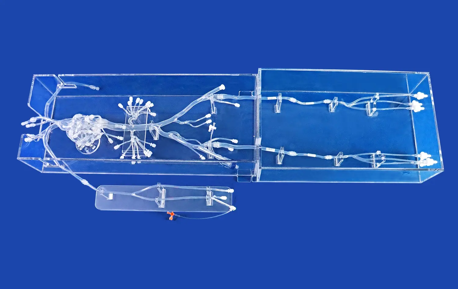
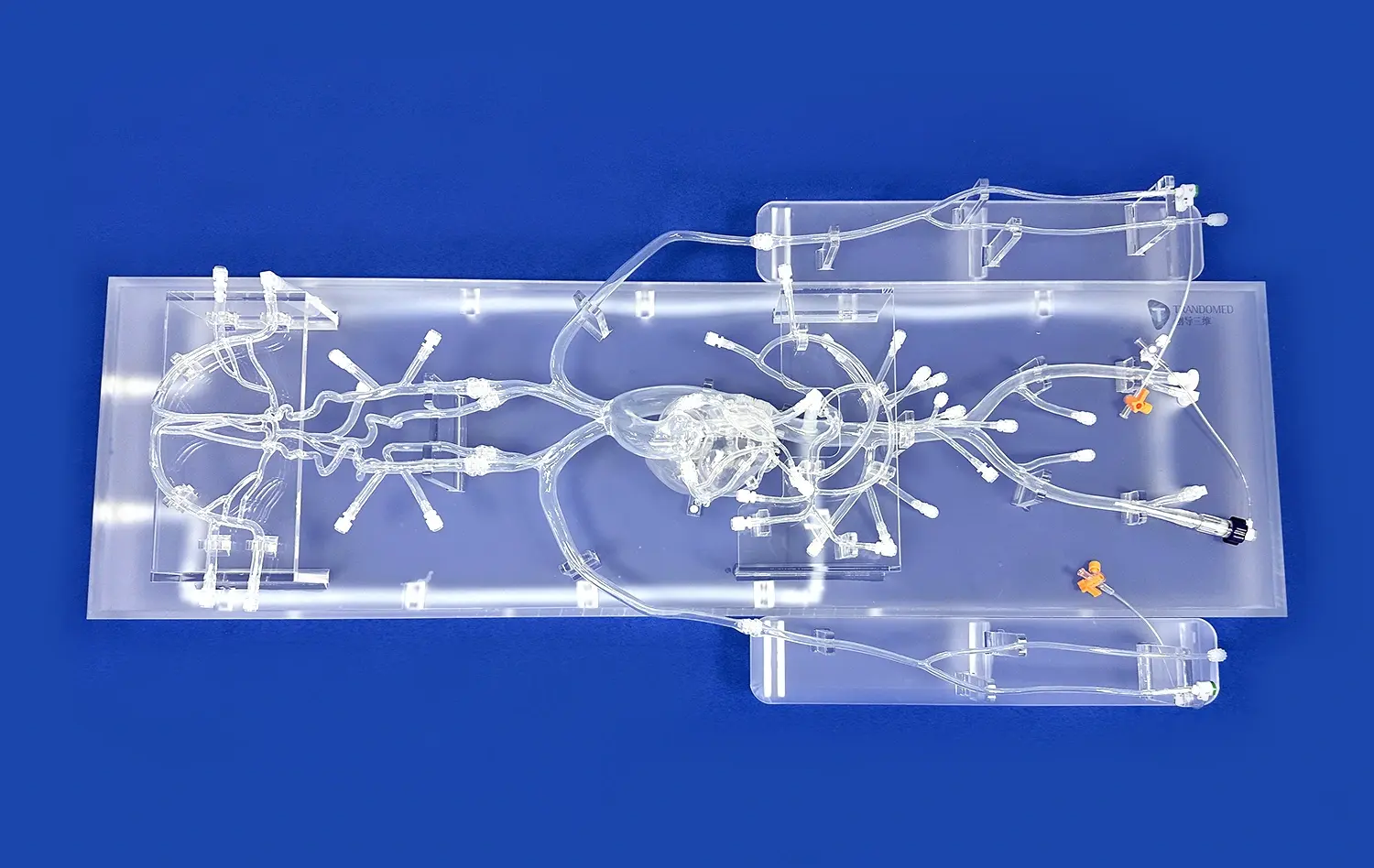
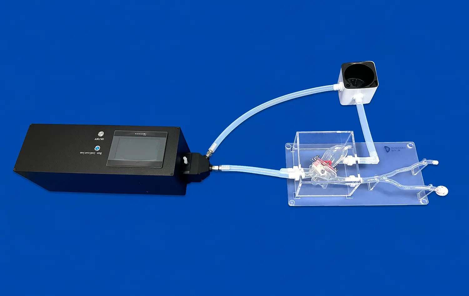
These design rules can be seen in the XXS004 venous cardiac electrophysiology model. This modeling tool is made from medical-grade silicone that is 40A hardness and gives real-life feedback when manipulating a catheter. The fully detailed model includes the inferior vena cava, superior vena cava, right atrium, right ventricle, and subclavian vein, which are all important structures that are reached during real EP treatments.
Evaluating and Selecting Venous Cardiac Electrophysiology Models
Procurement Criteria for Medical Institutions
When choosing simulation tools, medical schools, nursing schools, and clinical skills centers all have their own problems. Budget limits must be weighed against how well training works and how long models last. Models should be judged by how accurate they are in terms of anatomy, how long the materials last, how well the seller supports them, and how many ways there are to make them unique.
There are big differences between regular cardiac models and specialized venous electrophysiology simulations. Most traditional models center on the heart's external anatomy or the flow of blood through the arteries. Venous models focus on the pathways and structures that are used during EP procedures, which is useful for training programs and units that work with electrophysiology and cardiology.
Technical Validation and Clinical Relevance
Comparing dimensional values to reference anatomical data is one way to make sure that simulation models are correct. Advanced makers use reverse 3D reconstruction technology to get exact anatomical information from CT and MRI scans of real people. This method based on data makes sure that models reflect the real differences in anatomy and diseases that doctors see in reality.
Teams in charge of buying things should ask for proof that the model's dimensions match up with written studies on anatomy. Making the source data used in model development public shows that the company is serious about making and has a lot of technical know-how. When vendors give detailed technical specs and validation reports, it shows that they care about accuracy and how well their products teach.
Vendor Evaluation Considerations
To find trusted suppliers, you need to look at more than just the quality of the products they sell. Lead times affect how programs are planned. Manufacturers with 7–10 day production cycles make it possible to quickly apply curriculum. The terms of payment and the ability to ship goods internationally affect how the budget is managed and how the procedures are coordinated. When institutions need patient-specific models or unique anatomical variations, customization services that don't charge extra for the design work are very useful.
Trandomed's method to customization is a great example of service that puts the customer first. The company takes data files in CT, CAD, STL, STP, and STEP formats, which lets institutions make exact changes to models based on their needs. Being able to change the size of the foramen ovale and rebuild atrial segments using real patient data gives researchers a lot of freedom when they are preparing complicated cases.
Implementing Venous Cardiac Electrophysiology Models in Training Programs
Integration Framework for Educational Settings
Successful implementation of venous simulation models requires systematic planning across multiple operational dimensions. Training programs must establish clear learning objectives that align with procedural competencies required for clinical practice. Instructors need familiarization with model features and limitations to guide students effectively. Assessment protocols should measure skill acquisition objectively, documenting trainee progression from basic catheter navigation to complex mapping procedures.
The anatomical realism of silicone-based venous cardiac electrophysiology models enhances learning transfer from simulation to clinical environments. Trainees develop muscle memory and spatial awareness through repeated practice. Institutions report improved confidence levels among residents and fellows who trained extensively with realistic venous models before performing supervised procedures on patients.
Clinical and Research Applications
Beyond basic education, venous electrophysiology models serve multiple advanced applications. Preoperative planning benefits from patient-specific models that allow surgical teams to rehearse complex cases. Device manufacturers utilize these simulators for product testing and demonstration purposes, validating new catheter designs and ablation technologies before clinical trials. Research laboratories employ customizable models for biomechanical studies, investigating how different catheter tip designs interact with venous tissue during energy delivery.
Contract research organizations increasingly demand high-fidelity models that support regulatory submissions and product development milestones. The ability to modify anatomical features enables testing across diverse patient populations, ensuring device safety and efficacy across anatomical variations. This application area represents significant growth potential for manufacturers offering sophisticated customization capabilities.
Best Practices for Maximizing Learning Outcomes
Optimal utilization of venous cardiac models involves structured curriculum integration rather than isolated practice sessions. Progressive skill development should begin with basic catheter insertion techniques, advance through venous navigation and anatomical landmark identification, and culminate in complex mapping and ablation procedures. Simulation sessions benefit from debriefing protocols where instructors review technique, discuss alternative approaches, and connect simulation experiences to clinical evidence.
Durability considerations become paramount in high-volume training centers. The XXS004 model's robust silicone construction withstands repeated catheter insertions without significant degradation, maintaining anatomical fidelity across hundreds of training sessions. This durability translates to cost-effectiveness, reducing replacement frequency and supporting sustainable training programs.
Market Overview: Leading Venous Cardiac Electrophysiology Model Solutions and Providers
Current Landscape of Simulation Technology
The medical simulation market has expanded considerably, driven by patient safety initiatives and competency-based training requirements. Multiple vendors now offer cardiac electrophysiology models, though significant variation exists in anatomical completeness, material quality, and customization capabilities. Procurement professionals benefit from understanding vendor differentiation to match solutions with institutional needs.
Ningbo Trando 3D Medical Technology Co., Ltd stands out in this competitive landscape through its specialized focus on cardiac venous anatomy and extensive experience in medical 3D printing. As China's pioneering professional manufacturer in medical 3D printing, Trandomed brings over two decades of research and development expertise to cardiovascular simulation. The company's product portfolio encompasses vascular models, high-end simulators, and cardiovascular hemodynamics devices, reflecting comprehensive capabilities across cardiac training applications.
Pricing Considerations and Service Models
Investment in simulation technology involves multiple cost components beyond initial purchase. Institutions should evaluate total cost of ownership, including shipping, customization services, replacement parts, and technical support. Manufacturers offering transparent pricing structures and flexible payment terms facilitate budget planning and procurement approval processes.
Trandomed's business model emphasizes value through included services. The company absorbs design costs for customization projects, significantly reducing barriers for institutions requiring specialized models. Standard lead times of 7-10 days support rapid deployment, while established relationships with international shipping providers FedEx, DHL, EMS, UPS, and TNT ensure reliable delivery across the United States.
Emerging Partnership Trends
Forward-thinking institutions increasingly seek collaborative relationships with simulation manufacturers rather than transactional vendor arrangements. Strategic partnerships enable co-development of specialized venous cardiac electrophysiology models, input into product roadmaps, and priority access to emerging technologies. These relationships prove particularly valuable for research institutions and device manufacturers requiring ongoing innovation support.
The depth of technical capability matters significantly in these partnerships. Trandomed's proprietary 3D printing molding techniques and reverse 3D reconstruction technology represent substantial competitive advantages. The company's foundation in extensive real human CT and MRI data ensures that model development reflects authentic anatomical variations rather than idealized representations.
Future Trends and Innovations in Venous Cardiac Electrophysiology Modeling
Integration of Artificial Intelligence and Machine Learning
The next big thing in medical training is when physical modeling and computer intelligence come together. Artificial intelligence programs can look at how well trainees do in simulations, finding technical problems and suggesting specific activities for practice. Machine learning models that have been trained on a lot of procedural data can predict complications based on how the catheter is moved, and they get input in real time while they are training.
These technological improvements will change how institutions check for competency and how credential providers work. Objective performance metrics gathered from simulations provide standard evaluation criteria that make figuring out competence less subjective. More and more, regulatory bodies see simulation-based testing as valid proof of procedural proficiency. This is causing a need for advanced training platforms.
Next-Generation Materials and Fabrication Techniques
New developments in material science keep making simulations more realistic. Multi-durometer silicone mixtures make it possible for single models to represent different types of tissue with different levels of compliance. Adding sensors that are built in gives physiological input without changing the accuracy of the anatomy. Newer 3D printing technologies make it possible to make shapes that are more complicated, like small vascular branches and changes that are caused by disease.
Trandomed's dedication to material diversity puts the business in a good situation as these technologies develop. By choosing from different types of material, models can be made to fit specific training goals. For example, softer materials can be used to develop basic techniques, while more durable compositions are better for training centers with a lot of students. Finally, special recipes can be used for testing devices.
Strategic Implications for Procurement
Rapid changes in technology mean that procurement plans need to be able to balance what is needed now with what will be possible in the future. Institutions should give more weight to sellers who show they are committed to product evolution and ongoing research and development. Modular design methods that let you change parts make things last longer and protect the money you spent on training. When you work with makers who are on the cutting edge of innovation, you can get access to new features as they come out.
The regulatory context also affects choices about what to buy. Demand for validated, high-fidelity models is growing because certification programs are putting more and more stress on training through simulations. When vendors get involved with medical education groups and regulatory bodies, they help make the rules for simulation training, which puts their customers in a good situation as needs change.
Conclusion
Cardiac electrophysiology training has reached new levels of effectiveness through realistic venous cardiac electrophysiology models that accurately replicate human anatomy and tissue properties. These specialized simulators enable risk-free skill development, supporting medical education institutions, hospital training departments, device manufacturers, and research organizations. Careful evaluation of anatomical accuracy, material quality, customization capabilities, and vendor expertise ensures optimal selection decisions. As technology advances through AI integration and material innovations, strategic partnerships with experienced manufacturers will provide sustained competitive advantages in training program quality and clinical outcomes.
FAQ
What distinguishes venous cardiac electrophysiology models from standard heart models?
Venous models specifically replicate the pathways accessed during EP procedures—the superior vena cava, inferior vena cava, right atrium, and associated structures. Standard heart models often emphasize external anatomy or arterial circulation. Venous models provide the anatomical context necessary for practicing catheter navigation through venous access points, making them essential for electrophysiology training programs focused on arrhythmia diagnosis and ablation procedures.
Can these models accommodate institution-specific customization requirements?
Advanced manufacturers like Trandomed offer extensive customization capabilities, accepting patient data in CT, CAD, STL, STP, and STEP formats. Specific anatomical features such as foramen ovale dimensions and atrial geometry can be modified to reflect particular patient populations or pathological conditions. This flexibility enables research applications, complex case rehearsal, and specialized training scenarios that standard models cannot address.
What support services should procurement teams expect from reputable vendors?
Comprehensive vendor support includes technical consultation during model selection, customization design assistance, validation documentation, user training resources, and responsive after-sale service. Leading manufacturers provide detailed technical specifications, material certificates, and dimensional accuracy reports. Established logistics partnerships ensure reliable international shipping, while flexible payment terms facilitate institutional procurement processes. These service elements significantly impact the total value proposition beyond the physical product itself.
Connect With Trandomed for Advanced Cardiac Simulation Solutions
Elevating your cardiac electrophysiology training program requires partnership with an experienced venous cardiac electrophysiology model manufacturer. Trandomed combines two decades of medical 3D printing expertise with proprietary fabrication technologies and extensive anatomical databases. Our XXS004 model delivers exceptional realism through medical-grade silicone construction, complete venous anatomy, and comprehensive customization capabilities without design fees. Whether your institution requires standardized training tools or patient-specific models derived from CT data, our team provides responsive technical consultation and rapid 7-10 day production cycles. Contact Jackson Chen at jackson.chen@trandomed.com to discuss how our simulation solutions can enhance training outcomes, support device development, and advance your research objectives.
References
Josephson, M. E. (2016). Clinical Cardiac Electrophysiology: Techniques and Interpretations (5th ed.). Philadelphia: Lippincott Williams & Wilkins.
Zipes, D. P., & Jalife, J. (2018). Cardiac Electrophysiology: From Cell to Bedside (7th ed.). Philadelphia: Elsevier.
Huang, S. K., & Wood, M. A. (2015). Catheter Ablation of Cardiac Arrhythmias (3rd ed.). Philadelphia: Saunders.
McGivery, K., & Patel, A. (2019). Simulation-Based Training in Interventional Cardiology: Current Applications and Future Directions. Journal of Medical Education and Training, 13(4), 245-258.
Nielsen, J. C., Lin, Y. J., & de Groot, N. M. (2020). European Heart Rhythm Association Practical Guide on the Use of Cardiac Electrophysiology Models in Professional Training. Europace, 22(8), 1156-1169.
Darling, C. E., & Thakur, R. K. (2017). Fundamentals of Electrophysiology Training: The Role of Anatomical Simulation Models. Cardiac Electrophysiology Clinics, 9(3), 387-396.
1_1732869849284.webp)