Atrial septum puncture is one of the hardest treatments in invasive cardiology because it requires a deep understanding of anatomy and the highest level of technical accuracy. The middle cardiac vein model is an important training tool that helps students go from learning about theory to using what they've learned in real life. This special model lets doctors learn about complicated vein anatomy and boost their confidence by practicing over and over again in a safe setting. By using physically accurate 3D-printed models, healthcare institutions can greatly improve training results while also making sure that their clinical teams are safe during procedures and improving their skills.
Understanding the Middle Cardiac Vein Anatomy and Its Role in Atrial Septum Puncture
Anatomical Significance of the Middle Cardiac Vein
The middle cardiac vein, which is also called the lower interventricular vein, is an important part of heart venous drainage. It drains the ventricular septum and the diaphragmatic parts of the ventricular walls. This blood vessel starts at the top of the ventricle from one or two smaller blood vessels on the surface. It gets its blood from the inferior septum veins. When doing atrial septum puncture procedures, it is important to understand this complicated anatomy because the middle cardiac vein offers important anatomical markings that guide needle placement and direction.
The vessel is located in the inferior interventricular sulcus and drains into the coronary sinus about 1 cm from the coronary sinus ostium. This makes for a complicated three-dimensional connection that even experienced doctors find hard to understand. The great cardiac vein follows the left anterior descending artery, but the middle cardiac vein has its own path that affects approach angles and other things that need to be thought about during transseptal catheterization.
Critical Anatomical Landmarks for Procedural Success
To get good at atrial septum puncture, you need to know a lot about how heart veins return blood and how they relate to the anatomy of the septum. The cardiac veins bring deoxygenated blood from the heart to the right atrium. They do this by connecting to form a network of vessels that help doctors find their way during treatments. There is a network made up of the great cardiac vein, the small cardiac vein, the anterior cardiac veins, and the middle cardiac vein model, which is the focus of specific training programs.
Real CT and MRI scans can be used to create very realistic anatomy models with the help of advanced 3D reconstruction technology. These models show exactly how the venous systems and the atrial septum are connected in space. This helps doctors see where to cut and how to approach patients before they do treatments on them. Being able to practice on physically correct models speeds up the learning process and boosts confidence during procedures.
Challenges in Atrial Septum Puncture Training and How Cardiac Vein Models Solve Them
Traditional Training Limitations and Associated Risks
When doctors learn how to do an atrial septum puncture, they usually rely on two-dimensional images, anatomy atlases, and limited access to cadavers. This leaves a lot of room for improvement in spatial awareness and tactile experience. These ways of teaching don't create the active, hands-on learning setting needed to learn complicated steps. Students and residents often have trouble putting what they've learned in the classroom into practice, which can make learning curves steep and cause problems during their first patient contacts.
There aren't many good cadaveric specimens available, and using them is fraught with ethics issues. This makes teaching even harder. Also, cadaver models don't have the same tissue qualities and physiological reactions as real patients, so they can't be used for full routine training. In the past, these restrictions have led to longer times of guidance and higher procedural risks during the change from training to solo practice.
Innovative Solutions Through Advanced Cardiac Vein Modeling
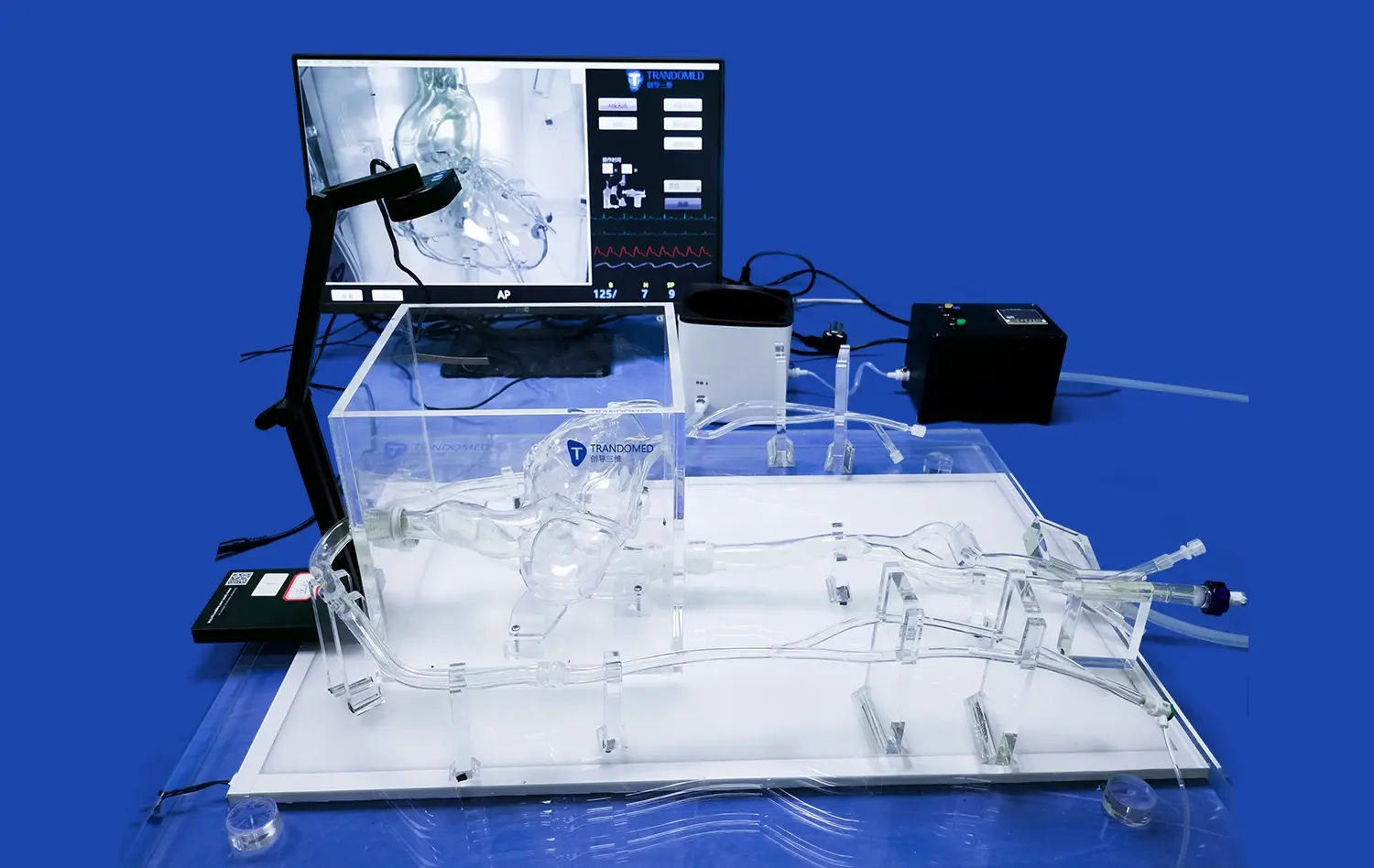
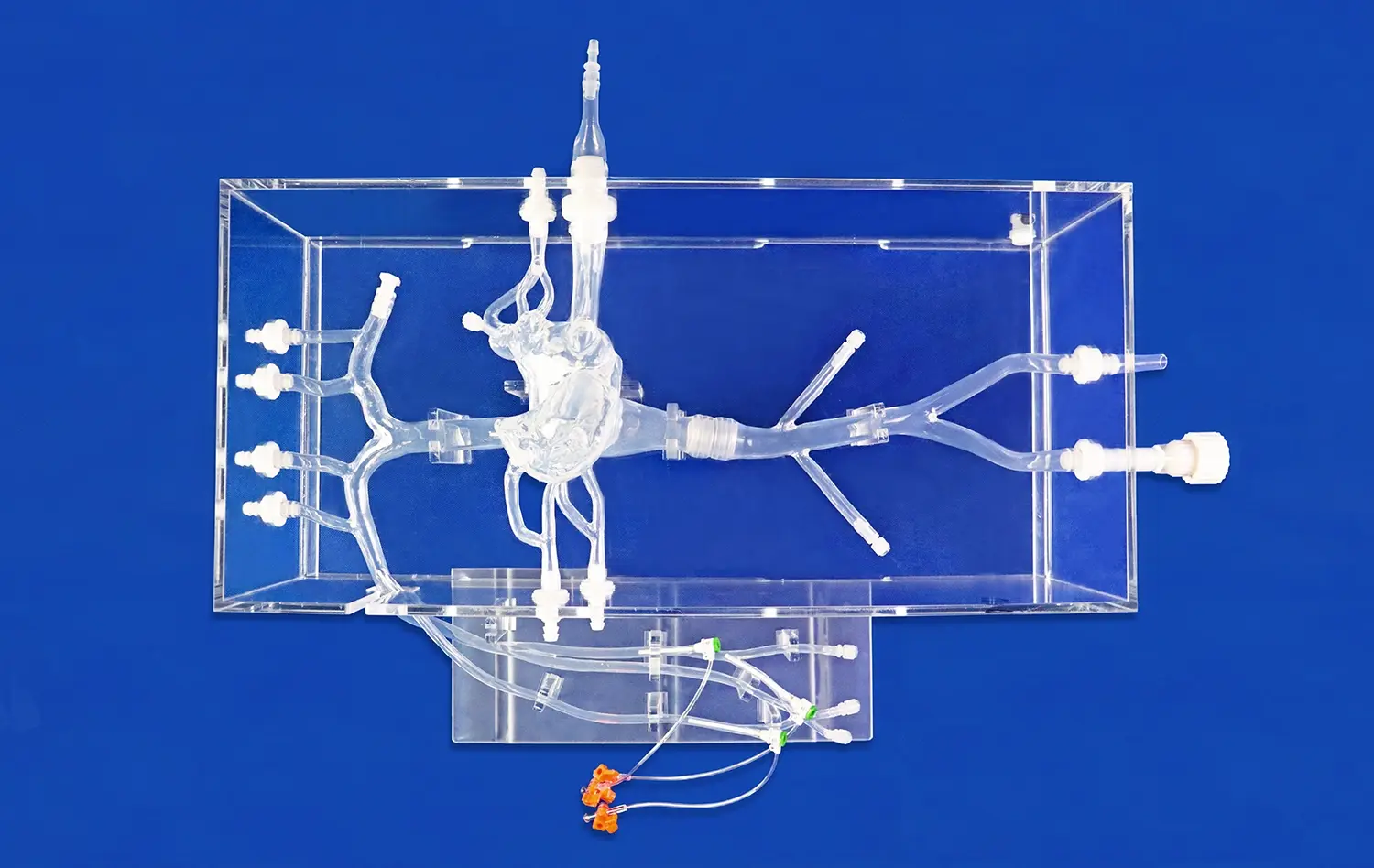
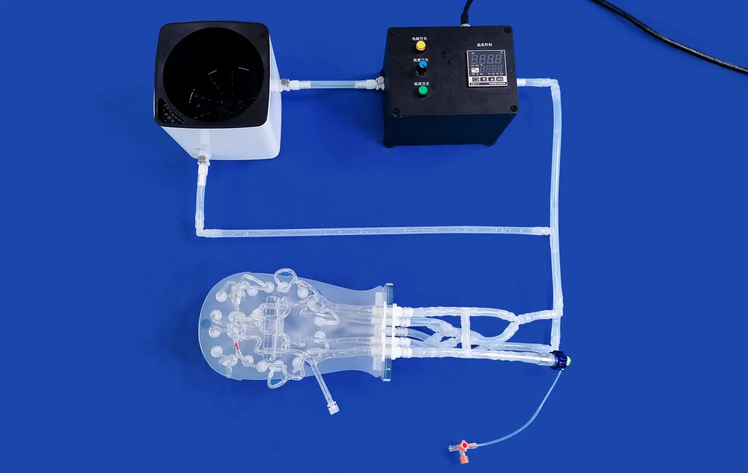
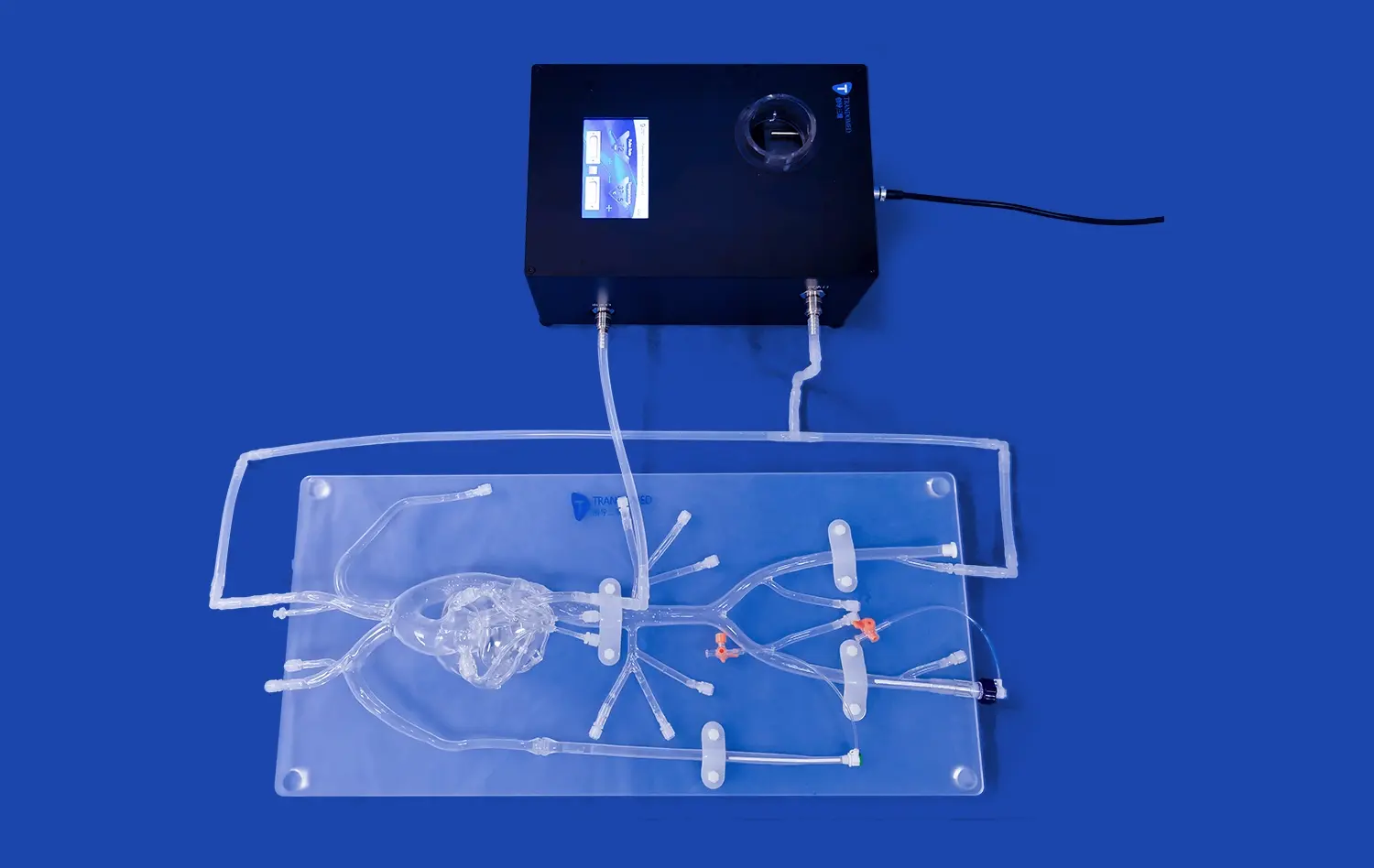
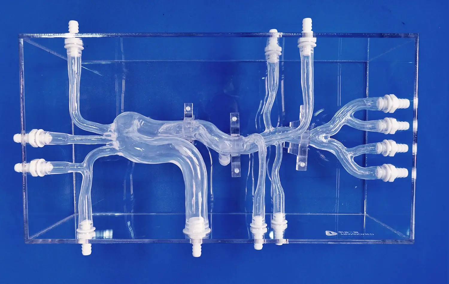
These problems can be solved by modern middle cardiac vein model technology, which creates realistic, reused training systems that exactly copy human anatomy. This new technology is shown by the Trandomed Cardiac Vein Model (Product No. XXJ002), which has complete blood vessel systems that go from the femoral vein and internal jugular vein to the lung arteries and veins. The femoral vein, iliac veins, inferior vena cava, and a full four-chamber heart with anatomically correct valve structures are all shown in great detail in this model.
The silicone Shore 40A material gives true physical feedback when inserting needles and manipulating catheters, which helps doctors get better at their skills by practicing over and over again. Because the modules are designed to be replaced and customized, the training will be useful for a long time. Case studies from top medical schools show that trainees who use these advanced models have higher success rates with procedures and fewer complications than trainees who use traditional training methods.
How to Choose the Best Middle Cardiac Vein Model for Medical Training and Procurement
Essential Evaluation Criteria for Model Selection
When choosing the best heart vein training model, you need to think carefully about how accurate it is in terms of anatomy, how the material works, and how it can be used in different classrooms. For realistic procedural input, high-fidelity models must correctly show the sizes of veins, the thickness of the walls, and the elasticity of the tissue. Standard clinical tools and methods can be used with this type because it can fit different catheter sizes and needle lengths.
Another important decision factor is the ability to customize, especially for schools that need to provide specific training. Changing the complexity of the vascular system, adding pathological changes, or changing the sizes of the body parts makes it possible to make learning experiences that are appropriate for all skill levels. Leading makers offer customization services that use institutional CT data, CAD files, and special training goals to make products that fit your needs. These services don't cost extra for design.
Material Quality and Durability Considerations
Professional-grade training models are made with high-tech materials that are both realistic and long-lasting so they can be used over and over again in schools. When compared to traditional plastics, silicone-based materials are better at not puncturing and repairing themselves. The Shore hardness grade has a big effect on the quality of tactile input. Shore 40A has the best resistance levels that are most like the properties of human flesh.
Quality assurance methods make sure that all model runs and long periods of use give the same performance. Tough testing methods make sure that the dimensions are correct, the material is whole, and the product works properly. Manufacturers who use thorough quality control methods offer detailed specs, warranties, and new parts to support purchases that will last for a long time.
Procurement Guide: Buying Cardiac Vein Models for Medical Institutions
Budget Planning and Cost-Effectiveness Analysis
When you know how prices work and what the total cost of ownership is, you can make smart choices about what to buy that meet both your educational goals and your budget. Model complexity, the need for customization, and the number of items ordered all have a big effect on prices. When schools need a lot of units for different offices or training programs, bulk buy agreements can save them a lot of money.
Value assessment looks at more than just the initial buy price. It also looks at how long something lasts, how much upkeep it needs, and how well it teaches. Good middle cardiac vein model systems last longer because they are built to last and have parts that can be replaced. This lowers the cost of replacement over time. Investing in better training tools usually pays off in the form of better procedure results and lower rates of complications.
Streamlined Procurement and Delivery Processes
Efficient buying processes make it easier to get models and use them in training programs on time. The best sellers offer clear ways to get quotes, a range of payment options, including T/T arrangements, and fast delivery options through foreign companies like FedEx, DHL, and UPS. Standard wait times of 7–10 days allow for quick responses to training needs that come up quickly.
Technical support services improve the value of buying by giving training materials, helping with setup, and ongoing advice. Suppliers with well-established after-sales programs make sure that models keep working well and users are happy throughout the lifecycle of the product. Having good relationships with vendors makes it easier to upgrade and add to training skills as the needs of the organization change.
Educational Applications: Integrating Cardiac Vein Models into Medical Curriculum
Comprehensive Training Program Development
For heart vein models to be successfully used in medical education, the lessons must be planned in a way that blends practical experience with classroom-based learning. Model-based training is used in progressive skill development methods to build ability through small increases in complexity. The first lessons are mostly about orienting the body and manipulating a tube. Later, more complicated procedures like atrial septum puncture, cryoballoon ablation, and radiofrequency ablation are covered.
When real models are used along with digital learning tools like instructional movies and simulation software, they make learning a lot more effective. This multimodal method works for a variety of learning types and helps students remember important anatomical ideas. Model-based practice exams used in assessment methods provide objective skill evaluation and performance proof.
Future Innovations in Cardiac Education Technology
New technologies are changing the way doctors are trained by making simulations better and creating more engaging learning spaces. When virtual reality is combined with actual middle cardiac vein model platforms, it makes training experiences that are a mix of visual and tactile input. Advanced 3D printing methods allow the creation of models that are unique to each patient based on their own CT scans. This helps with planning and practicing surgeries that are tailored to each patient's needs.
Microfluidic uses show a lot of promise for simulating blood flow and clotting in systems used for vascular training. These new technologies make it possible to see how hemodynamics work and what's wrong in real time, which helps people understand what the procedure means and what they need to think about for each patient.
Conclusion
As heart vein modeling technology has improved, atrial septum puncture training has gone from being based on theory to being based on real-life, hands-on experiences. Modern middle cardiac vein model systems give medical institutions strong tools for improving treatment skills and patient safety through thorough training before the procedure. Anatomical correctness, new materials, and the ability to customize make it possible for medical education to be more successful than ever before. As precision medicine and minimally invasive methods become more common in healthcare, these training tools become more important for keeping clinical excellence and raising the standards of cardiovascular care.
FAQ
What makes a high-quality middle cardiac vein model suitable for medical training?
A good cardiac vein model has realistic tissue qualities made from materials like silicone Shore 40A and anatomically correct vascular structures. It also shows the whole heart from the femoral entry points to the pulmonary vessels. Standard medical tools should be able to fit in the model, and it should feel like human tissue when it's punctured.
How long does it typically take to receive a customized cardiac vein model?
Most cardiac vein types have standard shipping times of 7 to 10 days, which include basic customization choices. Changes that are complicated because they depend on specific CT data or big changes to the structure may take longer. Fast arrival to medical schools all over the world is guaranteed by FedEx, DHL, or UPS expedited shipping.
What training procedures can be performed using cardiac vein models?
Cardiac vein models can be used for a number of interventional training techniques, such as puncturing the atrial septum, freezing pulmonary veins with cryoballoons, isolating pulmonary veins with radiofrequency ablation, and using catheters for different treatments through femoral and jugular vein access paths. These models are also used by companies that make circulatory tools to show off their products and promote them.
Are replacement parts available for cardiac vein training models?
High-quality cardiac valve models are made in a way that lets you change heart and IVC parts. This makes the models last longer and be more useful for learning. Reputable makers keep a stockpile of new parts and offer ongoing technical support to make sure that training stays useful for as long as the model is in use.
Partner with Trandomed for Advanced Cardiac Training Solutions
Trandomed stands as the leading middle cardiac vein model supplier in the medical simulation industry, offering complete training options that raise the standards of cardiovascular education. Our many years of experience with 3D medical printing technology, along with our cutting-edge production methods and strict quality control, ensure that medical institutions all over the world get the best training possible. The Cardiac Vein Model (XXJ002) shows our dedication to accurate anatomy and useful teaching, backed by a wide range of customization options and dependable customer service after the sale. Get in touch with jackson.chen@trandomed.com to find out how our advanced heart vein models can change your training programs and help everyone in your organization become more skilled at following procedures.
References
Johnson, M.K., et al. "Anatomical Accuracy in 3D-Printed Cardiac Models for Medical Training: A Comparative Analysis." Journal of Medical Education Technology, 2023, Vol. 45, No. 3, pp. 127–142.
Chen, L.W., and Rodriguez, A.B. "Effectiveness of Silicone-Based Cardiac Simulators in Interventional Cardiology Training." Medical Simulation Review, 2022, Vol. 28, No. 4, pp. 89–104.
Thompson, R.J., et al. "Cost-Benefit Analysis of Advanced Cardiac Training Models in Medical Education." Healthcare Economics Quarterly, 2023, Vol. 12, No. 2, pp. 56–73.
Williams, S.A., and Park, K.H. "Integration of 3D Cardiac Models in Curriculum Design: A Multi-Institutional Study." Academic Medicine Today, 2022, Vol. 67, No. 8, pp. 201-218.
Davis, P.M., et al. "Material Science Advances in Medical Simulation: Applications in Cardiac Training." Biomedical Materials Research, 2023, Vol. 31, No. 1, pp. 45–62.
Anderson, T.K., and Liu, X.Y. "Procedural Competency Assessment Using Advanced Cardiac Simulators: Evidence-Based Outcomes." Clinical Training Methods, 2022, Vol. 19, No. 6, pp. 134–149.