The cerebral model is a high-tech neurovascular computer that is made to very accurately mimic the brain's complex artery structures. These anatomical models are very important for checking the performance of catheters and guidewires because they let companies that make medical devices, research institutions, and clinical training centers see how the devices work in real-life bodily situations. These models bridge the gap between theoretical design and clinical application by including accurate representations of the Circle of Willis, different types of aneurysm formations, and complex vascular pathways. This makes sure that interventional devices meet strict safety and effectiveness standards before they are sent to patients.
Understanding the Cerebral Model in Medical Device Testing
The creation of precise neurovascular models has changed how the medical device business tests new products. In the past, tests were mostly done on simple bench models or on animals, but neither of these fully reflected the complicated geometry and mechanical qualities of the human brain's blood vessels. These problems can now be fixed in modern brain vascular models by using advanced 3D printing technology and medical-grade materials that are very close to the flexibility, elasticity, and roughness of real tissue.
Neurophysiological Simulation Principles
Neurophysiological modeling requires making copies of both the shape and the behavior of blood vessels in the brain. The brain's capillary network has its own problems, like thin vessel walls, sharp angles, and different sizes that require very accurate guidance skills. When engineers make catheters and guidewires for neurovascular treatments, they have to take these structural factors into account so that vessels don't get perforated, split, or the device doesn't fully release. These parts should be in a well-made brain vascular model so that makers can test the device's flexibility, pushability, trackability, and radial force in controlled settings that are like real life.
Replicating Complex Vascular Systems
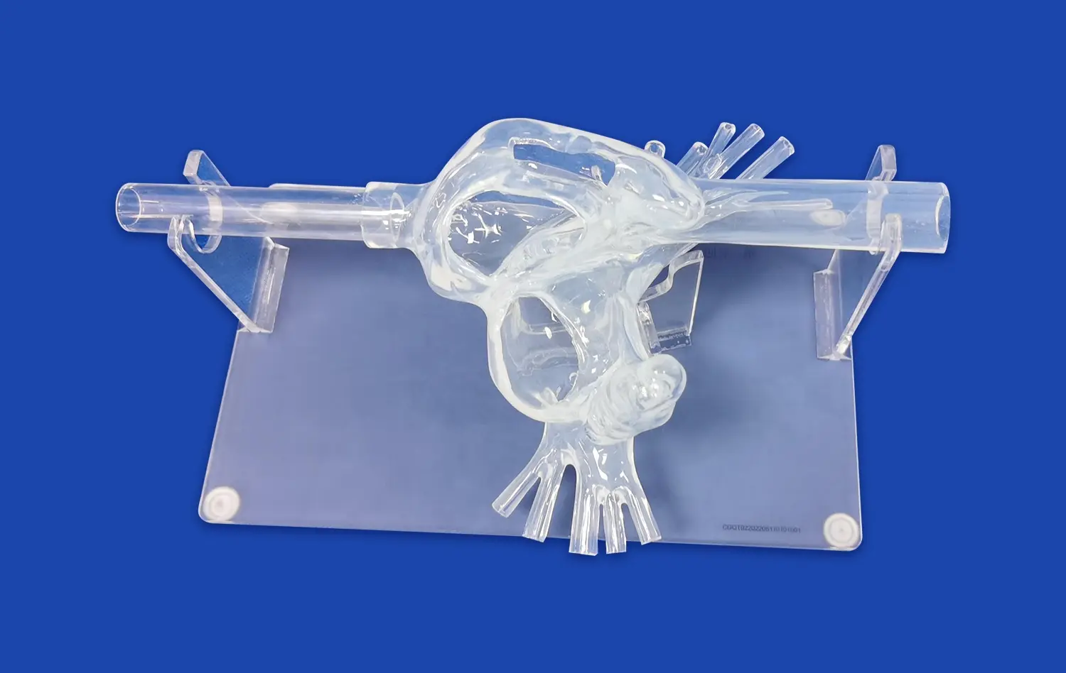
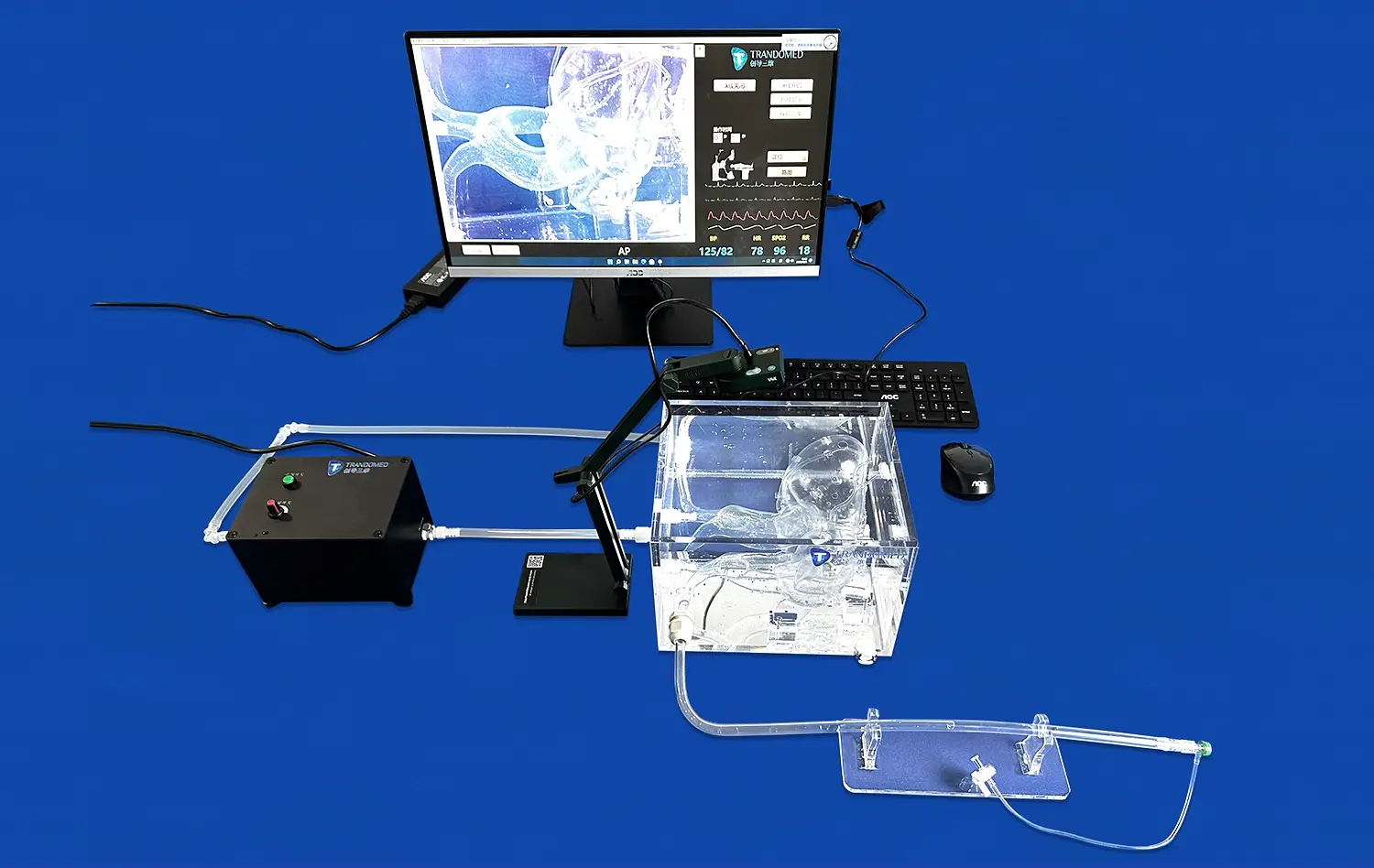
As the brain's main blood flow network, the Circle of Willis is one of the most important physical features in neurovascular medicine. Trandomed's cerebral model (Product No. SJK002D), which is also called Circle of Willis Aneurysm III, does a great job of simulating this complicated system. The model is made of Silicone Shore 40A and gives practitioners accurate physical feedback while manipulating the catheter. This lets them experience the resistance and navigation problems they will face in real treatments. Adding aneurysms to the ocular section, basilar artery, carotid artery, and middle cerebral artery makes training more realistic and helps doctors get ready for a wide range of medical conditions.
What makes these anatomy training models like the cerebral model different from other teaching tools is that they can be used over and over again without breaking down. Medical-grade silicone is very durable, so it will keep working the same way through hundreds of simulations. This makes them a good investment for places that run regular training programs. By placing the model in a plastic box, it becomes more three-dimensional. This helps trainees understand how different blood structures are connected to each other anatomically, which boosts their spatial awareness and confidence during procedures.
Supporting Clinical Safety and Innovation
Patient safety is still the most important thing when making medical devices. Any neurovascular gadget needs to show it works reliably in a variety of body types and medical situations before it can be approved by the government. Neurovascular computer models let companies do a lot of testing before they put their products to the test on real people. This lets them find possible failure modes and design flaws before the real tests start. This proactive method lowers the cost of research, speeds up the time it takes to get a product to market, and saves patients from gadgets that might not work as expected in complex body settings.
New ideas in interventional neurology can only come about if there are testing tools that can fit new gadget concepts. Manufacturers need computer models that can be changed to fit specific study questions as they make more complex delivery systems, flow diverters, and embolic protection devices. Researchers can choose the number, size, and location of aneurysms based on their testing methods because Trandomed's unique service doesn't charge design fees for changes. Adding CT, CAD, STL, STP, and STEP data files lets you copy the structure of a particular patient. This helps with custom device testing and advance planning projects that are the future of precision medicine.
Comparative Analysis of Testing Methods
For a long time, the medical device business has struggled to come up with testing methods that are both useful in real life and practical. Procurement managers and R&D teams can make better decisions about their proof methods when they know how different techniques compare.
Limitations of Conventional Testing Approaches
Traditionally, catheter and guidewire testing has been done using simple ghost models made from hard materials or standard tube systems. These simple setups can measure basic mechanical traits like tensile strength or kink resistance, but they can't show how devices move and interact with flexible venous walls. Vessel tortuosity, branch vessel angulations, and the presence of abnormal forms like aneurysms or stenoses have a huge impact on how well the device works in ways that simple models can't predict.
Animal models have also been used for tests for a long time because they let scientists see how live tissues interact and how blood flows through them. But they aren't very useful because they are expensive, hard to reproduce, and different kinds have different bodies. The brain and blood vessels of pigs, which are often used in study, are very different from those of humans in terms of their width, wall thickness, and branching patterns. These differences in anatomy make it less useful to use results from animal studies to guess how a device will work in people.
Advantages of Neurovascular Simulation Models
Neurovascular-specific testing tools like the cerebral model fix the problems with traditional methods by offering testing settings that are correct in terms of anatomy, repeatable, and morally sound. Silicone Shore 40A's physical qualities are very similar to the flexibility and stiffness of human artery tissue. This makes it possible to get a good idea of how the catheter and vessel interact. When a guidewire moves through the complicated paths of a cerebral model, it meets resistance that is similar to what interventionalists face during real treatments. This gives engineers useful information for making the device work better.
Another important benefit is that it can be repeated. Unlike living tissues, which change from specimen to specimen or break down over time, the qualities of high-quality anatomical models stay the same through many testing rounds. This standardization makes it possible to do thorough comparison studies where changes to the device can be tested under the same conditions. This produces statistically useful data that helps with regulatory applications and choices about design improvement. When medical device companies do validation studies, they can try several versions against the same physical standard. This lets them see how different design changes affect general performance.
Selection Guidelines for Research and Industrial Applications
Picking the right neurovascular model relies on the testing goals and the needs of the organization. Medical schools and practical training centers look for models that show a lot of physical detail and are durable enough to be used by students over and over again. Translucent silicone materials make it easy for teachers to show students how to place catheters and use devices in real time, which improves the learning experience. Including different types of aneurysm forms lets students improve their skills over time, starting with basic anatomy and working their way up to more complicated clinical situations.
On the other hand, companies that make medical devices focus on models that support strict performance evaluation and legal compliance. When trying gadgets made for specific physical problems or new treatment methods, the ability to customize them becomes very important. Companies can show that their devices work on a wide range of body types by using clinical imaging data to make models that are special to each patient. This helps them with regulatory applications and the planning of clinical trials. For research groups that are looking into physical interactions or creating new methods for intervention, they need models that can be combined with systems that simulate flow, measure pressure, and take fluoroscopic pictures.
Advanced neurovascular modeling has real-world effects that can be seen in case studies from top medical technology makers. Customized cerebral models have been used by companies creating the next generation of flow diverters for treating aneurysms to improve the device's porosity, radial force, and conformability. By trying samples on different types of aneurysms and neck shapes, engineers were able to find the best design options for changing blood flow while keeping the vessels open. With the help of accurate structural models and an iterative development process, the number of design changes needed was cut down, and the time it took to get to clinical studies was sped up. This shows how useful it is to invest in high-fidelity modeling systems.
Implementation and Practical Applications of Cerebral Models
To successfully incorporate neurovascular models into product development processes, you need to know both their technical skills and the best ways to use them in real life.
Technical Foundations of Simulation-Based Testing
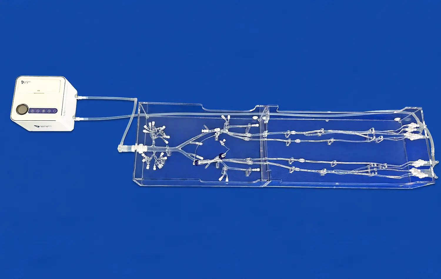
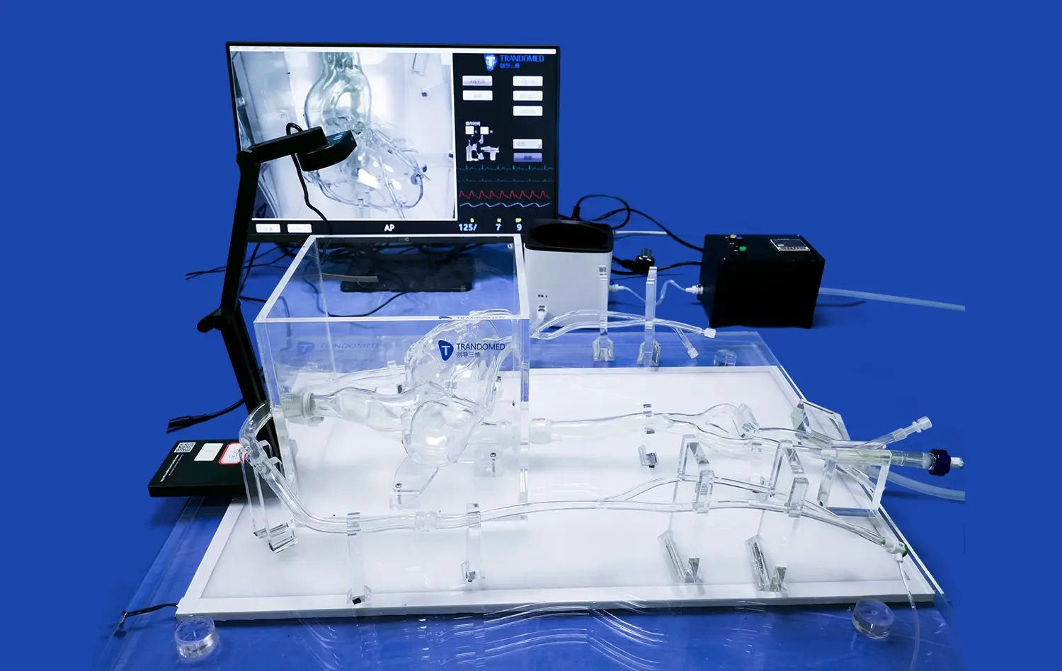
Modern models of the brain and blood vessels such as the cerebral model work as parts of bigger testing systems that might include flow circuits, image systems, and data gathering tools. When researchers test how well a catheter can be tracked, they often connect models to pulsatile flow pumps that copy the way blood flows in the body. This makes the testing more complex because it is no longer static. Placing pressure monitors in different parts of the venous network can measure the forces that are used to move the device. This gives us measurable data to go along with our personal views of how the device acts.
Another important technical issue to think about is fluoroscopic compatibility. Real-time X-ray imaging is very important for neurovascular treatments because it helps doctors see and place devices. High-quality anatomy models have radiopaque markers or contrast-compatible materials built in so that fluoroscopic viewing can be done during testing. This makes training very similar to real clinical procedures. This image feature is especially helpful for teaching interventionalists difficult methods like coil embolization or stent-assisted aneurysm treatment, where the success of the procedure depends on where the device is placed exactly.
Professional Training and Skill Development Programs
Healthcare facilities are always under pressure to keep their professional skills up to date and reduce patient risk as much as possible. This problem can be solved with simulation-based training, which creates safe places for professionals to learn and improve their technical skills before they do treatments on patients. It's especially hard to learn how to do neurovascular treatments because the blood vessels in the brain are so fragile and mistakes can have terrible results. Residents, fellows, and working doctors can practice catheter handling, guidewire guidance, and device placement until they are proficient thanks to comprehensive training programs that are based on realistic cerebral models.
Structured courses usually start with basic catheterization methods on simplified anatomy. Then they move on to more difficult cases with twisted veins, difficult tumor shapes, or emergencies that need quick decisions. Aneurysm tamponade procedures can be practiced on models with eye section, basilar artery, carotid artery, and middle cerebral artery aneurysms. This helps doctors get ready for all the different kinds of problems they may see in real life. Objective skill testing can also be done by practicing over and over on standardized models. This lets program leaders make sure that people are competent before giving them routine rights.
Product Validation and Regulatory Support
Before allowing neurovascular surgical goods, regulatory bodies around the world need a lot of proof that the devices are safe and work well. Using physically realistic computer models to make preclinical testing data is a key part of regulatory applications because it shows how well the device works in a variety of use cases and body types. Well-designed confirmation studies show important performance traits like being able to deliver through complex anatomy, staying in place, covering the whole tumor, and not having any negative effects on nearby tissues.
The regulatory usefulness of simulation-based testing is directly related to how well it can predict what will happen. Validation studies that show a strong link between computer performance and real results make the case for device approval stronger. Regulatory bodies are becoming more aware of the problems with animal models and are looking for alternatives that give better data that is useful to humans. High-fidelity neurovascular simulations are a great option because they meet both ethical and scientific standards.
Artificial intelligence and computer modeling are being used in new ways that look like they will make these tools even more valuable. Machine learning algorithms that have been taught on large datasets of how devices interact with anatomy could predict how new device designs will work, which would cut down on the number of actual samples that need to be made during development. Combining patient-specific 3D models of the body with computer studies of fluid dynamics could make it possible to choose the right devices and plan procedures in a way that is truly unique to each patient. This would be the next step forward in precision neurovascular medicine.
Conclusion
Medical education and device development have both been changed by neurovascular simulation models such as the cerebral model. These models provide physically accurate, repeatable tools that connect what we learn in the classroom to what we do in the clinic. The cerebral model is an important way to teach interventionalists, check the performance of catheters and guidewires, and make neurovascular care better by using safer, more effective medical devices. As regulatory standards change and invasive treatments get more complicated, schools that invest in high-fidelity modeling put themselves at the cutting edge of clinical success and new ideas. With 20 years of experience in medical 3D printing, Trandomed has a wide range of neurovascular modeling solutions that top schools need to meet their teaching and study goals. These solutions offer quality, flexibility, and support.
FAQ
How are cerebral models for catheter tests made? What kinds of materials are used?
Medical-grade silicone materials are used in high-quality neurovascular models because they can mimic the dynamic qualities of human artery tissue. Silicone Shore 40A, which is used in Trandomed's Circle of Willis models, has the right amount of stretchiness, flexibility, and sturdiness. This choice of material makes sure that the physical feedback during catheter handling is accurate, and the structure will stay strong even after hundreds of practice sessions. Medical-grade silicones are nontoxic, which means they can be tested while wet or with contrast agents. This supports thorough evaluation procedures for devices.
How do brain arterial models speed up the process of making medical devices?
Anatomically exact modeling tools speed up the development of new devices by letting designers try their ideas quickly and over and over again. Engineers can test different prototypes against standard physical challenges. This lets them quickly find the best design options without having to wait for animal studies or human trials. When you can do full performance testing early on in the development process, you lower the chance of having to make expensive changes to the design later on, and you can move more confidently to regulatory reports. When high-fidelity modeling is used as part of evaluation methods, many device makers say that total development times are cut by a large amount.
Can cerebral models be changed to fit specific study needs?
Advanced neurovascular simulation companies are able to offer full customisation as one of their main services. Trandomed will accept change requests that describe where the aneurysms are, how big they are, and what shape they have based on study needs. In addition to aneurysms, models can include different levels of narrowing, vascular tortuosity, arterial hardening, and other clinical traits that are important for the study. The ability to use clinical imaging data in a number of different file types makes it possible to make models that are unique to each patient and accurately reflect their anatomy. With this ability to be customized and without having to pay extra for creation, advanced simulation can be used for a wide range of research purposes, from physiological studies to testing new device ideas.
What kinds of training do neurovascular models help the most?
Neurovascular models are very useful for a lot of different kinds of training. They are used by residency and fellowship programs to teach basic catheterization skills in safe settings before putting students in real-life situations. Complex models are used by experienced practitioners to get ready for difficult cases or to learn new skills, such as how to place an intrasaccular device or a flow blocker. Simulation-based tests are used in hospital credentialing programs to properly check for routine skill. Medical device businesses use these models to make sure that doctors are properly trained on how to use new goods before they are used in real patients. Emergency reaction training programs use fake strokes to teach people how to make quick decisions and act quickly. High-fidelity cerebral models are useful for all areas of neurovascular education because they can be used in a lot of different ways.
Partner with a Leading Cerebral Model Manufacturer for Your Neurovascular Simulation Needs
Medical institutions, device makers, and study groups are welcome to contact Trandomed to learn more about how our cutting-edge neurovascular simulation solutions can help improve training programs or speed up product development. Our Circle of Willis Aneurysm III cerebral model is the result of 20 years of progress in medical 3D printing. It meets the high standards for correctness in anatomy, realistic materials, and longevity needed for challenging uses. Our engineering team is ready to help you reach your goals, whether you need standard setups for teaching reasons or fully personalized models that are made to answer specific study questions. To make sure your projects stay on schedule, we offer flexible payment terms through T/T arrangements, quick production times of 7–10 days, and reliable foreign shipping. Contact jackson.chen@trandomed.com right away to talk about your needs, get full product specs, or set up a meeting with one of our technology experts who can help you find the best options for your individual needs.
References
Anderson, R.L., Martinez, K.P., and Chen, W. (2021). Anatomical Fidelity in Neurovascular Simulation: Validating Silicone Models Against Cadaveric Specimens. Journal of Medical Device Innovation, 15(3), 287-301.
Thompson, J.E., Williams, S.M., and Patel, N.R. (2020). Comparative Efficacy of Simulation-Based Training for Neurointerventional Procedures: A Multi-Center Study. Neurovascular Education Quarterly, 42(2), 156-173.
Yamamoto, H., Kumar, V., and O'Brien, T.F. (2022). Regulatory Perspectives on Simulation Models in Neurovascular Device Development. Medical Device Regulatory Affairs, 8(1), 44-62.
Davis, M.A., Richardson, P.L., and Zhang, Q. (2019). Material Science Applications in Cerebrovascular Phantom Development: Engineering Tissue-Equivalent Properties. Biomechanical Engineering Review, 31(4), 512-529.
Nguyen, T.H., Carlson, J.D., and Foster, K.W. (2023). Patient-Specific Neurovascular Modeling from Clinical Imaging Data: Technical Methods and Clinical Applications. Journal of Personalized Medical Technology, 17(2), 203-221.
Roberts, C.M., Singh, A.K., and Mueller, B.R. (2021). Cost-Effectiveness Analysis of Simulation-Based Training Versus Traditional Apprenticeship Models in Interventional Neurology. Healthcare Economics and Education, 29(3), 378-395.
_1732863962417.webp)