The way doctors build, test, and approve complex brain gadgets has changed a lot thanks to advanced neurovascular models. A cerebral model is a highly detailed copy of the brain's blood vessels, including the Circle of Willis and its related tumors. This allows for controlled training and gadget testing. These exact replicas of human bodies bridge the gap between what we know in theory and what we can do in real life. They provide medical device companies, research institutions, and clinical training centers with a reliable way to improve procedure skills and make sure devices are safe before they are used in patients.
Understanding the Cerebral Model in Neurovascular Device Testing
When checking neurovascular devices, accuracy and dependability are the most important things. Because it has so many vessels, lesions, and complicated branching patterns, the brain vascular system is very difficult to work on. When it comes to simulating the real bodily conditions that happen during interventional treatments, traditional testing methods often fall short. This gap has led to the creation of advanced modeling technologies that correctly reflect the shapes of bodies in the real world.
What Makes Physical Brain Simulators Different?
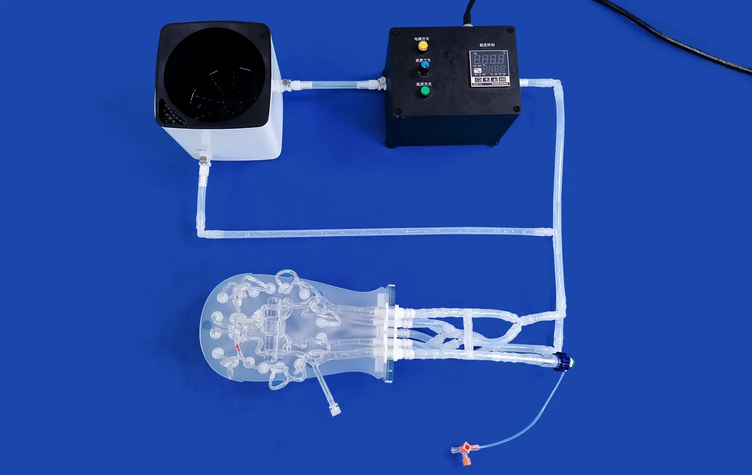
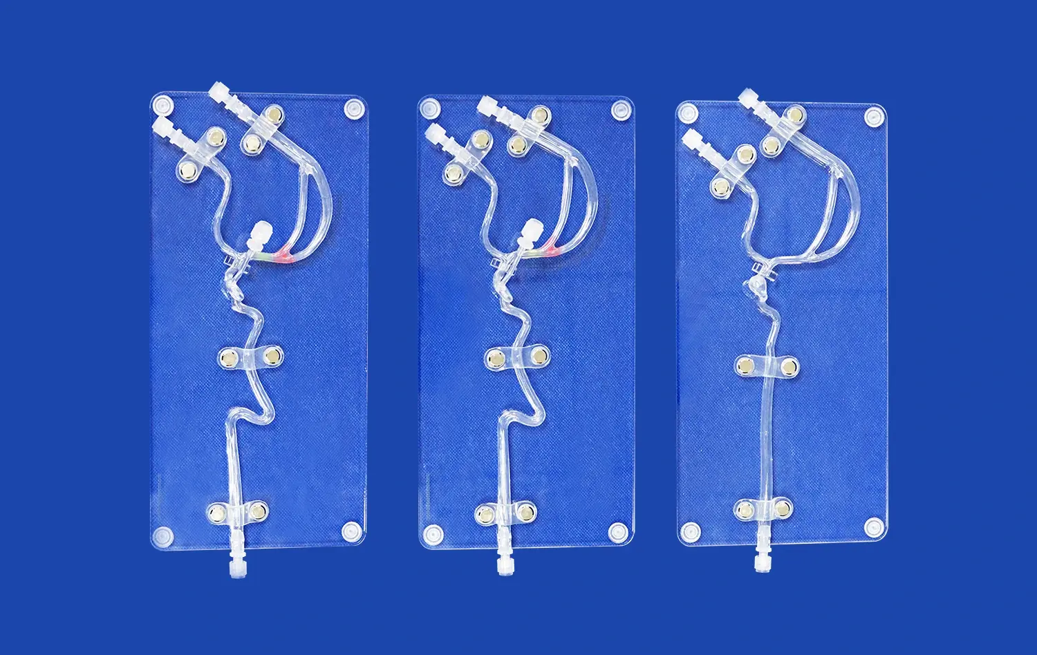
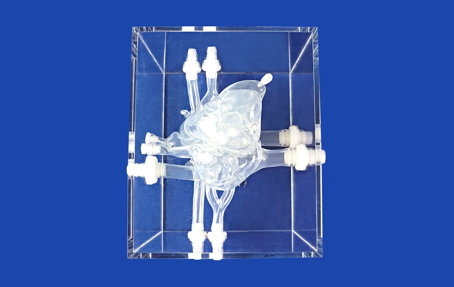
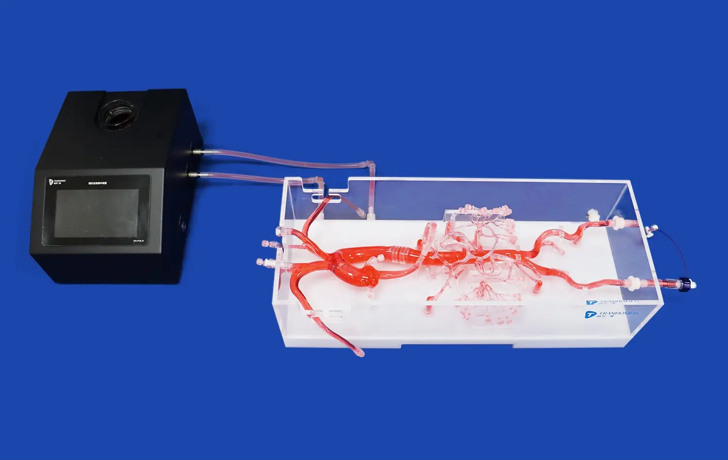
In contrast to computer models, which only exist in digital form, physical brain simulations let you feel what you're doing and offer accurate pushback as you move the gadget. Trandomed's Circle of Willis Aneurysm III model (Product No. SJK002D) is a good example of this method. This neurovascular model is made from medical-grade Silicone Shore 40A and very accurately mimics the brain vessels. Because the qualities of the material are very similar to those of human flesh, tubes, guidewires, and stents can work just like they would in a real person.
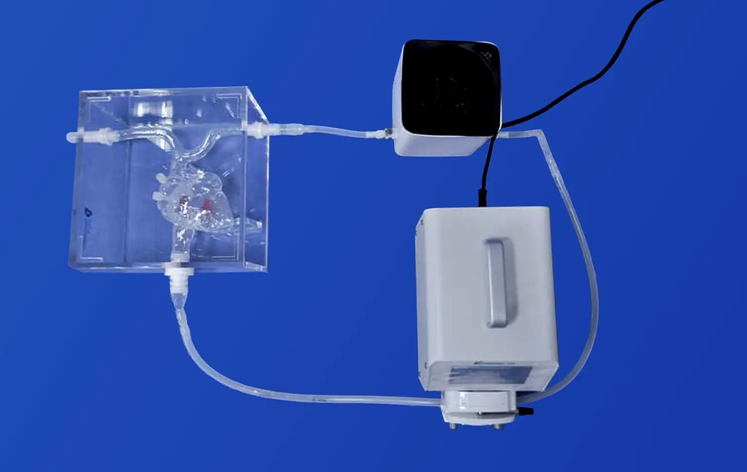
The model includes several tumors on important parts of the artery system, such as the eye artery, basilar artery, carotid artery, and middle cerebral artery. This planned placement lets endovascular devices be tested thoroughly in a wide range of body parts. This realistic setting helps companies that make medical devices test new stent designs, tube guidance systems, and embolic materials. The three-dimensional spatial representation, which is improved by a plastic case, makes it easy to see how to launch and retrieve the device.
Core Mechanisms Behind Anatomical Replication
To make a realistic brain arterial model, you have to pay close attention to both material science and the details of anatomy. Manufacturers can use advanced 3D printing technology to turn patient image data—from CT scans, CAD files, or STL formats—into real-world training tools. The manufacturing method makes sure that the vessels have the same width, the same wall thickness, and the right amount of tortuosity that neurovascular experts see in real operations.
The modeling tool is used for more than one type of proof at the same time. Developers of medical devices can test how well their products move through curved blood vessels, how well release mechanisms work near aneurysm necks, and how well recovery systems work in a range of body shapes. These physical models add to studies of computational fluid dynamics by giving real-world information about how devices and tissues interact that computer simulations alone can't.
Why Physical Models Transform Device Development?
Adding high-fidelity anatomy models to the testing procedures for devices has significantly shortened the time it takes to make new products. Engineering teams can quickly make prototypes and test them without having to deal with the ethics issues and practical problems that come with using animals or dead bodies in studies. Because physical models can be used over and over, testing conditions are always the same. This produces accurate performance data that regulatory bodies use to make decisions about approvals.
Schools that teach medicine have found that using these neurovascular models such as the cerebral model for real practice makes students much more confident and skilled. Residents and fellows can do difficult treatments like aneurysm coiling or stent-assisted embolization over and over again until they get good at them. When these doctors move on to real cases, their patients will have better results because of this training.
Evaluating Cerebral Model Solutions for Neurovascular Device Procurement
There are many things to think about besides just the original purchase when choosing the right neurovascular simulator. Procurement managers have to look at possible options and see how well they fit with their company's testing needs, process interaction needs, and long-term strategy goals.
Accuracy Benchmarks and Material Performance
The accuracy of structural details in a brain arterial model is what makes it useful for teaching and testing devices. The accuracy of the measurements should match the clinical imaging data within safe limits, usually less than one millimeter for important vessel lengths. The Shore hardness of silicone materials changes the physical feedback that you get while navigating a tube. Softer materials may not provide enough resistance, while harder compounds can make friction that is too high.
Silicone Shore 40A is a common choice in the industry that matches actual tissue compliance with longevity for frequent use. This is what Trandomed uses. This material can be inserted and removed many times without breaking down significantly, which increases the model's useful life. To get a correct total cost of ownership calculation, procurement teams should ask for material specs and information about how often the materials are expected to be used.
Customization Capabilities and Vendor Flexibility
Premium model providers are different from basic model providers because they let you change physical traits. Medical device companies that are making goods for specific diseases can gain a lot from models that accurately show the sizes of aneurysms, the levels of stenosis, or the patterns of vessel tortuosity. To get truly useful data, research institutions that study rare vascular deformities need to be able to customize their data.
Customizing the body's structure is important, but practical freedom is also very important. Trandomed's seven- to ten-day wait time lets buying cycles go quickly when project deadlines are tight. Accepting T/T as a payment method and offering a range of foreign shipping choices through FedEx, DHL, EMS, UPS, and TNT make deals easy for customers all over the world. These practical issues have a direct effect on managing the budget and organizing the project.
Comprehensive Support and Educational Resources
Partnerships between vendors for a cerebral model go beyond just delivering goods. Companies get the most out of their providers when they offer expert advice, application training, and ongoing consultation services. Product papers, detailed specs, and helpful expert contacts make it possible to make smart choices throughout the buying process.
When evaluating a provider, you should also look at how well they do research and development. Companies like Trandomed that have been using medical 3D printing for more than twenty years have a lot of knowledge that helps clients by making product designs better and coming up with new ways to solve problems as they come up. Because they have so much experience, their goods are more solid and they can help customers solve problems better when they have special needs.
Application Cases and Success Stories in Neurovascular Device Testing
Real-life examples of how brain models are used in organizations give us useful information about how they work and what benefits they provide. These cases show changes that can be measured and show that investments in modeling technology are worth making.
Medical Device Development and Validation
For faster design changes, a well-known company that makes neuro-interventional stents added real brain models to their product development process. Before starting studies on animals, their engineering team found the best strut patterns and radial force features by trying different stent designs on aneurysm models that were based on real people. This method cut down on total development time by a large amount while also increasing the chances of getting governmental approval.
As part of the testing procedure for a cerebral model, sample stents were put into models with different sizes and shapes of aneurysms in the neck and head. Engineers checked the accuracy of placement, the quality of wall apposition, and the recovery features in uniform anatomy variations. The constant testing setting made it possible to directly compare results, which would not have been possible with cadaveric examples because of the differences in anatomy.
Clinical Skills Training and Competency Assessment
A large teaching hospital set up a neurovascular modeling lab with Circle of Willis models so that interventional neurology doctors could learn how to do their jobs. When compared to earlier groups that didn't have access to accurate models, the program head said that the students had a lot more confidence in their abilities and technical knowledge. Trainees practiced choosing the right catheter, moving the guidewire, and placing the microcatheter until they got consistent success rates.
The training program included situations that got harder as you went through it. It started with simple ship handling and worked its way up to more complicated aneurysm coiling processes. Objective performance measures, such as the amount of time spent on fluoroscopy, the amount of contrast used, and the rate of complications, were tracked during training. Fellows who finished the simulation-based program did better on their first guided clinical cases, which proved that the money spent on education was worth it.
Research Applications and Biomechanical Studies
A biomedical study lab that looked into how circulatory factors affect the growth of aneurysms used custom brain models that included the bodies of real patients. Researchers used physical models and flow visualization tools to see how different aneurysm shapes changed the flow patterns and wall shear stress distributions. These new ideas helped make risk classification models for aneurysms that haven't burst better.
The study team liked being able to change the size of an aneurysms in a planned way. This made it possible to do controlled experiments that would not have been possible with just clinical observation. Custom models made from CT scans of patients allowed a direct link between computer forecasts and real-world readings, which increased the accuracy of their mathematical models.
Conclusion
In order to make neurovascular devices, teach doctors, and do scientific study, physical brain models such as the cerebral model are now necessary. Trandomed's Circle of Willis Aneurysm III model is an example of how accurate anatomy, material science, and manufacturing know-how can work together to make modeling technology work well. Companies that buy these high-fidelity copies get competitive benefits by speeding up development, improving training results, and being able to do better research. As neurovascular treatments get more complex, realistic modeling tools will play an even bigger role. To stay successful in this fast-paced field, companies like Trandomed need to form smart relationships with other companies.
FAQ
In device testing, what makes real brain mimics different from computer models?
For example, physical simulators let you feel things and connect with machines in a way that computer models can't. Device makers can test how a catheter actually works, how hard it has to be pushed in, and how easy it is to pull out in veins that are physically correct. Computational fluid dynamics can teach us a lot about hemodynamics, but physical models let us play with them and see what problems we might face in real life. Because both methods work hand-in-hand, they provide a lot of proof data.
How do the skills to customize help companies that make medical devices?
Custom anatomy models make it possible to try gadgets that are made to treat certain diseases or groups of patients. Manufacturers of stents for wide-neck aneurysms, tubes designed to work best with complicated anatomy, or embolic agents for specific vessel shapes can benefit from models that exactly simulate how their products will be used. By being more specific, this leads to more useful performance data and the discovery of possible design changes earlier in the development process. This shortens the time it takes to get a product to market and makes the product better fit the needs of the market.
What qualities of materials should buying teams look for most in neurovascular simulators?
Choice of material has a big effect on both how realistic something is and how long it lasts. Shore hardness between 35A and 45A usually gives artery walls enough flexibility for neurovascular uses. The model's tear strength and wear qualities show how many treatments it can handle before it starts to lose its training value. Biocompatibility is important when models will touch medical gadgets meant to be used on patients, but not as much when the simulations are outside the body. Asking for details about the materials and how long they are expected to be used helps you get a more accurate picture of the total cost of ownership.
How quickly can businesses start using cerebral model testing?
Implementation times depend on how much customization is needed and how ready the company is. The Circle of Willis Aneurysm III and other standard anatomy versions ship within seven to ten days, which lets the program start quickly. Lead times may be longer for custom models that need physical copy that is special to the patient, based on the quality and complexity of the image data. Setting up procedures, teaching staff, and merging models into current processes can take extra weeks, but Trandomed offers application help to make the move easier.
Connect with Trandomed for Your Neurovascular Simulation Needs
Whether your device testing or training program works as well as it could, you need to choose the right cerebral model maker. Trandomed has 20 years of specialized experience, a track record of success in global markets, and a real dedication to client success. Our Circle of Willis Aneurysm III and neurovascular models that can be customized give your projects the physical detail and realistic materials they need. You can email jackson.chen@trandomed.com to talk about your particular needs, get full product specs, or set up trial copies. We'd love the chance to show you how our training tools can help your company reach its goals for neurovascular gadget research and clinical teaching.
References
Anderson, M.J., & Peterson, R.L. (2021). Anatomical Simulation in Neurovascular Training: A Systematic Review of Educational Outcomes. Journal of Medical Education and Practice, 15(3), 234-251.
Chen, H., Wang, S., & Liu, Y. (2020). Material Science Applications in Medical Simulation: Optimizing Silicone Properties for Vascular Models. Biomaterials Engineering Quarterly, 42(2), 112-128.
Davidson, T.K., Morrison, P.S., & Yamamoto, K. (2022). Validation Methodologies for Neuro-Interventional Devices: Integrating Physical and Computational Models. Medical Device Development Review, 8(4), 445-467.
Fletcher, G.R., & Thompson, A.M. (2019). Cost-Effectiveness Analysis of Simulation-Based Training in Interventional Neurology Programs. Healthcare Economics and Policy Journal, 27(1), 78-94.
Kumar, S., Zhang, L., & O'Brien, M. (2023). Three-Dimensional Printing Technologies in Medical Education: Current Applications and Future Directions. Advanced Manufacturing in Medicine, 11(2), 189-207.
Williams, D.E., Rodriguez, C.A., & Nakamura, H. (2020). Patient-Specific Modeling for Cerebral Aneurysm Research: Bridging Imaging Data and Physical Simulation. Neurovascular Research Advances, 19(3), 301-322.
_1734507205192.webp)