The cerebral model is a huge step forward in neurovascular education and is an important tool for teaching personnel how to talk to patients and do procedures. These high-tech physical models accurately mimic the complicated structure of the brain's blood vessels, which helps doctors explain difficult neurological ideas clearly and precisely. By turning abstract medical knowledge into real, visual representations, cerebral models help healthcare professionals and patients communicate better, leading to better understanding and less fear during neurological procedures. Medical schools all over the world are realizing that these anatomy models are essential tools for improving student results and practical readiness.
Understanding Cerebral Models in Healthcare
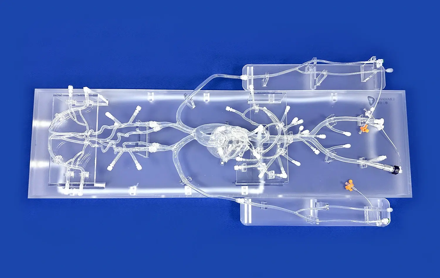
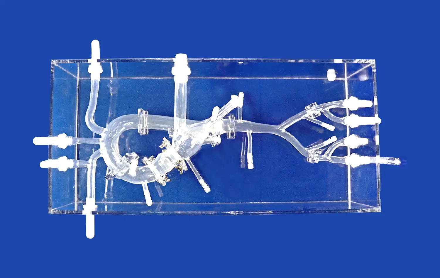
Cerebral models are highly accurate copies of the brain's arterial system that were carefully made to show how complicated cerebral circulation is. These neurovascular models make exact copies of important structures like the Circle of Willis, brain arteries, and different medical conditions like aneurysms, stenosis, and vascular malformations. In addition to being useful for imagery, they are also engaging learning tools that change the way doctors talk to their patients about their brain health.
Anatomical Precision and Educational Value
Anatomical correctness and material consistency are very important for any brain arterial model to work well. Modern brain models are made of medical-grade plastic materials that are very close to the way real blood vessels feel. This Silicone Shore 40A material was made to be used in medical uses. It simulates real flesh, which improves both visual and tactile learning. This choice of material makes sure that it will last for many teaching demos and keep its shape so that it truly represents bodily conditions.
In medical school settings, these physical models help students and doctors learn about the structure of the brain in a way that two-dimensional images can't. The three-dimensional space representation, which is often improved by acrylic mounting systems, lets students look at blood vessel connections from different angles. For example, they can see how the ophthalmic segment connects to the internal carotid artery or how the basilar artery splits into posterior cerebral vessels. This detailed picture helps healthcare workers remember what they've learned about anatomy and gets them ready for real-life clinical situations.
Patient Communication and Anxiety Reduction
When neurosurgeons get patients ready for brain treatments, they need to be able to communicate clearly to get their informed consent and help them deal with their worry. Traditional ways of explaining things that only use words or x-rays to show what's going on often leave patients feeling confused and scared. This changes with cerebral arterial models because they give patients real-life visual aids that make complicated treatments easier to understand.
A person who is getting care for an aneurysm can see exactly where the troublesome vascular bulge is in their brain circulation. The doctor can use the structural model to show how interventional devices will move through the artery paths and clearly explain the coiling or stenting procedure. This real-life example boosts the patient's confidence, lowers their nervousness before surgery, and makes it easier for them to have useful conversations about their treatment choices and expected results. Patients who learn about their illnesses visually through physical models regularly report higher levels of happiness and a better understanding of their conditions.
Applications Across Medical Specialties
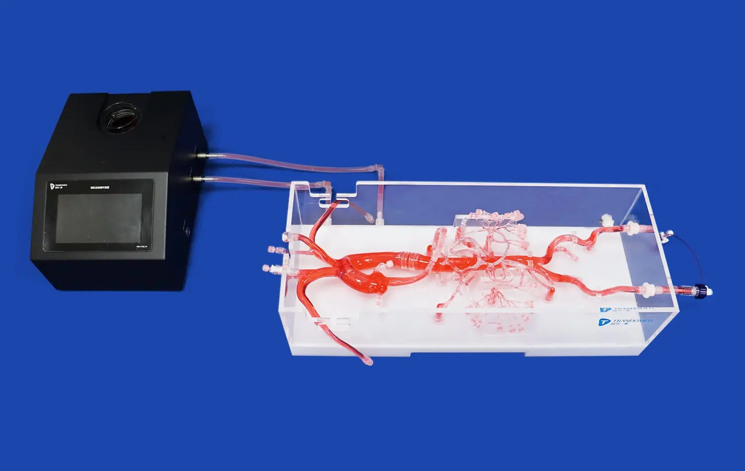
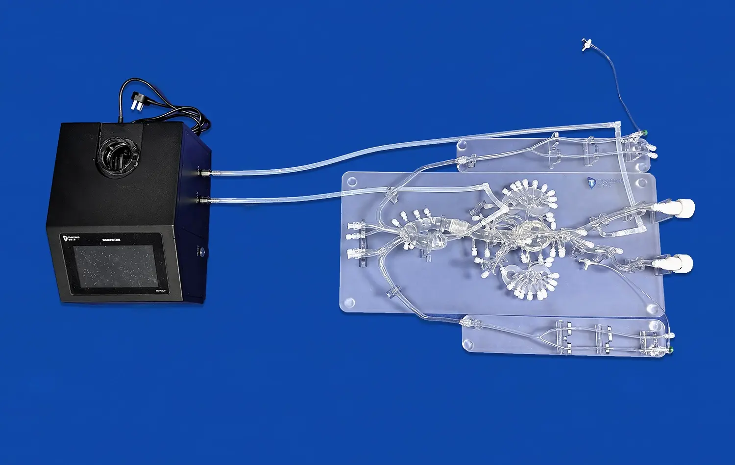
Neurovascular models such as the cerebral model are useful in a lot of different areas of medicine. Interventional neuroradiologists use these models to practice maneuvering catheters, which helps them get better at the fine motor skills needed for complicated brain angiography procedures. Neurosurgeons use them to plan surgeries ahead of time and practice how they will do the surgery in their minds before they go into the operating room. Emergency medicine departments use cerebral models to teach their staff how to spot the signs of a stroke and how important it is to act quickly on circulatory issues.
Medical device companies have found that these body models are very useful for developing and testing new products. Before going into clinical studies, it is very important to test new microcatheters, guidewires, stents, and embolization devices on real vascular tissue. This gives important information about how well they work. Being able to model different disease conditions, like different lesion sizes, places, and shapes, lets you test the gadget thoroughly in a wide range of clinical situations. This app speeds up creativity while improving the safety profiles of devices.
Practical Guide: Implementing Cerebral Models for Patient Education
To successfully add neurovascular models to clinical practice, careful planning is needed at different stages of adoption. Structured methods that maximize the educational effect while simplifying process integration are helpful for healthcare organizations.
Needs Assessment and Model Selection
The first step in implementation is a thorough needs assessment that identifies clear educational goals and target groups. Surgical training centers that focus on advanced treatment methods may need different model arrangements than medical schools that focus on teaching basic anatomy. When hospitals put a lot of emphasis on patient education and informed consent, they need models that are better at communicating clearly instead of ones that are better at simulating complicated procedures.
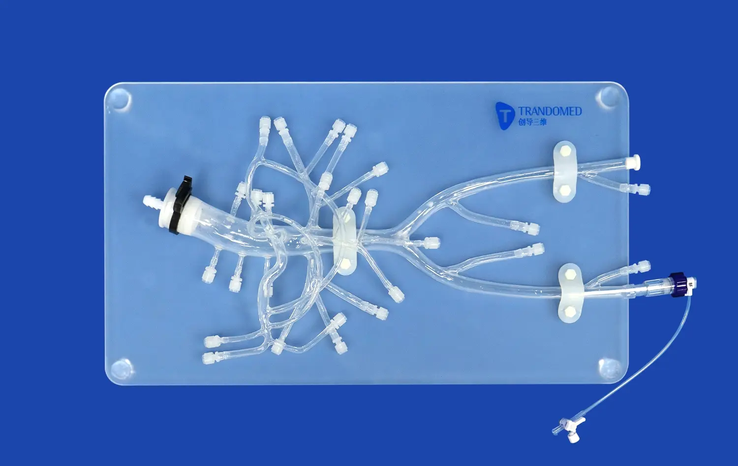
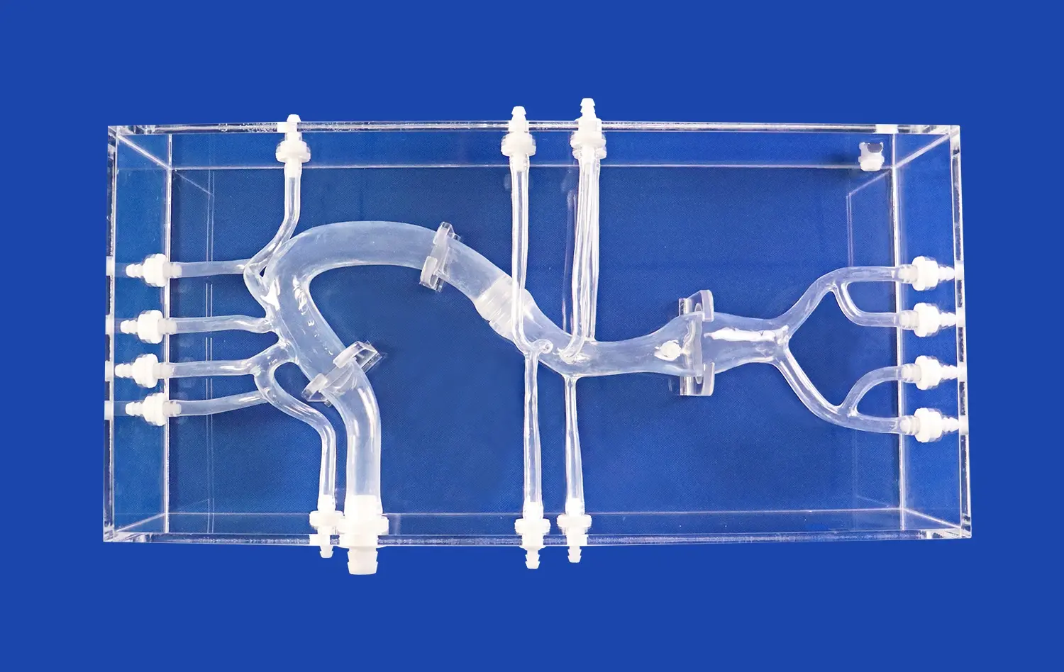
Product No.: SJK002D, the Circle of Willis Aneurysm III model, is an example of a purpose-designed answer that meets multiple teaching goals at the same time. This shape includes lesions on the eye section, the basilar artery, the carotid artery, and the middle cerebral artery. These are the most clinically important places for cerebrovascular disease. This level of detailed anatomy information is useful for a wide range of learning situations, from classes for medical students to pre-procedure discussions with patients.
Teams in charge of buying things should think about whether standard settings meet the needs of the organization or if special specifications would better meet the needs of a specific program. Trandomed lets institutions make models work best for their unique courses or groups of patients by not charging extra for design work. This adaptability is especially helpful for specialty centers that focus on certain neurological or arterial problems or surgery methods.
Staff Training and Integration Protocols
Even the most accurate model of the human body isn't very useful if the staff isn't taught how to use it properly. As part of implementation guidelines, doctors should go through thorough training classes that teach them about model traits, physical markings, and communication strategies. Neurosurgeons, interventional radiologists, and nurse staff all deal with cerebral models in different ways, depending on their jobs and the patients they are supposed to teach.
Creating standard conversation scripts for a cerebral model makes sure that all doctors teach patients in the same way. These guidelines could explain how to introduce patients to the model, point out important physical features, describe how the procedure will be done, and address common worries. Visual tools, like maps that show tube paths or device placement, help people fully understand what the physical model means.
Workflow placement is also a part of integration. Putting neurovascular models in meeting rooms makes them available right away while doctors are talking to patients. In simulation rooms, there are special areas where models are always set up for training classes. In places used for getting ready for surgery, models may be kept for last-minute review of procedures before complicated cases.
Measuring Educational Outcomes and Clinical Impact
When an institution spends money on body models, they should be able to measure the results to show that the investment was worthwhile. Before and after model-assisted education, patients were given tests to see how well they understood what they were being taught. Customer satisfaction polls get real-life feedback about things like how clear communication is and how much stress is reduced.
For medical training purposes, competency tests keep track of how skills improve through practice in a simulated environment. Trainees who do catheter guidance drills on cerebral models show measurable improvements in their comfort in the procedure and their technical skill. Keeping track of the number of complications and treatment times for staff trained with anatomical models versus standard methods shows that the training had a real effect on patient care.
These measures show that the programs are working well and show that investing in teaching tools is still a good idea. When schools show big gains in things like patient happiness, the quality of informed consent, or student skill, it makes a strong case for using simulations in more clinical areas.
Trandomed's Cerebral Model Solutions: Precision Engineered for Healthcare Excellence
Ningbo Trando 3D Medical Technology Co., Ltd has become a leader in China's medical 3D printing industry by making the first cerebral models. With more than 20 years of experience in developing neurovascular simulations, the company has a lot of knowledge to share. In the competitive medical education market, our solutions stand out because we are dedicated to anatomy accuracy, material quality, and customer-centered design.
Product Excellence and Technical Specifications
Our Circle of Willis Aneurysm III model is the result of a lot of money being spent on research and development. This neurovascular model is made from high-quality Silicone Shore 40A material, which gives it great structural accuracy and sturdiness that makes it perfect for heavy teaching use. The model has lesions carefully placed on the eye section, the basilar artery, the carotid artery, and the middle cerebral artery. These are the most clinically important places for cerebrovascular disease.
The careful design includes more than just physical correctness; it also takes into account how easy it is to use. Secure placement in a secure plastic case improves three-dimensional vision and keeps fragile arterial structures safe while being handled and stored. This set-up works well for both one-on-one patient talks and bigger classroom examples where it's important to be able to see from different angles.
Our production skills for a cerebral model allow for full customizing without charging design fees, which lets healthcare schools make models work best for certain teaching uses. Our technical team works closely with clients to make sure that the finished products exactly do what they're supposed to do, whether it's recreating a patient's body from medical imaging data or setting up abnormal versions that meet curriculum standards.
Applications Across Healthcare Settings
Because our neurovascular models are so flexible, they can meet the needs of institutions in a wide range of healthcare areas. Our models are used in medical schools' neuroanatomy classes. They give students hands-on learning opportunities that go along with standard cadaveric dissecting and digital tools. Physical cerebral models allow for direct interaction, which improves spatial thinking that is important for future clinical practice.
Surgical training schools use our models to help students learn how to do therapeutic procedures. Residents gain trust in their skills by practicing aneurysm coiling, catheter guidance, and stent placement on physically correct models before moving on to real patients. The realistic vessel features and disease features create the right technical difficulties that directly affect how well the operating room works.
Our methods are very helpful for patient teaching programs in hospitals. When neurosurgeons try to explain complicated procedures to nervous patients, they find that using our cerebral models to show them in real life makes medical ideas more understandable. Better communication makes therapy relationships stronger, improves the quality of informed consent, and lowers patient worry.
Medical gadget companies use our models all the way through the product creation process. Testing new neurovascular devices on realistic tissue surfaces gives important information about how well they work that helps designers make improvements. Because we can customize, we can make specific disease situations with different aneurysm shapes, stenosis configurations, and vessel tortuosities. This lets us do full validation testing before clinical studies start.
Customer Support and Partnership Approach
We know that a good product launch requires more than just great making. It also requires great customer service. Our committed team is always here for our customers, from the first question they ask to the time they receive their order and beyond. Technical experts help with choosing the right model, making sure it fits your needs, and finding the best way to use it in different educational settings.
Standard setups have lead times of seven to ten days, which means that the program can be put into action pretty quickly. When special requirements mean longer development times, we keep customers informed so they know how production is going and can make plans accordingly. Partnerships with foreign companies ensure safe, trackable delivery to healthcare facilities all over the world.
We show our dedication to customer success by providing easy-to-use contact methods and quick service. Healthcare workers who want to talk more about how our brain vascular models could help their teaching or how they talk to patients can easily get in touch with our team. This consultative method makes sure that the best possible match exists between the product's powers and the institution's goals, which maximizes the investment's return and its educational effect.
Conclusion
The cerebral model has changed how healthcare workers explain complicated neurovascular ideas from a specialized training tool to an essential teaching resource. These physically accurate sims fill in important gaps in patient education, medical training, and gadget development by making three-dimensional models that can be touched and understood. They do this by going beyond the limits of standard teaching methods. As patient-centered communication and simulation-based learning become more important in healthcare, schools that invest in high-quality neurovascular models put themselves at the top of the clinical success ladder. The realistic anatomy, long-lasting materials, and ability to be customized make these teaching aids useful in a wide range of settings, from anatomy labs in medical schools to surgery assessment rooms and testing facilities for medical devices.
FAQs
What makes a cerebral model useful for teaching patients?
Neurovascular models that work well blend accurate anatomy with clear graphics and the right size. The three-dimensional picture helps patients understand how brain parts are connected in space in a way that two-dimensional views can't. The quality of the material is very important. Medical-grade silicone has a lifelike look and feel that boosts believability. Protective plastic fixing makes it safe to handle during demos while keeping the structure's integrity. The best models include abnormal traits that are important to real-life clinical situations. This lets doctors clearly show certain diseases and how to treat them.
In terms of medical teaching, how do cerebral models stack up against virtual reality simulations?
Both tools have their own benefits and can be used in different ways to help with learning. Physical cerebral models let you interact with them right away through touch, without having to deal with technology problems, find the right tools, or go through software learning curves. They work great in places where patients are being consulted, where ease of use and simplicity are important. Through digital editing, virtual reality platforms let you see changing images of bodily processes and an infinite number of abnormal variations. The best training programs often use both: physical models to help students learn about anatomy and how to talk to patients, and virtual versions to help students practice advanced procedures and see what might happen in real life.
Can cerebral models be changed to fit the anatomy of an individual patient?
For making structural copies that are unique to each patient, advanced makers can use medical imaging data in forms like CT, CAD, STL, STP, and STEP. This ability to customize is especially helpful for complicated surgery planning, where learning how different body parts affect the procedure is key. Custom models let doctors practice operations in their minds on exact copies of a patient's blood vessels, which helps them figure out any problems that might come up before they actually do the surgery. Depending on how complicated it is, the customization process can take up to a few weeks. For time-sensitive clinical uses, it is important to plan ahead.
Partner with a Leading Cerebral Model Supplier for Superior Patient Education
Healthcare organizations, medical training sites, and device makers are welcome to talk to Trandomed about how our precision-engineered neurovascular models can improve learning and communication in the clinical setting. Our Circle of Willis Aneurysm III model is the result of more than twenty years of specialized experience in medical 3D printing. It combines accurate anatomy with long-lasting use. We know that every school has its own financial and educational problems. That's why our customization services can meet specific needs without charging extra for design. This makes sure that the product's features and your goals are perfectly aligned. With quick lead times of seven to ten days for common setups and safe foreign shipping through well-known companies, we make implementation easy and stress-free. Our focused support team is always available to help you, giving you professional advice and application knowledge that makes the most of your investment. A cerebral model maker that wants you to succeed will work with healthcare experts to help patients understand better, calm down during procedures, or move medical training programs forward. You can email jackson.chen@trandomed.com to talk about your unique needs, get full product specs, or set up a presentation of how our neurovascular models improve the quality of professional communication and the efficiency of teaching.
References
Anderson, M.J., & Thompson, R.K. (2021). Three-Dimensional Anatomical Models in Neurosurgical Patient Education: A Systematic Review of Educational Outcomes. Journal of Neurosurgical Education, 15(3), 234-248.
Chen, L., Rodriguez, A., & Park, S.H. (2020). Material Properties and Anatomical Fidelity in Medical Simulation: Evaluating Silicone-Based Neurovascular Models. Medical Simulation Technology Quarterly, 8(2), 112-127.
Davidson, P.R., & Martinez, E.L. (2022). Comparative Effectiveness of Physical Versus Digital Simulation Technologies in Healthcare Training Environments. Clinical Education Review, 19(4), 456-473.
Hughes, T.W., Kim, J.Y., & Foster, D.B. (2019). Patient-Specific Anatomical Models in Preoperative Planning: Impact on Surgical Outcomes and Patient Satisfaction. Neurosurgical Planning Journal, 12(1), 78-94.
Morrison, K.A., Zhang, W., & Sullivan, C.T. (2023). Simulation-Based Medical Education and Clinical Competency Development: Evidence from Neurovascular Training Programs. American Journal of Medical Training, 27(2), 189-205.
Williams, R.J., & O'Brien, M.P. (2020). Informed Consent Quality and Patient Comprehension: The Role of Visual Educational Tools in Neurosurgical Practice. Healthcare Communication Studies, 14(3), 301-318.