The way doctors prepare for complicated treatments has changed because of neurovascular modeling. A high-fidelity cerebral model is an important part of cerebral angiography and percutaneous cardiac intervention (PCI) modeling because it shows the brain's anatomy in a way that is true to life. These high-tech neurovascular models accurately reproduce the complex structure of the brain's blood vessels, such as the Circle of Willis, different tumor sites, and artery paths. More and more medical schools are realizing that hands-on practice with physically correct models lowers the risks of procedures and boosts doctor confidence. Incorporating these kinds of training tools into school programs is a big step forward for patient safety and improving practical skills in both neurology and interventional cardiology.
Understanding Neurovascular Simulation in Medical Training
The Science Behind Anatomical Replication
To make an accurate copy of the brain arteries, you need to know a lot about neuroanatomical features and hemodynamics. Modern neurovascular simulators use medical-grade plastic materials that feel like human blood vessels. This gives the simulators real resistance and flexibility while the tube is being moved. Shore 40A silicone, which is used in advanced models, is strong and still has the realistic tissue flexibility that lets you practice for a long time without much wear and tear.
Some of the physical features that are built into these models are the internal carotid artery, the basilar artery, the middle cerebral artery, and the anterior communicating artery. The three-dimensional spatial links in these models help trainees build muscle memory for moving the guidewire and placing the catheter. Physical models, unlike simple sketches or digital images, use more than one sense. They help you learn by giving you direct feedback and visual proof.
Integration with Clinical Imaging Modalities
Physical simulations and medical scan data can work together, which is good for modern training programs. Modern makers can make changes based on CT, MRI, and digital subtraction angiography (DSA) files. This turns anatomy specific to each patient into real training tools. This feature is especially helpful for surgery teams getting ready for difficult cases with complicated venous structures or multiple diseases.
Imaging data types like STL, STP, and STEP files can be easily turned into real models. This makes it easy for medical teams and training facilities to work together. Before going into the operating room, interventional neurologists can practice procedures on copies made from scans of real patients. This lets them find problems and come up with solutions before they actually do the surgery. This practice makes people much more confident and cuts down on the time it takes to do procedures during real treatments.
Benefits for Clinical Decision Support
Simulation-based training isn't just for learning basic skills; it can also be used for strategy planning and evaluating risks. When doctors can move tubes through a model with tumors on the eye section, basilar artery, and middle cerebral artery, they get a better sense of how to approach and choose the right device. This practical training leads straight to better results during real treatments.
High-fidelity neurovascular models like the cerebral model are used in training programs that show real changes in the success rates of procedures and a drop in complications. Being able to practice stent placement, coil embolization, and aneurysm tamponade procedures in a controlled setting consistently improves skills. Schools know that spending money on good training equipment pays off by making graduates more prepared and improving the school's image.
Advanced Applications in Cerebral Angiography and Interventional Training
Preoperative Planning and Risk Stratification
Anatomical models are being used more and more by medical teams as part of their preoperative planning routines. The Circle of Willis Aneurysm III type, which is marked with Product No. SJK002D is an example of this tool because it shows in great detail where aneurysms are located, which is the hardest part of the process. Before cutting or inserting tools, surgeons can look at the vessel tortuosity, the shape of the aneurysm neck, and the parent artery connections.
This preparation work is especially helpful when dealing with cases that have odd features or have had surgery in the past. Being able to move tools around physically through a patient-matched model shows approach limits and best entry routes that might not be clear from flat imaging alone. When thorough simulations are done before real procedures, surgical teams say that operations take less time and have fewer problems during the processes.
Device Testing and Validation
Neurovascular models are used by companies that make medical devices at all stages of the product development process, from the first prototypes to the final evaluation tests. Engineers can test how stents are deployed, how well catheters can track, and how well guidewire tips respond in conditions that are very close to real-life situations. Before expensive clinical studies start, these tests find problems with the design and limits on how well it works.
Simulation data that shows how well the gadget works in different physical situations is useful for regulatory paperwork. Customizing models with different amounts of stenosis, embolism sites, and vascular tortuosity makes it possible to fully assess the device's strengths and weaknesses. These models help companies that are making neuro-interventional equipment like microcatheters, micro guidewires, and embolic agents come up with new ideas faster while still meeting safety standards.
Patient Education and Informed Consent
Neurovascular models are useful for more than just training professionals; they are also great for communicating with patients during appointments. Clinicians can show patients how planned procedures will be done by using physical models that they can see and touch. This turns abstract medical terms into clear visual concepts. This better understanding makes it easier to talk about informed consent and makes patients less anxious about future treatments.
Because these models are three-dimensional, they help patients understand how aneurysms and related structures fit into space, why certain treatments work, and what problems might happen. Using physical models in educational meetings has been linked to higher patient happiness scores and better treatment compliance, since people are more involved in choices they fully understand.
Trandomed's Circle of Willis Aneurysm III: Precision Engineering for Medical Excellence
Technical Specifications and Material Innovation
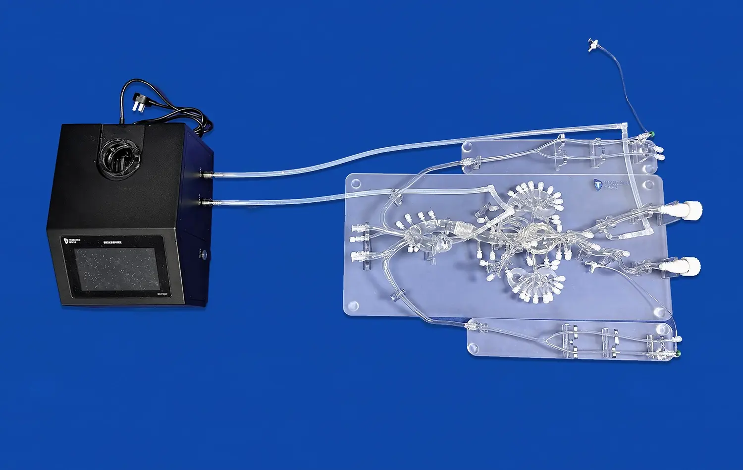
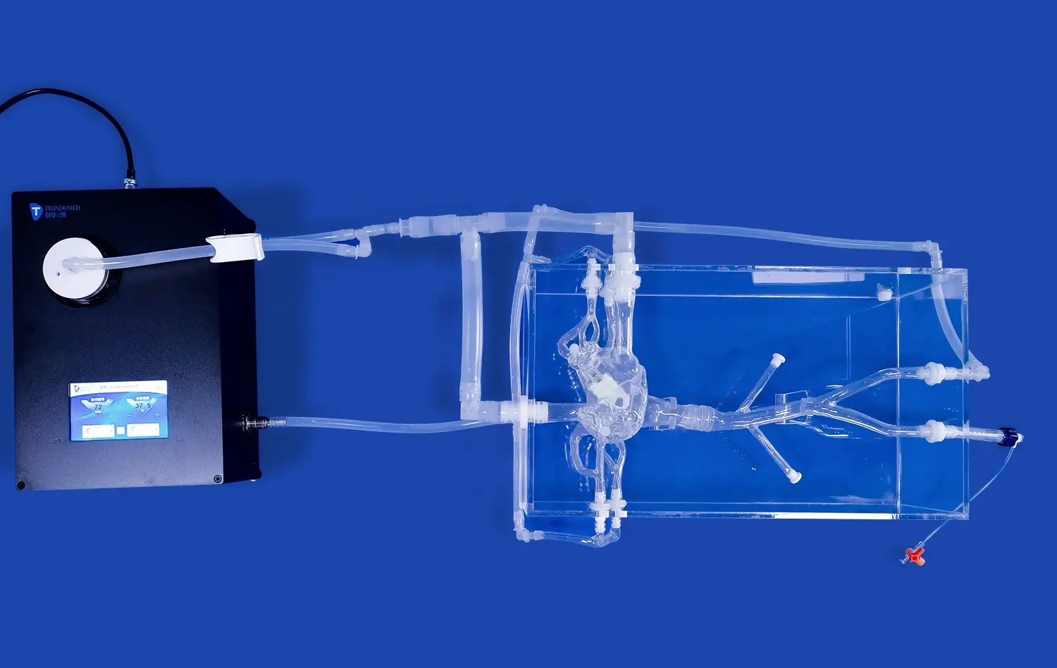
Ningbo Trando 3D Medical Technology Co., Ltd has become a leading company that makes cerebral models by constantly improving materials science and the correctness of their models' anatomy. The Circle of Willis Aneurysm III model is the result of more than 20 years of study and development that aimed to accurately replicate neurovascular structures. This model is made from medical-grade Silicone Shore 40A and gives interventionalists feedback that they say feels very real.
The choice of materials shows that professional needs were carefully thought through. Shore 40A silicone strikes the perfect mix between being durable enough to be used over and over again and having true tissue compliance when the catheter is being moved. This model can be used for hundreds of training classes without losing its structure integrity or physical correctness. It is a good investment for schools that need to manage a lot of workers over long periods of time.
Each model goes through strict quality control steps that make sure the dimensions are correct and the material features are always the same. Aneurysms in clinically important places, like the eye section, basilar artery, carotid artery, and middle cerebral artery, were added based on feedback from working interventional neurologists who chose the best training situations. The model is in a clear plastic box that lets you see the placement of instruments and the paths for movement without any problems. This makes the learning experience better.
Customization Capabilities Without Design Fees
Trandomed knows that different schools and fields have different clinical training needs, so they offer full customization services without asking extra for design. Medical schools that train residents to be general neurovascular competency might ask for standard aneurysm shapes. On the other hand, advanced training programs that focus on complicated diseases can ask for specific lesion amounts, sizes, and anatomical positions that are best for learning.
The customization process works with different types of data forms, like CT, CAD, STL, STP, and STEP files, so it's easy to connect to university image libraries. Because of this, surgery teams can order models based on real patient cases, which creates training chances that are a perfect reflection of upcoming treatments. Researchers working on new ways to treat illnesses can try out different designs over and over to test their ideas and make the methods better before using them in patients.
Customization goes beyond aneurysms and includes a number of different clinical conditions, such as narrowing with varying levels of severity, thromboembolism, and vascular tortuosity. Because of this, a single computer platform can be used for a full training program that covers everything from learning basic skills to mastering advanced techniques. Procurement teams like that they can choose options that meet the needs of the business without having to deal with complicated price systems or long negotiation processes.
Logistics and Support Infrastructure
Trandomed has set up fast delivery processes for a cerebral model that are meant to cut down on the time it takes from placing an order to starting training. Standard wait times of seven to ten days make it possible to start the program quickly, and relationships with FedEx, DHL, EMS, UPS, and TNT for global shipping make sure that schools all over the world get their packages on time. This ability to handle logistics is especially helpful when training plans need quick access to specialized tools or when large programs need new units.
Payment terms work with standard transfer methods to suit university buying procedures. This makes budget management and approval routines easier. The company's customer service doesn't stop when the sale is made; they also offer technical help with integrating models into current training programs and advice on how to maintain simulators in a way that makes them last as long as possible.
Procurement Considerations for Medical Training Equipment
Evaluating Total Cost of Ownership
The most obvious cost is the original purchase price, but a full buying study must also take into account long-term practical factors. Durable construction with medical-grade rubber means longer service life, which means fewer replacements and the paperwork that comes with them. When compared to lower-quality options that need to be replaced often, ones that can handle hundreds of practice sessions without losing much of their performance are a better deal.
Customization options that don't cost extra for design get rid of unexpected costs that can put a strain on training budgets. Institutions can choose exactly designed models that meet their teaching needs without having to deal with complicated price levels or minimum order numbers. This makes it easier to plan and approve budgets, especially at university medical centers that have to deal with a lot of different funding sources and stakeholders' needs.
Shipping with reputable foreign companies cuts down on arrival delays and the chance of damage during transport. When you have reasonable wait times and reliable arrangements, you can accurately schedule programs and avoid costly delays or changes to the curriculum. These operating savings add real value above and beyond the base product price when you figure out the total cost of ownership.
Aligning Product Capabilities with Institutional Objectives
To successfully buy a simulator like the cerebral model, training goals and target skill levels must be clearly stated. Institutions that focus on teaching basic catheter navigation skills might choose models with standard vascular anatomy. On the other hand, programs that prepare experts for complex aneurysm treatments benefit from simulations that include difficult diseases and differences in anatomy. This range of needs can be met by Trandomed's customization framework, which makes sure that product specs and educational results are in line with each other.
The link between modeling and clinical practice is made possible by the ability to send image data from a patient for model building. Surgical teams can practice specific procedures ahead of time, getting used to the unique features of each body part before they go into the operating room. With this feature, simulation goes from being a general way to improve skills to an exact way to get ready for a procedure, which has a direct effect on patient safety and procedure speed.
When research schools want to make new devices or techniques, they need things that aren't needed in regular clinical training programs. Being able to choose specific tumor features, blood vessel arrangements, and material qualities makes it possible to do experiments that would not be possible or practical with human subjects or animal models. Teams in charge of buying things should make sure that the sellers they've chosen can meet these specific needs by having the right technical knowledge and industrial skills.
Vendor Reputation and Industry Experience
As China's first professional producer of medical 3D printing, Trandomed has a lot of technical know-how and experience in the field. Twenty years of focused development in this specialized field shows long-term dedication and a wealth of knowledge that newcomers to the market can't match. This knowledge means that they have a better understanding of healthcare needs, better production methods, and a history of making customers happy.
More and more, institutional buying policies stress the importance of stable and long-term vendors. This is because training equipment needs ongoing support and may need to be expanded in the future. Startups that don't have proven business plans or a lot of technical knowledge are riskier than well-established makers that have been in business for a long time. Referrals from current customers and approval from well-known professional trainers give buyers even more faith in their choices.
Conclusion
When high-fidelity neurovascular models such as the cerebral model are used in medical education, they make a huge difference in how healthcare workers learn how to do interventions. These high-tech training tools close the important gap between knowing things in theory and being able to do them in practice. They let professionals improve their skills in controlled settings before they use them on real patients. The Circle of Willis Aneurysm III model is an example of this technology because it accurately copies the anatomy, is built to last, and can be customized in a lot of ways to meet the needs of different institutions. As competency-based frameworks and simulation-based learning become more common in medical education, investing in high-quality anatomical models becomes not only a good idea but also a must for schools that want to provide the best practical training and keep patients safe.
FAQ
In what ways is a professional-grade cerebral model different from simple anatomy training tools?
Professional neurovascular simulations use medical-grade materials that mimic the qualities of real tissue. This gives users accurate feedback when manipulating catheters and guiding wires. Instead of hard plastic models that are only used to show how the body works, modern simulations like the Circle of Willis Aneurysm III are made of Shore 40A silicone, which is flexible enough and durable enough to be used for multiple procedures. These models show abnormalities like aneurysms in clinically important places. This way, trainees can learn skills that can be used right away in complex treatment situations instead of just remembering how parts of the body fit together.
How does training through simulations improve the results of brain angiography procedures?
By practicing with high-fidelity models, interventionalists can build muscle memory for controlling catheters, get better at choosing the right device, and get used to difficult physical differences before they have to deal with real patients. Studies show over and over that practitioners who have been trained in simulations have faster procedure times, lower rates of complications, and more confidence when dealing with difficult cases. You can practice certain techniques, like tamponade aneurysms or stent placement, in a safe place, which helps you learn faster and keeps patients from getting hurt while you're learning.
Can neurovascular models be changed to fit the anatomy of a specific patient?
To make copies that are unique to each patient, advanced makers like Trandomed can use medical imaging data in forms like CT, MRI, STL, STP, and STEP files. With this customization feature, surgery teams can practice future procedures on models that exactly match the anatomy, blood structure, and diseased conditions of real people. This method works especially well when dealing with complicated cases that need careful planning before surgery, tissue that isn't normal, or changes made by surgery in the past. Custom models change modeling from a general way to learn skills to a way to practice specific surgeries, which has a direct effect on how well the surgery goes.
What maintenance do silicone neurovascular simulators require?
Medical-grade silicone models like those built from Shore 40A material show excellent longevity with minimal upkeep needs. As part of normal maintenance, it should be cleaned gently with mild detergents after each use, dried completely before being stored, and kept out of direct sunlight and high temperatures. Storing things correctly in the plastic case that comes with it keeps things from getting damaged or contaminated. If you handle these models the right way, they can last through hundreds of training lessons without losing their accuracy or material qualities. Most of the time, manufacturers give specific care instructions that are best for their products and building methods.
Contact Trandomed: Your Partner in Advanced Neurovascular Training
If medical schools really want to improve their therapeutic training programs, they should look into Trandomed's full range of neurovascular modeling options. Our Circle of Willis Aneurysm III model and wider range of products are the result of a lot of study, working together with doctors, and making sure the products are of the highest quality. To get more information about our full line of anatomy models and to set up a meeting, please email jackson.chen@trandomed.com. This is for procurement managers, clinical trainers, and research leaders. As one of the biggest companies that makes cerebral models, we want to help you with your educational goals by giving you the best goods, the fastest service, and the best prices.
References
Dawson, S. (2006). Procedural simulation: a primer. Journal of Vascular and Interventional Radiology, 17(2), 205-213.
Robison, R. A., Liu, C. Y., & Apuzzo, M. L. (2011). Man, mind, and machine: the past and future of virtual reality simulation in neurologic surgery. World Neurosurgery, 76(5), 419-430.
Spiotta, A. M., Rasmussen, P. A., Masaryk, T. J., Benzel, E. C., & Schlenk, R. (2013). Simulated diagnostic cerebral angiography in neurosurgical training: a pilot program. Journal of NeuroInterventional Surgery, 5(4), 376-381.
Cooke, M., Irby, D. M., & O'Brien, B. C. (2010). Educating Physicians: A Call for Reform of Medical School and Residency. San Francisco: Jossey-Bass.
Bambakidis, N. C., Selman, W. R., & Sloan, A. E. (2013). Surgical rehearsal platform: potential uses in microsurgery. Neurosurgery, 73(Suppl 1), 122-126.
Chaer, R. A., DeRubertis, B. G., Lin, S. C., Bush, H. L., Karwowski, J. K., Birk, D., ... & Kent, K. C. (2006). Simulation improves resident performance in catheter-based intervention: results of a randomized, controlled study. Annals of Surgery, 244(3), 343-352.
_1736214519364.webp)
_1734507415405.webp)
_1732863713705.webp)