Choosing the right cardiac electrophysiology training model takes careful thought about how accurate it is in terms of anatomy, how long the material will last, and how it can be used in real life. A well-made venous cardiac electrophysiology model lets doctors practice ablation treatments, catheter navigation, and mapping techniques in a controlled but realistic setting. These training tools help people go from knowing a lot about anatomy to being good at it in real life, especially when the models look just like the inferior vena cava, right atrium, superior vena cava, and subclavian vein. The best option relies on the training goals, patient safety goals, and difficulty of the procedures your team needs to learn at your institution.
Understanding Venous Cardiac Electrophysiology Models
What Sets Venous Models Apart from Arterial Counterparts?
Even though cardiac electrophysiology includes both arterial and venous pathways, most EP treatments are still done through veins. When doctors put catheters into the heart chambers through the femoral or subclavian veins, they use models that exactly copy the path that the tubes take through the body. Unlike arterial models, which focus on the heart's arteries or overall circulation, venous EP models look at the heart's right side, including the tricuspid valve area, coronary sinus ostium, and important conduction paths.
It's important for staff who are being trained on techniques like implanting a pacemaker, placing a defibrillator lead, or using radiofrequency ablation to treat atrial fibrillation to understand this difference. The venous method has its own problems with vessel tortuosity, valvular navigation, and avoiding perforation risks. Good training models should accurately replicate all of these aspects.
The Heart's Conduction System and Model Relevance
The heart's transmission system starts in the sinoatrial node, goes through the atrioventricular node, and then spreads through the His and Purkinje fiber bundle. When this electrical pathway doesn't work right, it can lead to arrhythmias, which are irregular heartbeats that electrophysiologists have to identify and treat. When training models include anatomical landmarks that are important to these electrical processes, they do their job best.
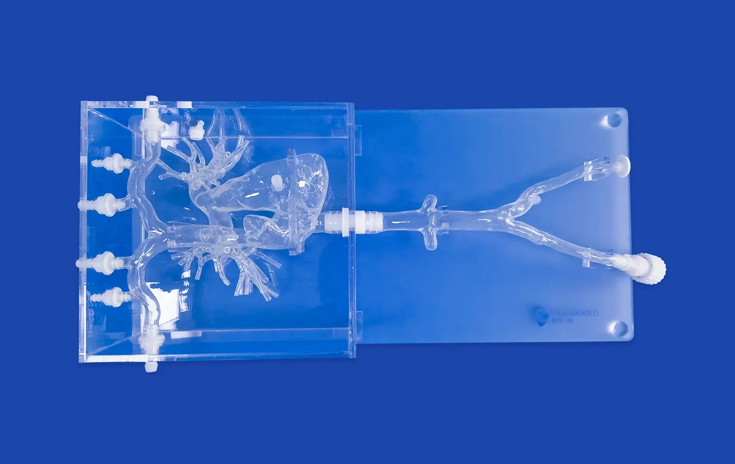
When medical schools teach students about the triangle of Koch, which is an important part of the heart anatomy that holds the AV node, they need models that show how this area fits in relation to the coronary vein and tricuspid annulus. In the same way, when hospitals teach cardiologists how to use catheters to treat atrial flutter, the model must clearly show the cavotricuspid isthmus, which is where ablation lines are usually made.
Integration of Imaging Data and Manufacturing Precision
With today's production methods, data from imaging patients can be turned into real training tools. Manufacturers use reverse 3D reconstruction technology to get detailed information about the body from CT and MRI pictures. This method lets people make models that show how people really are, not just how they should look according to a guide.
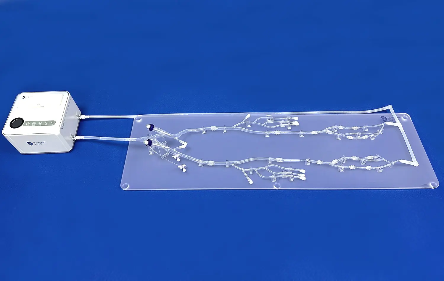
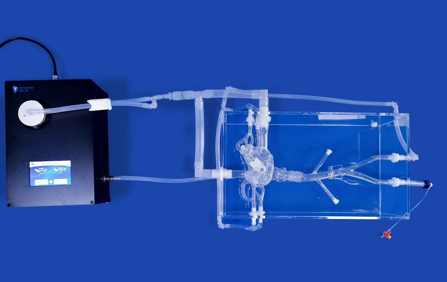
The XXS004 Venous Cardiac Electrophysiology Model is made by Trandomed using special 3D printing techniques and precise molds to make it out of medical-grade silicone (Shore 40A). This choice of material gives physical feedback that is similar to real tissue, which helps trainees learn how to properly manipulate catheters. The manufacturing process also allows for customization based on specific CT data. This means that institutions can ask for models that show certain pathologies or differences in anatomy that their staff often sees.
Clinical Applications Driving Model Development
There are more training uses than just learning basic anatomy. Surgical teams use these models to plan ahead for surgery when there are complicated cases with unusual vein anatomy or when patients are born with birth defects that make it hard to use normal access routes. Anatomical models are used by device makers to try new catheter designs, ablation tools, and mapping systems on them before they are used on humans.
Standardized venous cardiac electrophysiology models are needed for controlled tests in research labs that are looking into new ablation energy sources, catheter materials, or navigation technologies. Having consistent anatomical platforms makes it possible to compare things correctly across different experimental situations and speeds up the process of putting lab results into clinical practice.
Key Criteria for Choosing the Right Model
Assessing Anatomical Accuracy and Fidelity
Accurate knowledge of anatomy is the basis of good teaching. Models should not only copy big structures, but also small features that affect how well a process works. In real life, catheters behave differently depending on the width of the coronary sinus ostium, the angle between the superior vena cava and the right atrium, and the thickness of the atrial wall.
Teams in charge of buying things should ask for confirmation data that shows how models compare to real human bodies. The model's measurements should match up with published anatomical studies about the size of the chambers, the diameters of the vessels, and the links between spaces. When institutions look at the XXS004 model, they will see that it has five important structures: the inferior vena cava, the right atrium, the right ventricle, the superior vena cava, and the subclavian vein. These structures are placed based on data from several patient scans instead of a single idealized design.
Material Properties and Durability Considerations
Choose the right materials for realism and durability. Different types of silicone materials are hard, flexible, and don't react well to being put in a catheter over and over again. Shore 40A durometer strikes a mix between a realistic tissue feel and structural integrity that doesn't break down after hundreds of workouts.
Training places that offer a lot of courses need models that don't lose their anatomical accuracy after being used a lot. Poorer materials might tear where the catheter goes in, become permanently deformed, or lose their tactile qualities after only a few uses. When asking for product specifications, you should ask manufacturers how long the product is expected to last under certain conditions of use and whether broken parts can be changed one at a time instead of the whole model having to be replaced.
Customization Capabilities and Technical Flexibility
Standardized models are good for general training, but they often need to be changed for more specific programs. Some hospitals specialize in atrial fibrillation ablation, which needs more attention to the pulmonary vein ostia and left atrial appendage. Others focus on ventricular tachycardia treatments that need exact copies of the papillary muscles and trabeculations.
Different body types should not be the only things that can be customized; pathological situations should also be included. Models of dilated cardiomyopathy, birth flaws like atrial septal defects, or anatomy after surgery help doctors get ready for all the different kinds of cases they might see. Trandomed can make changes to orders without asking extra for design work. They use data files from customers in formats like CT, CAD, STL, STP, and STEP. Because of this, institutions can create training scenarios that are perfect for their specific clinical group and procedure focus.
Being able to change certain traits, like the size of the foramen ovale or the thickness of the atrial wall, lets models be made that look like pediatric patients, bariatric patients, or people with certain body types. This amount of oversight makes sure that training stays relevant to real clinical practice and not just general situations.
Integration with Existing Training Infrastructure
Models work best when they work well with modeling software that is already in use. The training value goes beyond simple catheter handling when it is compatible with fluoroscopy simulation systems, intracardiac echocardiography platforms, or electroanatomical mapping systems.
Some advanced training programs use these anatomical models along with electrophysiology recording systems. This lets trainees practice figuring out what signals mean while they are moving devices around. How useful models are for full training programs depends on whether they can be put in standard positions, linked to flow circuits that simulate cardiac output, or used in hybrid modeling situations.
The technical specs should make it clear if the models work with standard catheterization table setups, if they can handle different access site configurations, and how they connect to imaging equipment that your school already has. The total cost of ownership goes down and the return on investment goes up with seamless merging.
Market Leaders and Product Offerings
Evaluating Manufacturer Credentials and Track Record
The medical simulation industry includes both established corporations and specialized manufacturers focused exclusively on cardiovascular training tools. Manufacturer experience matters particularly when requesting custom modifications or troubleshooting integration challenges.
Trandomed brings over two decades of focused expertise in medical 3D printing technology, positioning the company among China's pioneering professional manufacturers in this sector. This extensive background translates into refined manufacturing processes, comprehensive understanding of clinical training requirements, and accumulated expertise in problem-solving across diverse institutional settings. When selecting a supplier, examine their portfolio of completed projects, client testimonials from similar institutions, and evidence of ongoing innovation in materials and manufacturing techniques.
Manufacturers demonstrating commitment to the medical education sector typically maintain relationships with training institutions, participate in simulation conferences, and publish validation studies demonstrating their products' effectiveness. These indicators suggest a partner invested in long-term product development rather than transactional sales relationships.
Product Specifications and Performance Characteristics
The XXS004 Venous Cardiac Electrophysiology Model represents the intersection of anatomical accuracy and practical durability. Constructed from medical-grade silicone with Shore 40A hardness, this model provides realistic resistance during catheter advancement while maintaining structural integrity across repeated training sessions. Its design incorporates precise replication of the inferior vena cava, right atrium, right ventricle, superior vena cava, and subclavian vein—the complete venous pathway used in most electrophysiology procedures.
Production efficiency supports training program timelines, with lead times ranging from seven to ten days from order confirmation to delivery. International shipping through established carriers including FedEx, DHL, EMS, UPS, and TNT ensures reliable delivery to institutions throughout the United States. Payment through standard T/T terms aligns with institutional procurement processes while providing transaction security for both parties.
Support Services and Customer Relationship Management
Beyond the physical product, manufacturer support services significantly impact long-term satisfaction. Responsive technical assistance helps troubleshoot integration challenges, optimize model positioning for specific procedures, or adapt training protocols as clinical techniques evolve.
Comprehensive support should include detailed user documentation, training for simulation center staff on model maintenance and optimal usage, and availability of replacement components when needed. Some manufacturers offer periodic model inspection services, providing objective assessment of when models should be refurbished or replaced to maintain training quality.
Establishing clear communication channels from initial inquiry through post-delivery support ensures smooth project execution. Direct contact with technical specialists rather than layered sales organizations typically results in more accurate needs assessment and tailored solutions.
Practical Steps to Implementing Venous Cardiac EP Training Models
Conducting Needs Assessment and Defining Training Objectives
Successful implementation begins with clear understanding of training goals. Different specialties within your institution may have varying requirements—internal medicine residents need basic catheter navigation skills, while practicing electrophysiologists require models supporting advanced ablation technique refinement.
Gathering input from multiple stakeholders produces a comprehensive requirements document. Educational leaders identify curricular objectives, procedural areas requiring additional training emphasis, and assessment criteria for trainee competency. Clinical department heads contribute perspective on specific procedures their staff perform most frequently and emerging techniques requiring new training approaches. Simulation center directors evaluate technical requirements for model integration with existing equipment and facility capabilities.
This collaborative assessment clarifies whether your institution needs a single versatile model, multiple specialized models for different procedures, or customized versions representing specific patient populations your clinicians serve.
Technical Validation and Quality Verification
Systematic validation makes sure the model meets the standards after delivery. Measurements of length, width, and height make sure that the chamber sizes, vessel diameters, and spatial relationships are correct and match the details given during purchase. Clinicians with a lot of experience should use the model to test catheter navigation and see if the tactile feedback, anatomical landmarks, and procedural problems are the same as what they've seen in real life.
Writing down this process of validation sets baseline performance standards that can be used in the future and helps figure out when models need to be fixed or replaced. Photographic records, measurement logs, and feedback forms for clinicians all provide objective proof that helps with ongoing quality assurance efforts.
When using custom models made from institutional CT data, checking the end product against the source image makes sure the manufacturing process was done correctly. This step of verification makes sure that trainees practice on physically correct models so that problems aren't found after the training has started.
Developing Training Protocols and Curricula
For training to be effective, it needs to follow an organized plan that builds skills from the most basic to the most advanced. During the first lessons, the patient may learn basic skills like how to handle a catheter, set up a vein access, and recognize important body landmarks. In intermediate training, certain techniques are taught, such as coronary sinus cannulation or mapping of the right atrium. Advanced sessions are hard for skilled operators because they have to deal with complicated ablation patterns or problems that come up during the procedure.
Protocols should be very specific about what the learning goals are for each level of training, how to measure progress, and how long you should practice for. There is evidence that simulation training improves procedural outcomes when it uses deliberate practice principles, such as focused repetition of specific skills with instant feedback and increases in difficulty.
These models have the most educational effect when they are added to existing fellowship programs, continuing medical education courses, or hospital privilege processes. Scheduling regularly ensures consistent access instead of random use that hinders skill development.
Maintenance, Longevity Planning, and Performance Monitoring
Models last longer and learn better if they are properly maintained. Setting up cleaning procedures for each use stops material degrading caused by body fluid simulants or catheter residue. Silicone keeps its traits when it is stored in a controlled environment with a steady temperature and humidity. Regular checking finds early signs of wear that mean a part needs to be replaced before the accuracy of the anatomy is lost.
Tracking usage gives you useful information about how long a venous cardiac electrophysiology model will last in your institution's specific conditions. Keeping track of the number of procedures, the types of catheters used, and any damage events helps with planning replacements and finding the best time to buy them. This information also helps with budget reason when asking for money to update models or add more features to simulations.
Reevaluating training goals on a regular basis makes sure that models keep meeting the needs of institutions as clinical methods change. To keep training useful, new methods or device technologies might mean that models need to be changed or extra gear needs to be bought.
Conclusion
Selecting the right cardiac electrophysiology training model demands thorough evaluation of anatomical accuracy, material quality, customization capabilities, and manufacturer support. The venous approach to EP procedures requires models that faithfully replicate the complete pathway from peripheral access through right-sided cardiac chambers, incorporating landmarks essential for safe catheter navigation and effective ablation. Beyond physical specifications, implementation success depends on proper validation, structured training protocols, and ongoing maintenance programs. When institutions invest in high-fidelity simulation tools backed by experienced manufacturers, they create learning environments that accelerate skill development, reduce patient risk during early-career procedures, and foster continuous quality improvement across their electrophysiology programs.
FAQ
What advantages do venous cardiac electrophysiology models offer compared to other cardiac training tools?
Venous-focused models provide specialized training for the most common access route in electrophysiology procedures. Unlike general cardiac anatomy models, these simulators emphasize right-sided structures, venous pathways, and catheter navigation challenges specific to EP interventions. The targeted design allows concentrated practice on skills directly transferable to clinical procedures.
How can institutions evaluate model accuracy before purchasing?
Request detailed specifications including dimensional measurements compared to published anatomical data, validation studies demonstrating correlation with actual patient anatomy, and testimonials from similar training programs. Arrange demonstrations where your clinical staff can test catheter manipulation and assess tactile feedback realism before committing to purchase.
What customization options typically support specialized training needs?
Manufacturers like Trandomed can modify anatomical dimensions, incorporate pathological variations, adjust material properties in specific regions, and create models from institution-provided CT data. These modifications enable training scenarios reflecting your specific patient population demographics, congenital variations, or post-surgical anatomy encountered in your practice.
What factors influence model longevity and replacement timing?
Material quality, usage frequency, catheter types employed during training, maintenance protocols, and storage conditions all affect lifespan. Quality silicone models withstand hundreds of procedures when properly maintained, though high-traffic simulation centers may require replacement or component refreshing more frequently than programs with occasional usage patterns.
Partner with a Trusted Venous Cardiac Electrophysiology Model Manufacturer
Trandomed specializes in developing anatomically precise cardiac training simulators that elevate medical education standards. Our XXS004 Venous Cardiac Electrophysiology Model combines two decades of medical 3D printing expertise with advanced silicone materials, delivering training tools that withstand rigorous repeated use while maintaining exceptional anatomical fidelity. We manufacture each model using reverse 3D reconstruction technology applied to extensive human CT and MRI databases, ensuring accuracy that reflects genuine anatomical variation rather than simplified representations.
Our customization services accommodate your institution's specific requirements without additional design fees, allowing modifications based on your provided imaging data in multiple file formats. Whether your training program emphasizes basic catheter skills, advanced ablation techniques, or device testing protocols, we collaborate with your team to create optimal solutions. Contact Jackson Chen at jackson.chen@trandomed.com to discuss your training objectives, request detailed specifications, or arrange a demonstration of our venous cardiac models. We support medical education institutions, research laboratories, and device manufacturers throughout the United States with reliable shipping, responsive technical support, and products manufactured to exacting quality standards.
References
Josephson, M.E. (2016). Clinical Cardiac Electrophysiology: Techniques and Interpretations (5th ed.). Philadelphia: Wolters Kluwer Health.
Zipes, D.P., Jalife, J., & Stevenson, W.G. (2018). Cardiac Electrophysiology: From Cell to Bedside (7th ed.). Philadelphia: Elsevier.
Calkins, H., Hindricks, G., Cappato, R., et al. (2017). "2017 HRS/EHRA/ECAS/APHRS/SOLAECE expert consensus statement on catheter and surgical ablation of atrial fibrillation." Heart Rhythm, 14(10), e275-e444.
Huang, S.K.S., & Wood, M.A. (2015). Catheter Ablation of Cardiac Arrhythmias (3rd ed.). Philadelphia: Saunders Elsevier.
McGaghie, W.C., Issenberg, S.B., Cohen, E.R., Barsuk, J.H., & Wayne, D.B. (2011). "Does simulation-based medical education with deliberate practice yield better results than traditional clinical education? A meta-analytic comparative review of the evidence." Academic Medicine, 86(6), 706-711.
Al-Khatib, S.M., Stevenson, W.G., Ackerman, M.J., et al. (2018). "2017 AHA/ACC/HRS guideline for management of patients with ventricular arrhythmias and the prevention of sudden cardiac death." Circulation, 138(13), e272-e391.
_1734507815464.webp)