Circle of Willis Brain Model for Endovascular Procedure Practice
2026-01-30 18:16:56
The circle of Willis brain model is an important part of contemporary neurovascular education and practical training because it gives doctors a way to learn endovascular procedures that is accurate in terms of anatomy. This special modeling tool creates a copy of the cerebral artery network that keeps blood flowing to the brain. This lets students practice difficult procedures like aneurysm coiling, thrombectomy, and stent placement in a safe, controlled setting. Investing in high-fidelity anatomical models has become necessary to bridge the gap between academic knowledge and hands-on surgery skills in neurovascular care as healthcare institutions around the world focus on competency-based training and patient safety.
Understanding the Circle of Willis Brain Model and Its Clinical Importance
The Circle of Willis is the brain's main arterial safety network. It is placed at the base of the head in a way that makes sure brain blood flow keeps going even if one or more arteries become blocked. This amazing piece of anatomy links the front and back circulation systems with a network of vessels that talk to each other. These vessels provide important extra blood flow during a stroke, aneurysm burst, or surgery. Medical workers need to have a deep understanding of this framework in order to predict problems and come up with effective treatment plans.
Anatomical Foundation and Vascular Architecture
The cerebral artery circle is made up of seven main blood channels that work together to send fresh blood to all parts of the brain. The front part of the body is made up of the anterior communication artery and the A1 segments of the two anterior cerebral arteries. At the same time, the internal carotid arteries change into the middle cerebral arteries at their terminal segments. They also send communication branches backwards to join the posterior cerebral arteries at their P1 segments. This complicated setup makes sure that there are multiple sources of blood flow, which protects nerve tissue from ischemic damage when one or more blood veins become blocked or narrow.
According to research, only twenty to twenty-five percent of people have a full, uniform arterial circle. This is because some people have abnormalities in their anatomy, such as parts that aren't developing properly or no connecting arteries at all. Because these differences have such a big effect on surgery planning and patient results, correct physical models are needed for training programs that prepare doctors for the complexity of the real world. Advanced cerebral modeling tools can copy these differences, so trainees can see a range of body structures before they go into the surgery room.
Clinical Relevance in Endovascular Training
Endovascular treatments require keen spatial awareness, the ability to manipulate catheters, and the ability to make decisions while using a fluoroscope to guide the process. The circle of willis brain model is an important part of training because it helps doctors build muscle memory that helps them find their way around blood vessels that are twisted and turned, choose the best access routes, and carefully place invasive devices. Simulation-based practice shortens the time it takes to learn a procedure and keeps patients from having to deal with problems caused by the trainee during the early stages of learning the skill.
Because this capillary network is in the interpeduncular cistern of the subarachnoid space and surrounds important structures like the optic chiasm and pituitary infundibulum, it is important to be very careful during therapeutic treatments. Good anatomical models accurately show these spatial relationships, which helps trainees understand how close important nerve and arterial structures are and how they need to be protected during catheter progress and device placement. This understanding of the situation is very helpful when practitioners move from simulations to real-life patient care.
Top Circle of Willis Brain Models for Endovascular Procedure Practice: Comparison and Selection Criteria
To choose the right neurovascular modeling tools like the circle of willis brain model, you need to carefully consider the training goals, the material qualities, and the realism of the anatomy. There are a lot of different choices on the market for medical simulations, from hard plastic displays of the body to complex silicone models that mimic how tissues bend and behave under fluoroscopic imaging. To get the best return on educational investment, procurement workers have to balance training efficiency with limited budgets.
Material Science and Tactile Realism
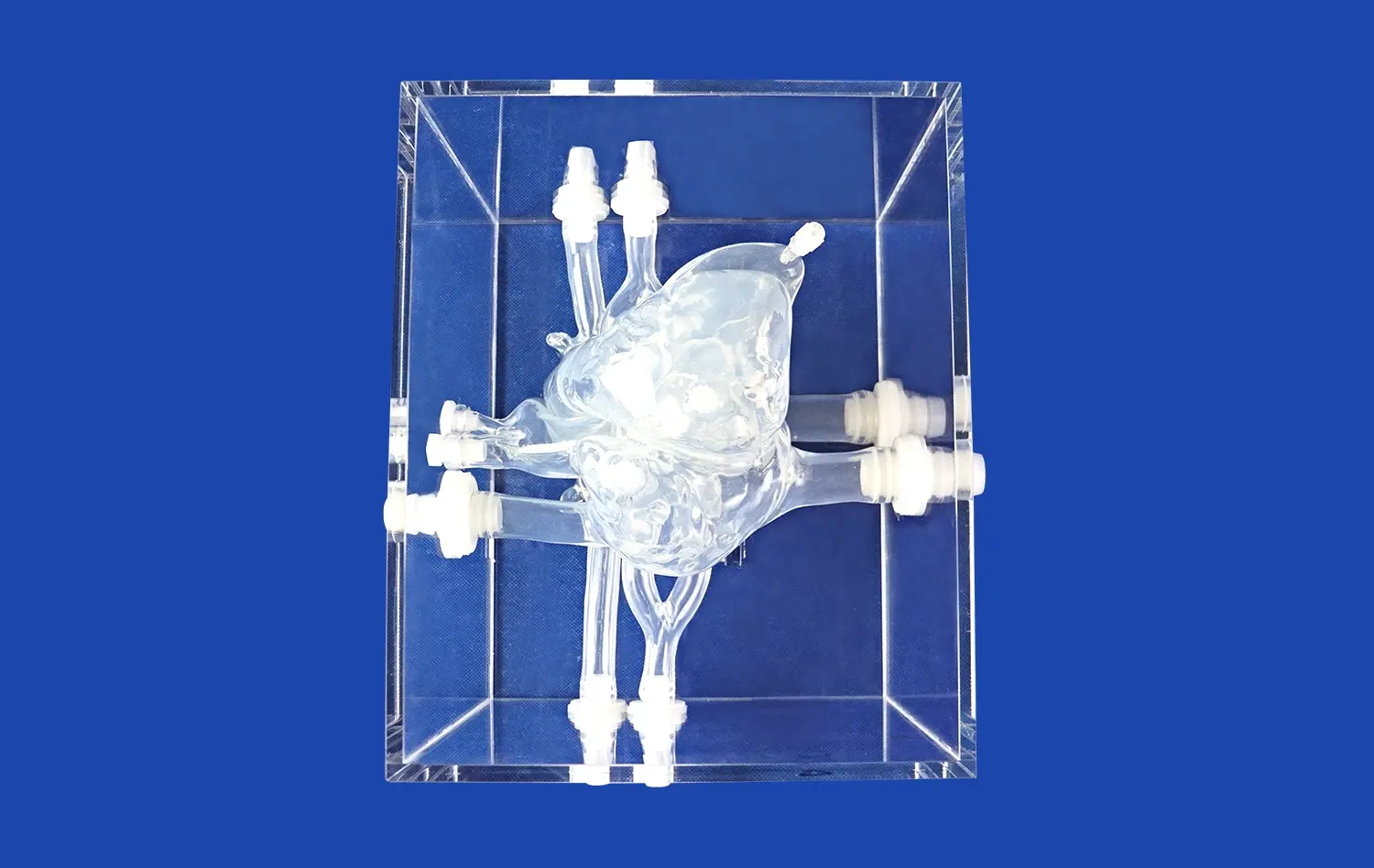
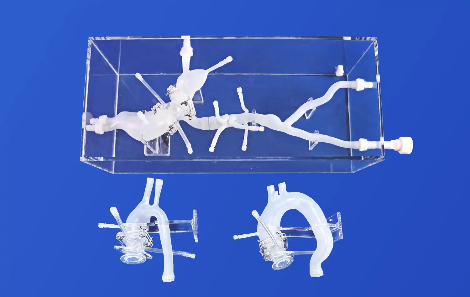
Cerebrovascular models made of silicone have become the best choice for endovascular training because they can mimic the dynamic qualities of live tissue. It uses medical-grade Silicone Shore 40A in the Trandomed Circle of Willis Aneurysm II (Product No. SJL001D), which gives it realistic vessel compliance that feels a lot like guiding tubes through human arteries. For developing the fine touch needed for wire progress and catheter setting, this physical feedback is important. Rigid plastic options are not good for developing these skills.
Material choice has a direct effect on how long a model lasts and how useful it is for teaching over time. High-quality silicone versions can handle multiple catheter insertions, balloon inflations, and device launches without breaking down significantly. This means that educational institutions can get the most use out of their devices before they need to be replaced. For long-term training, a durometer value of forty on the Shore A scale is the best mix between how realistically the vessel can bend and how strong it is.
Anatomical Accuracy and Pathological Features
To get complete neurovascular training, you need to learn about both normal structure and common abnormal conditions that doctors see in patients. A carefully planned disease feature on the Trandomed cerebrovascular simulator includes an M1 segment stenosis on the right middle cerebral artery and three strategically placed aneurysms affecting the basilar artery, the ophthalmic segment of the left carotid artery, and the left middle cerebral artery. Because of this variety in pathology, trainees can practice focused treatments for a range of lesion kinds and sites during the same training lesson.
Anatomical precision includes more than just the overall shape of the body. It also includes accurate vessel width, branch angle, and tortuosity patterns that match the real body of the patient. When compared to general estimates, models made from real CT and MRI scan data using reverse three-dimensional reconstruction technology are more accurate in terms of anatomy. This data-driven design method makes sure that trainees learn skills that can be used right away in clinical situations, instead of just applying techniques they learned on too-simplified examples.
Customization Capabilities and Training Flexibility
To meet the needs of different skill levels and educational goals, different training programs need different abnormal situations. Customizable neurovascular models let schools change the number of aneurysms, how they are spread out in size, and where they are located in the body based on the needs of the curriculum. Modern makers can work with clinical imaging data in a variety of forms, such as CT, CAD, STL, STP, and STEP files. They can then use this information to turn patient-specific anatomy into physical training models that help doctors get ready for planned treatments or to practice difficult cases.
Adding more disease traits like cerebral edema, varying stenosis severity, and vessel dissection to training makes it useful for more than just learning basic skills. People who make decisions about purchases should give preference to sellers that offer full customization services without charging too much for design work. This allows for long-term usefulness in training as educational needs change and new treatment techniques appear.
Practical Guide to Purchasing Circle of Willis Brain Models for B2B Clients
To get specialized medical training tools like the circle of willis brain model, you need to know what the provider can do, what the quality assurance standards are, and how to deal with logistics that affect the total cost of ownership. When you buy something, you should make sure that the product specs match the goals of the institution's training while also building trusting relationships with vendors that help the growth of ongoing educational programs.
Identifying Qualified Manufacturers and Suppliers
There are well-known names like 3B Scientific and GPI Anatomicals in the global medical simulation market, as well as niche makers that only make neurovascular training tools. Ningbo Trando 3D Medical Technology Co., Ltd. is a leader in three-dimensional printed medical models, and they have over 20 years of experience in research and development in the field of cerebrovascular simulation. Their technical base is made up of a lot of real human imaging data and their own additive manufacturing methods. This makes sure that the quality of their products always meets strict educational standards.
When analyzing possible sources, buying workers should look into their manufacturing skills, such as how well they use three-dimensional printing technology, the range of materials they offer, and their quality control standards. Companies with strong technical skills usually keep their certifications up to date, release validation studies, and back up their claims of physical accuracy with clear paperwork. Direct interaction with technical teams through product demos and facility visits builds trust in the supplier's abilities and sets up lines of communication that are necessary for the success of special projects.
Logistics, Lead Times, and Global Shipping
International purchasing makes shipping rules, clearing customs, and delivery times more complicated, which affects when training programs are put into action. Reliable sellers have long-term partnerships with major companies like FedEx, DHL, EMS, UPS, and TNT. This makes sure that high-value packages are transported safely and can be tracked in real time. Standard models have typical production wait times of seven to ten days, which allows for quick order completion. However, based on the complexity of the design, special setups may need longer manufacturing periods.
Telegraphic transfer agreements are a popular way to make payments. They protect both parties and make foreign deals easier. Before signing buy deals, procurement experts should make sure that they understand the guarantee coverage, return policies, and after-sales technical support clauses. These things have a big impact on the total owning experience and long-term happiness with simulation investments.
OEM Partnerships and Bulk Ordering Advantages
Original equipment maker partnerships can help medical device businesses, large-scale training programs, and healthcare systems that are spread out by offering unique solutions at low prices for large orders. These working together ties make it possible to create customized training models that are in line with certain sets of devices, methods of procedure, or teaching styles. When suppliers offer free design advice, it makes modification easier and encourages new ideas in simulation-based learning.
Buying in bulk can save you money through economies of scale and make sure that products are always available for training programs that take place in more than one location. To get the most out of their relationships with vendors, procurement managers should discuss detailed deals that cover everything from pricing levels and delivery dates to technical help sharing and the availability of new parts.
Conclusion
Buying good cerebral modeling tools is a long-term investment to providing the best care and keeping patients safe in neurovascular care. The circle of willis brain model is an important part of training for building arterial skills. It lets professionals get good at their job through careful practice before taking on care of patients. When choosing modeling partners, procurement workers need to look at how accurate the anatomy is, how good the materials are, how much customization is possible, and how reliable the suppliers are. With its scientific know-how, cutting-edge production, and customer-focused service, Trandomed is the perfect partner for schools that want to improve neurovascular education through simulations based on evidence. As medical education moves toward competency-based testing and hands-on skill verification, physically accurate models will continue to be important tools for connecting what is learned in the classroom with what is done in the clinic.
FAQ
What makes good circle of willis brain models different from simple physical displays?
The best neurovascular computer models are different in a number of important ways that have a direct effect on how well training works. Anatomical accuracy based on real patient imaging data makes sure that trainees see real arterial design instead of rough estimates. Choosing the right material has a big effect on the physical input. For example, medical-grade silicone formulations give accurate vessel compliance, which is important for learning how to manipulate catheters correctly. Pathological traits like aneurysms, stenosis lesions, and structural differences that show clinical variety should be included in high-quality models. Durability under frequent use keeps things from breaking down too quickly, protecting establishment investments and making sure that all learners have the same training experiences.
How do cerebral models improve the effectiveness of training for endovascular procedures?
Using physically correct models in simulation-based education speeds up the learning of skills by creating safe spaces for focused practice that don't put patients at risk. Trainees learn important skills like how to move a guidewire through a blood path that is curved, how to place a catheter in the right place, how to read an angiographic picture, and how to use an interventional device. Hands-on practice is the only way to build muscle memory and trust in how to do something, as classes and video examples can't. When practitioners move from practice scenarios to clinical settings, they can quickly become proficient because they have practiced difficult situations many times. Studies show that computer training leads to better results for procedures, fewer problems, and higher standards of patient safety.
What customization choices are there for large buys from institutions?
Leading makers, such as Trandomed, offer a wide range of customization options that can be adapted to specific training needs and program goals. To meet learning goals, institutions can define the number of aneurysms, how they are distributed in size, and where they are located in the body. Based on what students need to learn, more clinical traits can be added, such as the intensity of the stenosis, the patterns of vessel tortuosity, cerebral edema, and dissection. Using clinical imaging data to create patient-specific models lets doctors practice surgery before it happens on complicated cases or make standard training scenarios based on interesting pathology. When you place a bulk order, you can usually get free consultations to help with unique design, better prices, and synchronized delivery dates for applications in more than one location.
Why is the Circle of Willis so important for planning surgery and understanding how the brain works?
The Circle of Willis is the brain's main secondary circulation system. It provides other ways for blood to flow when one or more arteries become blocked, narrow, or surgically interrupted. This extra protection keeps brain tissue safe from ischemic damage during strokes, aneurysm surgery, and planned vessel sacrifice during tumor removal. It's important to know about each person's unique anatomy when planning surgery, because missing or uneven artery rings change collateral capacity and affect how risky the procedure is. Neurosurgeons and interventionalists need to know a lot about this vascular network in order to predict problems that might happen, choose the best ways to treat patients, and put safety measures in place during complicated procedures.
How many people have full physical rings, and why is that important?
Clinical study shows that only twenty to twenty-five percent of people have full, uniform Circles of Willis. The rest of the population has different anatomy, such as hypoplastic parts, missing communication arteries, or strange branching patterns. This variety in anatomy has a big effect on collateral flow capacity during vascular failure and on the safety of the procedure during neurovascular interventions. This variety must be taught to medical professionals as part of their training, which is why anatomy models that copy common differences are so important for education. By looking at a patient's body before surgery, doctors can predict problems and change their treatment plans to deal with them, which leads to better results and fewer complications.
Partner With a Trusted Circle of Willis Brain Model Manufacturer
Trandomed wants people who work in procurement, medical education, and clinical training to look into how our cerebrovascular modeling solutions like the circle of willis brain model can improve programs that teach people about the brain and blood vessels. Our expert support team is ready to offer one-on-one meetings to talk about special training goals, customization needs, and financial concerns. We recommend setting up product demos so that you can see for yourself how accurate the physical details are, how good the materials are, and how well our models work compared to other options.Get in touch with our experts at jackson.chen@trandomed.com to talk about the benefits of buying in bulk, get detailed specs, or set up sample packages for review by your school. Trandomed's production experience and dedication to customer success make sure that the best solutions are found for improving clinical skill in endovascular neurosurgery, whether it's standard training tools or patient-specific models for preparing complicated cases.
References
Alper, F., et al. "Anatomical Variations of the Circle of Willis: A CT Angiography Study in Turkish Population." Journal of Neurological Sciences, vol. 34, no. 2, 2017, pp. 156-163.
Cohen, E.R., et al. "Cost Savings from Reduced Catheterization Laboratory Time After Simulation-Based Education: A Health Economic Analysis." Simulation in Healthcare, vol. 13, no. 1, 2018, pp. 21-27.
Kraima, A.C., et al. "Three-Dimensional Printed Anatomical Models for Neurovascular Training: A Systematic Review of Educational Effectiveness." Medical Teacher, vol. 42, no. 8, 2020, pp. 891-899.
Lippert, H., and Pabst, R. Arterial Variations in Man: Classification and Frequency. Munich: J.F. Bergmann Verlag, 1985.
McGaghie, W.C., et al. "A Critical Review of Simulation-Based Medical Education Research: 2003-2009." Medical Education, vol. 44, no. 1, 2010, pp. 50-63.
Shapiro, M., et al. "Simulation-Based Teamwork Training for Emergency Department Staff: Does It Improve Clinical Team Performance When Added to an Existing Didactic Teamwork Curriculum?" Annals of Emergency Medicine, vol. 43, no. 6, 2004, pp. 641-651.
 (SJ001D)_1734504338727.webp)