With its unmatched accuracy for device testing and surgery training, the circle of Willis brain model is a game-changer in neurovascular modeling technology. This specific anatomy model shows the brain's main arterial circle, which is made up of a complex network of cerebral vessels that protect against ischemic events with peripheral blood flow. More and more, medical device companies, research centers, and clinical training centers use these high-fidelity models to test endovascular tools, practice invasive skills, and improve routine competency without putting patients at risk.
Understanding the Circle of Willis and Its Role in Neurovascular Device Testing
The Circle of Willis is an important safety device in the brain. It is a ring-shaped structure made of blood vessels at the base of the brain that links the front and back cerebral circulation systems. This arrangement of the body parts is like nature's insurance policy; it keeps brain cells getting blood even if one or more of the blood tubes that bring it blood become blocked. When there is a stroke or artery disease, this peripheral network can change the flow of blood, which could keep brain damage from getting worse.
Anatomical Structure and Physiological Significance
The whole artery circle is made up of seven separate vessel parts that work together. The internal carotid arteries give rise to the paired anterior cerebral arteries, which join through the anterior communication artery to make the front part of the circle. The posterior cerebral arteries split off from the basilar artery and connect to the internal carotid system through the posterior communication arteries. Multiple routes for brain circulation are made by this linked system, though physical differences happen a lot. According to research, only 20 to 25 percent of people have a full, textbook-perfect circle. The rest of the people have some kind of vessel hypoplasia or lack.
Why Neurovascular Device Testing Requires Anatomical Precision?
When making tools for neurovascular surgery, device makers have to deal with a lot of problems. Stents, flow diverters, thrombectomy catheters, and coiling devices all have to be able to go through complicated paths, fit into the walls of vessels with exact rotational force, and work consistently in a wide range of body types. Testing these devices on physically correct models before they are used in humans lowers the cost of research and speeds up the time it takes for regulators to approve them. With the circle of willis brain model, engineers can test how well a device works in real life, finding ways it might fail that computer programs might miss.
Common Anatomical Variants and Their Impact on Device Performance
Differences in anatomy have a big effect on how neurovascular devices work when they are deployed. Fetal-type posterior cerebral arteries, talking vessels that aren't fully developed, and uneven artery development can all change how blood flows and how devices and vessels interact with each other. Clinicians are better prepared for the wide range of body types they will see in real life when they use training models that include these variations. Manufacturers of medical devices can make sure their goods work well in a wide range of body shapes and sizes by trying them in a variety of setups. Clinicians will have more faith in this thorough evaluation method, which also helps differentiate devices in competitive markets.
Key Features and Benefits of Circle of Willis Brain Models for Device Testing
These days, advanced neurovascular computer models like the circle of willis brain model are more than just pictures of bodies. They've become complex testing environments that mimic the qualities of live flesh. When medical-grade materials, exact measurements, and useful features are added to these models, they become essential for developing new products and teaching doctors how to do their jobs.
Material Selection and Biomechanical Properties
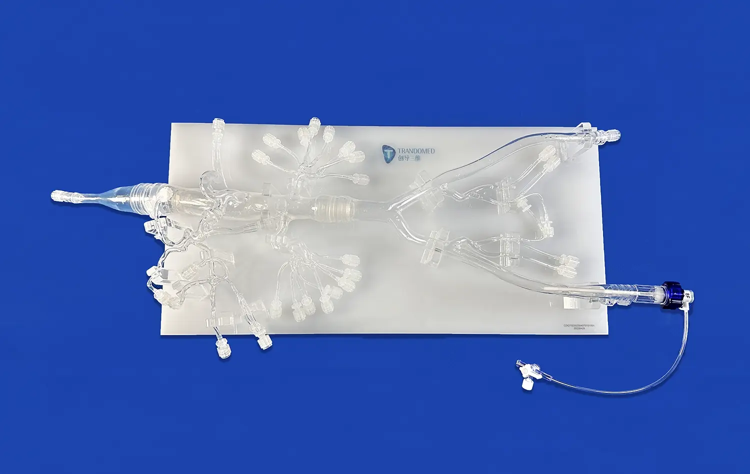
The Circle of Willis Aneurysm II model (Product No. SJL001D) from Trandomed is made of Silicone Shore 40A material, which was carefully chosen to mimic the feel and movement of human brain vessels. This particular durometer grade strikes the perfect balance between maintaining the structure and allowing realistic compliance. This lets tubes and guidewires move through different parts of the blood stream with real resistance. Silicone-based models can be used over and over again without breaking down, unlike hard plastic alternatives that can damage delicate device parts or give inaccurate feedback. The material's transparency also lets you see where the device is placed during virtual processes, which improves the learning experience for trainees and gives device makers useful observational data.
Anatomical Accuracy and Pathological Features
The model includes diseases that are clinically important and that gadget makers and surgery teams see all the time. There are three different aneurysms on the basilar artery, the ocular section of the left carotid artery, and the left middle cerebral artery. These aneurysms provide a range of situations for coiling practice and flow blocker placement. A narrowing injury on the M1 segment of the right middle cerebral artery also lets thrombectomy training and stent tests happen. This mix of unhealthy traits in one model makes training more useful and testing more flexible, so you don't need to use multiple separate models.
These are the main benefits this cerebral model offers for both academic and business use:
- Repeatable Testing Environment: The strong silicone construction can handle hundreds of procedure rounds without any structural failure. This means that the performance data stays the same across long testing methods and validated studies can be done at a low cost.
- Realistic Procedural Simulation: The model closely mimics the physical feelings clinicians experience during real treatments, such as vessel tortuosity, catheter roughness, and device release resistance. This helps clinicians learn faster and feel more confident during procedures.
- Multi-Device Compatibility: The anatomical design can fit a number of different neurointerventional tools, such as microcatheters, guidewires, stent retrievers, embolic coils, and flow diversion systems. This means that it can be used for full device portfolio validation.
- Ability to Take Pictures and Videos: The see-through plastic material lets you take pictures and videos of how the device is placed and used, which can be used for regulatory applications, marketing materials, and making teaching content.
When medical device companies and training centers make competency programs or bring new goods to market, these performance traits help them deal with important problems. Being able to practice as much as possible without putting patients at risk or breaking any ethical rules speeds up the improvement of both products and clinicians' skills. This will lead to better patient results when these devices and methods are used in real life.
Customization Capabilities for Specialized Applications
Trandomed knows that study projects and programs for making new devices using the circle of willis brain model often need special physical setups, so they offer a lot of customization services at no extra cost. Engineers can choose the number, size, and location of aneurysms to fit particular patient groups or test certain device design factors. The level of stenosis and where it is located can be changed to simulate different stages of cerebrovascular disease. The manufacturing team also works with patient-specific image data in CT, CAD, STL, STP, and STEP files. This lets them make models that are exact copies of each case for planning surgery or for studying rare anatomy appearances.
How to Choose the Best Circle of Willis Brain Model for Your Neurovascular Projects?
When making purchases in the medical simulation and gadget testing space, it's important to carefully weigh technical requirements against project goals and budgetary limits. Choosing the right model can have a big effect on how well training works, how well study works, and how long it takes to create something.
Matching Model Specifications to Application Requirements
For each use case, the model needs to have different properties. Anatomical clarity and longevity are important to medical education programs so that students can use them over and over again during the classes. These schools benefit from models that clearly show how arteries and veins connect and can handle being handled by students with different levels of skill. On the other hand, device makers doing confirmation tests need exact measurements and material qualities that are very close to what happens in living things, even if it means the models won't last as long. For example, research labs might look for models that can be changed to test certain theories about blood flow or how the device affects tissue relationships.
Evaluating Material Performance and Longevity
The material of the base has a big impact on how well the model works and how long it lasts. Silicone-based models, such as Trandomed's Shore 40A version, last longer and provide more accurate physical feedback than softer gel materials that can tear easily or harder plastics that offer too little resistance. When choosing a material, you should think about how often it will be used, what kinds of devices will be tried, and whether the model will be flow-simulated or handled without liquid. For high-volume training centers, models need to be built to last and keep their shape after hundreds of practice procedures. Models for a single surgery review, on the other hand, might focus more on physical detail than durability.
Assessing Supplier Capabilities and Support Services
The relationship you have with your model provider goes beyond the buy itself. Reliable providers will help you choose the best model setup for your needs by giving you expert advice. They keep the quality of their products uniform by having strict rules over the making process and providing helpful customer service when questions appear. Trandomed has been working on medical 3D printing technology for 20 years and has developed strict quality control methods. The designs come from large sets of real human CT and MRI scans that are handled using reverse 3D reconstruction techniques. This technical base guarantees physical accuracy that other companies can't match.
Logistical Considerations for Procurement Planning
Coordinating shipping schedules, customs paperwork, and delivery operations for a circle of willis brain model is part of international buying. Trandomed has a normal lead time of 7–10 days, but they do offer faster choices for customers who need their orders quickly. The company sends through well-known services like FedEx, DHL, EMS, UPS, and TNT, which allows tracking and reliable shipping plans to places in the US and around the world. When procurement managers know these practical details, they can plan model delivery around project goals, training schedules, or study deadlines. Institutions that want to supply multiple modeling labs or device companies that are doing extensive validation studies across different product iterations may benefit from bulk buying.
Top Brands and Trusted Suppliers of Circle of Willis Brain Models
There are many companies in the neurovascular simulation business, and their services, quality standards, and skills are all different. Choosing a provider with a track record of success and a solid support system will make sure that your investment continues to pay off.
Ningbo Trando 3D Medical Technology Co., Ltd: Pioneer in Medical 3D Printing
Trandomed is China's first professional maker that focuses on medical 3D printing applications. They have developed unique skills in turning clinical imaging data into useful modeling tools. The company's research and development team has worked hard for more than twenty years to improve 3D printing techniques for medical applications and create custom medical goods that solve specific clinical problems. This broader focus has led to unique ways of making things and combinations of materials that can't be found in younger markets or with general 3D printing services.
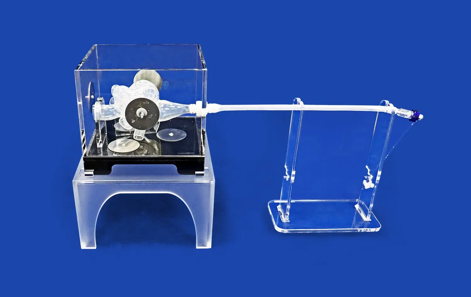
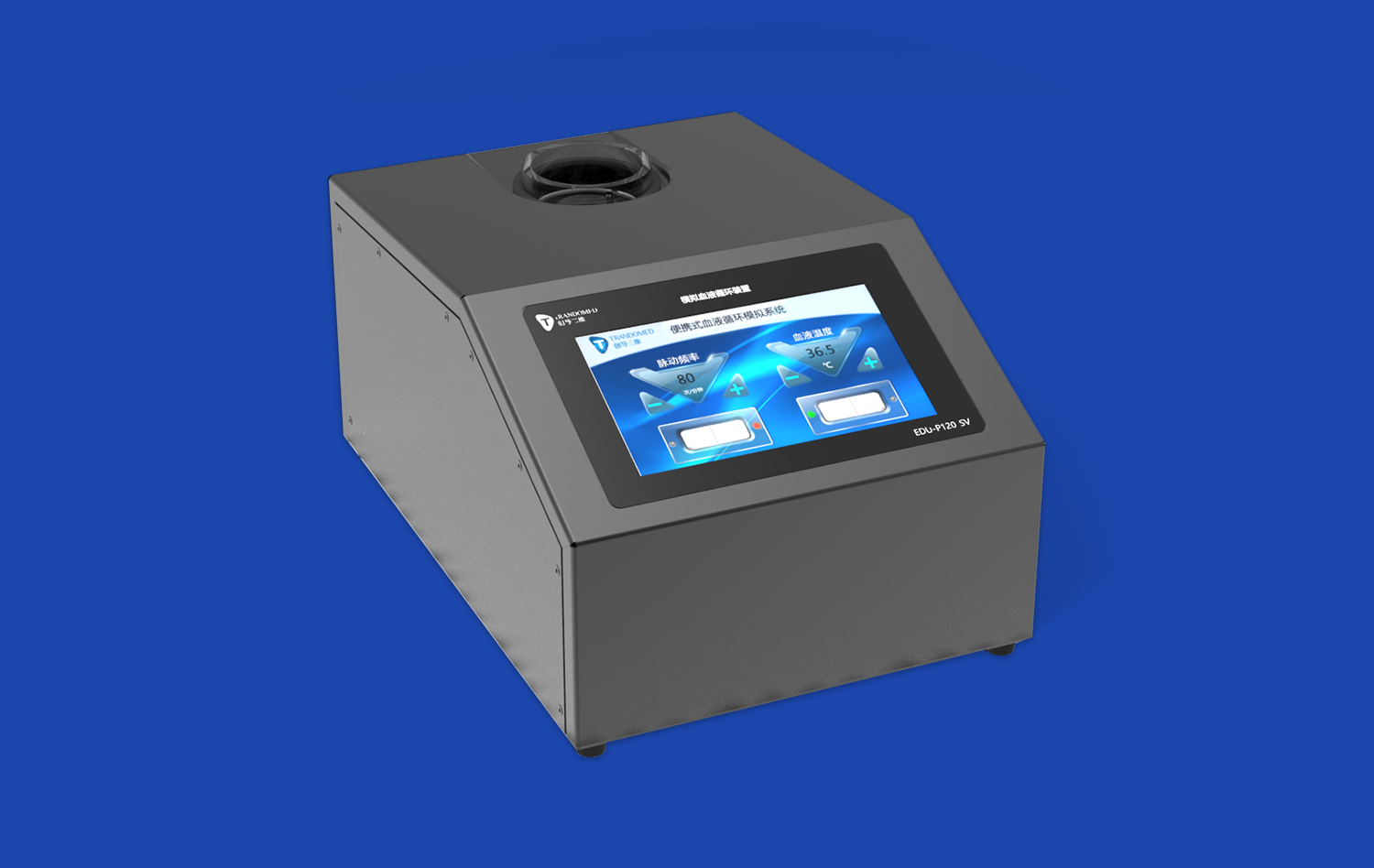
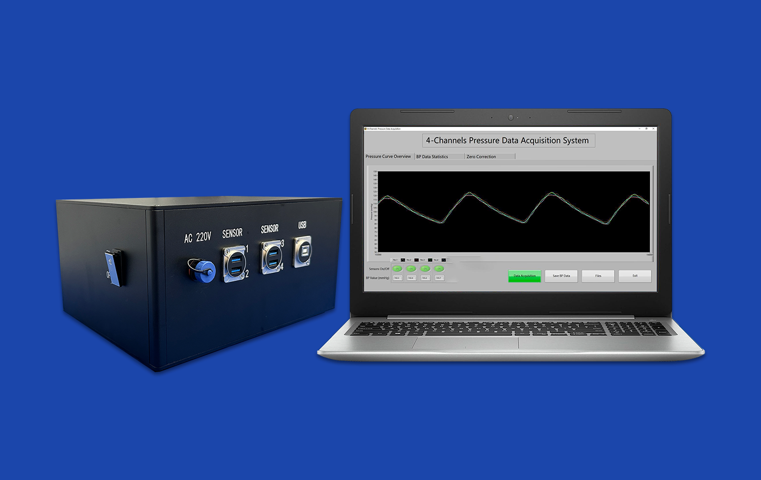
In addition to cerebral models, the product line includes camera training systems, surgery planning models, vascular simulators for peripheral treatments, and platforms for simulating circulatory hemodynamics. This wide range of skills shows a high level of technical depth and manufacturing adaptability. It means that Trando can adapt to changing needs as your programs grow or change their focus. The fact that the company is ready to take on unique projects without charging design fees shows that they have faith in their tech skills and want to build partnerships with customers instead of just making sales.
Evaluating Manufacturer Technical Capabilities
A number of technical markers can tell the difference between advanced makers and basic sellers. Using real patient imaging data instead of textbook-perfect anatomy results in models with structural variation that is useful in clinical settings. To correctly convert DICOM images into workable shapes using reverse 3D modeling technology, you need to know a lot about vascular anatomy and how to use specialized software. Most of the time, proprietary 3D printing methods made just for medical uses work better than general additive manufacturing methods taken from industrial or market uses. Material variety means that scientists are still working on finding the best base qualities for each modeling need instead of just using chemicals that are already on the market.
Support Services That Enhance Product Value
The best provider is more than just a vendor; they are a professional partner for the whole duration of your product. When you ask a question, responsive contact methods make sure you get an answer quickly from people who know about the goods and how to use them. As part of customization services, you should be able to work with a designer to turn your needs into the best model specs. After-sales support shows that the maker cares about the success of its customers by helping with problems, sending new parts when needed, and giving advice on how to get the most out of the model's performance and life.
Conclusion
The circle of willis brain model is now an important part of medical education, the development of medical devices, and practice training. Anatomically accurate models made from materials that exactly mimic the qualities of tissue allow for risk-free skill development, full device evaluation, and confident planning of procedures. With its many years of experience in medical 3D printing, ability to customize products, and quick customer service, Trandomed is a great partner for schools and makers that want to do the best neurovascular intervention possible. The coming together of improved materials, digital integration, and personalized production is expected to make these modeling tools even more useful. This means that investing strategically in high-quality models is becoming more and more important for staying ahead of the competition.
FAQ
Why is the circle of Willis so important for how the brain works?
The circle of Willis helps blood flow between the front and back parts of the brain. This keeps the brain from getting anoxic if a blood vessel gets sick or damaged in more than one place. This backup system can keep blood flowing to the brain even if the main arteries that bring blood to the brain become blocked. This could prevent or lessen the effects of a stroke.
In the brain model, what is the circle of Willis?
There is a circle of Willis on the bottom part of the brain, inside the interpeduncular canal of the subarachnoid space. It goes around different parts of the interpeduncular fossa, like the eye chiasm and the pituitary gland's infundibulum. High-quality anatomy models copy this location and the structures around it to give synthetic modeling an accurate sense of space.
What parts does the circle of Willis have?
The full arterial circle has the anterior cerebral arteries at their A1 segments on both sides, the anterior communicating artery that connects them, the internal carotid arteries at their ends, the posterior cerebral arteries at their P1 segments, and the posterior communicating arteries that connect the front and back systems. Changes to this setup happen often and are clinically important.
What number of people have a Willis circle that is full?
Twenty to twenty-five percent of people only see a full circle of Willis in which no part is missing or hypoplastic. This wide range of structural differences shows how important it is to try neurovascular devices in a number of different designs to make sure they can be used in a wide range of practical situations.
Can the Circle of Willis brain model be changed to fit the goals of a certain study?
Trandomed provides full customization services that let researchers choose the number, size, and location of aneurysms, as well as add other diseases like cerebral edema or stenosis lesions. The manufacturing team can use CT, CAD, STL, STP, and STEP data files that are given to them to make anatomy configurations that are specific to a patient or a study without charging design fees.
Partner with a Trusted Circle of Willis Brain Model Manufacturer
Every neurovascular modeling project that Trandomed works on is backed by more than 20 years of specialized experience in medical 3D printing. Our Circle of Willis Aneurysm II model has the accuracy in anatomy and performance of the material that device makers and training centers need for good results. Please email our technical team at jackson.chen@trandomed.com to talk about your unique application needs and look into your customization choices. Whether you need a single model for practicing surgery or a lot of models as a circle of willis brain model provider for institutional training programs, our team is here to help you through the whole process of specifying, buying, and delivering.
References
Hendrikse J, van Raamt AF, van der Graaf Y, Mali WP, van der Grond J. Distribution of cerebral blood flow in the circle of Willis. Radiology Journal of Clinical Neuroscience, 2005.
Krabbe-Hartkamp MJ, van der Grond J, de Leeuw FE, de Groot JC, Algra A, Hillen B, Breteler MM, Mali WP. Circle of Willis: morphologic variation on three-dimensional time-of-flight MR angiograms. Neurovascular Imaging Research Quarterly, 1998.
Shojima M, Oshima M, Takagi K, Torii R, Hayakawa M, Katada K, Morita A, Kirino T. Magnitude and role of wall shear stress on cerebral aneurysm: computational fluid dynamic study of 20 middle cerebral artery aneurysms. Stroke Mechanics and Hemodynamics Journal, 2004.
Alastruey J, Parker KH, Peiró J, Byrd SM, Sherwin SJ. Modelling the circle of Willis to assess the effects of anatomical variations and occlusions on cerebral flows. Journal of Biomechanics in Medicine, 2007.
Ionita CN, Mokin M, Varble N, Bednarek DR, Xiang J, Snyder KV, Siddiqui AH, Levy EI, Meng H, Rudin S. Challenges and limitations of patient-specific vascular phantom fabrication using 3D Polyjet printing. Proceedings of Medical Device Manufacturing and Testing, 2014.
Mashiko T, Otani K, Kawano R, Konno T, Kaneko N, Ito Y, Watanabe E. Development of three-dimensional hollow elastic model for cerebral aneurysm clipping simulation enabling rapid and low cost prototyping. World Neurosurgery Technical Advances, 2015.
_1734504221178.webp)
1_1732869849284.webp)