Circle of Willis Brain Model in Aneurysm Clipping and Coiling Simulation
2026-01-27 09:00:03
The circle of Willis brain model is a big step forward in neurovascular training because it gives medical schools and healthcare workers a way to practice clipping and coiling aneurysms that is accurate in terms of anatomy. This special cerebral model makes a copy of the complicated artery network at the base of the brain, complete with abnormalities like aneurysms and stenosis lesions. As neurosurgical methods get more complex, the need for high-fidelity modeling tools has grown incredibly in medical schools, hospital training units, and research labs that want to improve procedural skills without putting patients at risk.
Understanding the Circle of Willis Brain Model and Its Clinical Significance
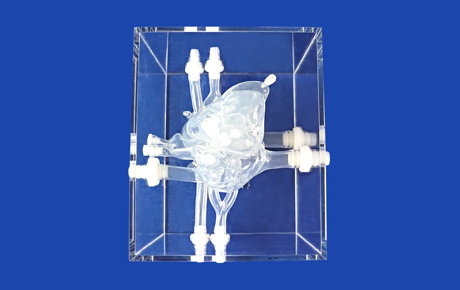
The Circle of Willis is a unique ring of arteries at the base of the brain in the interpeduncular canal. It is the most important part of cerebrovascular anatomy. This vascular structure is like nature's insurance policy; it provides backup circulation that keeps brain tissue safe from ischemic damage when the main blood vessels become damaged. Understanding this structure isn't just useful for school; it has a direct effect on how well neurovascular treatments go during surgery.
Anatomical Components and Functional Importance
The artery circle is made up of several important blood channels that work together to keep blood flowing to the brain. The anterior circulation is made up of the anterior communication artery and the two anterior cerebral arteries at their A1 parts. At their ends, the internal carotid arteries contribute, and the posterior circulation is made up of the posterior cerebral arteries at their P1 parts. These arteries connect to the anterior system through the posterior communication arteries. This arrangement makes a backup that saves lives when a blood vessel is blocked or during surgery.
The fact that this tissue is different for each person makes it especially useful for modeling training. According to research, only 20 to 25 percent of people have a full, textbook-perfect Circle of Willis. The rest of the people have some degree of hypoplasia or lack in one or more parts. These differences have a big effect on the risk of aneurysm growth and the choice of surgery method. So, for advanced cerebral models to prepare doctors for all the different kinds of clinical situations they will see, they need to include a wide range of accurate body parts.
Role in Aneurysm Formation and Intervention
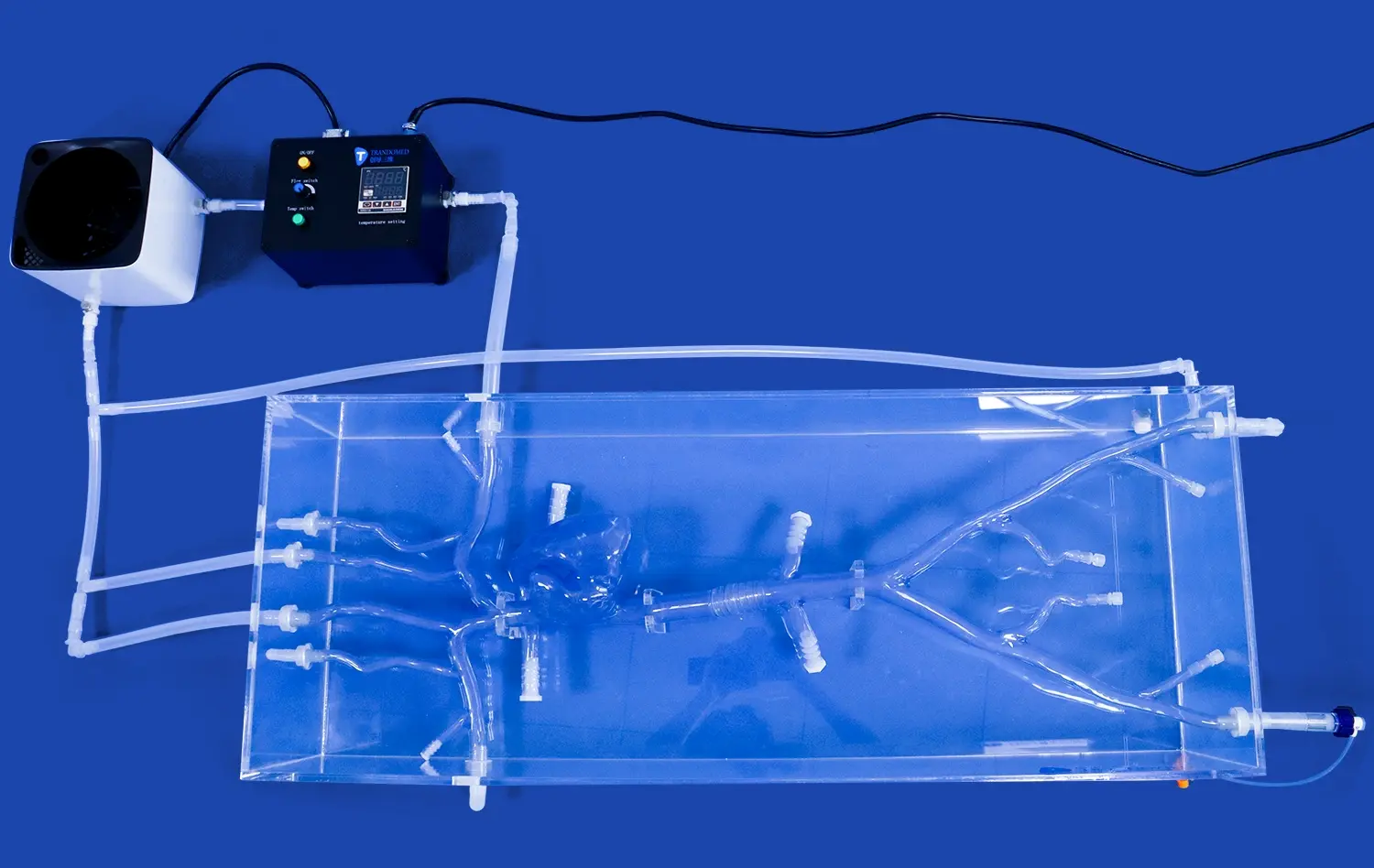
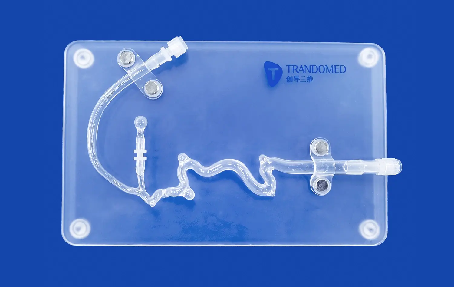
Aneurysms form in certain parts of this network of arteries, usually where blood flow is concentrated at the places where two arteries meet. Aneurysms often form at the split in the middle cerebral artery, the junction of the anterior communicating artery, and the start of the posterior communicating artery. Our neurovascular training model has three aneurysms carefully placed on the basilar artery, the eye section of the left carotid artery, and the left middle cerebral artery. These aneurysms are based on real-life clinical appearances.
The model has an M1 segment stenosis injury in the right middle cerebral artery, which makes it more complicated and more like a real disease. This stenotic part lets trainees practice thrombectomy treatments along with aneurysm therapies, which broadens the learning beyond situations with just one disease. This kind of thorough modeling turns regular practice into skill development that is more complex, connecting academic knowledge to real-life clinical use.
Simulation Advantages for Surgical Planning
Neurosurgeons use patient-specific models such as the circle of willis brain model more and more to plan and practice their procedures before they go into the operating room. The cerebral artery computer model lets you feel and see things in three dimensions that two-dimensional images can't do. In a safe setting, surgeons can try out different clip placement angles, figure out the best way to do a craniotomy, and plan for possible structural problems. This planning immediately leads to shorter operations, fewer problems, and better results for patients. These are very important measures for hospital quality offices and surgery training programs.
Procurement Guide: Selecting and Purchasing Circle of Willis Brain Models
It takes careful planning and a good idea of what the organization needs to get through the buying process for specialized medical training tools. This part gives useful advice for people who are in charge of buying neurovascular training equipment, such as training leaders, procurement managers, and clinical engineers.
Defining Core Requirements
A full needs assessment with key partners from across your school is the first step to successful buying. The neurosurgery staff should decide what specific procedural skills the model needs to support, such as anterior circulation aneurysms, basilar apex disease, or full covering. Simulation center leaders give advice on how to deal with limited room, upkeep needs, and connecting new systems to current training systems. The budget officials set the financial parameters that determine which vendors to use and how much customization is possible.
When you look at the Trandomed neurovascular training model, you should think about how its features fit with the learning goals of your program. The three separate aneurysms help with graduated learning, and the combined stenosis lesion increases the ability to train people in thrombectomy. Institutions that focus on research uses may put an emphasis on customization features that let models be changed to fit the anatomy of specific patients or the testing needs of new devices. Making these goals clear makes it easier for vendors to talk to you and makes sure that the solutions they offer meet your specific needs instead of just having general features.
Cost Optimization Strategies
Budgets always play a role in purchasing choices, but cost-effectiveness is more than just unit price. Comparing list prices alone doesn't give you as much financial information as looking at the total costs of owning. Medical-grade silicone is used to make durable products that don't need to be replaced as often, spreading out the cost of the original purchase over a longer period of time. Models that can be used for multiple training purposes, like clipping, coiling, and thrombectomy, get rid of the need to buy multiple models for each reason, which saves money and space.
Larger schools or healthcare systems with multiple locations can save even more money by using volume purchasing agreements. Trandomed can handle large orders and offers good terms because they know that economies of scale are good for both the producer and the customer. When you work with other institutions or coordinate buying across linked training centers, you make enough orders to get special treatment. These joint methods to buying make it easier to negotiate and build professional networks where people can share training materials and data on how well they worked.
Ordering Process and Logistics
Figuring out the transaction process helps you set reasonable deadlines for execution and keep everyone's expectations in check. Our expert team can talk to you about customization options, such as changing the number, size, or position of an aneurysms, at no extra cost. This is an extra service that sets our customer approach apart.
After an order for a circle of willis brain model is confirmed and payment is processed through a foreign bank transfer, the production wait time is usually between 7 and 10 days. This quick turn-around shows how well our manufacturing has been streamlined and helps us keep key materials in stock. International shipping with well-known companies like FedEx, DHL, EMS, UPS, and TNT guarantees arrival and lets you see where the package is at all times. Transit times to places in the United States are usually between 5 and 7 working days, but this can change depending on how the customs process goes. Buying tools at least one month before it needs to be deployed gives you plenty of time in case something goes wrong.
Customization and After-Sales Support
One thing that makes Trandomed stand out is that we can customize products without making the costs too high. We can work with medical imaging data in a number of different forms, including CT, CAD, STL, STP, and STEP. This data is then turned into models that are specific to the patient or the disease. This feature is very helpful for research groups looking into certain artery combinations, device makers making sure their goods work on different body types, and training programs focusing on uncommon appearances.
In addition to delivering the goods, we are committed to providing full help after the sale. Technical questions about how to set up the model, the best way to use it, or the best way to do care are quickly answered by experts. Even though we have strict quality control, if problems do happen, we stand behind the performance of our products with a strong guarantee and quick problem settlement. This service infrastructure shows that we are more interested in long-term partnerships than in being a business provider. It helps customers succeed throughout the lifecycle of a product.
Enhancing Aneurysm Clipping and Coiling Training with Anatomical Models
The best way to judge any modeling technology is by how well it helps students learn and how much it helps them improve their practical skills. High-fidelity neurovascular models have changed how schools teach practical skills, switching from learning from patients when they are available to organized, competency-based courses.
Educational Impact on Skill Development
In the past, neurosurgery training was mostly based on watching others and taking on more authority while being supervised. This is called an internship approach, which doesn't work well in today's healthcare setting. The time it takes to become procedurally independent has been shortened because of concerns about patient safety, fewer hours for residents to work, and more complicated cases. Simulation-based training gets around these problems by giving people endless chances to practice in safe places where mistakes help them learn instead of hurting them.
Medical teachers who use physically accurate cerebral models say that their students' work improves in a number of skill areas. When compared to just classroom teaching, organized modeling practice makes a big difference in improving technical skills like microcatheter navigation, coil release precision, and clip application accuracy. Learners' cognitive skills also improve as they learn to recognize patterns in the different shapes and sizes of aneurysms and make decisions about the best ways to treat them. These benefits are still seen when the same patients go through the same treatments, which proves that simulations are a good way to build portable skills.
Real-World Application Success Stories
Some of the best medical schools have added realistic neurovascular modeling such as the circle of willis brain model to their regular training programs, and the results are truly amazing. Before trainees could work on aneurysm cases, the neurosurgery training program at a big university hospital made them do simulated rotations using detailed models of the brain arteries. During the two-year review period, the program saw a 40% drop in mistakes made during treatments and a 25% drop in the average length of time young doctors spent on each case. These results show that better simulation training has led to real improvements in patient safety and business efficiency.
In the same way, device makers have used anatomy models to create and test their products. To test a new flow diversion system, a business that makes arterial devices used special Circle of Willis models that looked like difficult body parts. Being able to try out different designs and see how well they work in different artery setups sped up the development process and helped find problems before they happened in clinical studies. This way of doing pre-clinical testing lowers the risks of getting regulatory approval and builds more proof that the gadget is safe and works.
Emerging Technologies and Future Directions
Simulation technology is moving in a direction where real models will be more and more integrated with digital improvement systems. Augmented reality platforms add step-by-step instructions on top of anatomy models, making training experiences that are a mix of real-life input and physical realism. Models with sensors can keep track of where instruments are placed and give objective performance measures, which supports competency assessment processes that are better than subjective evaluation methods.
New discoveries in material science keep pushing the limits of how well tissues can be replicated. New silicone mixtures that are being made promise to be even more like the mechanical qualities of a live artery, such as its pulsatile flow and dynamic valve compliance. These new developments will make the gap between simulation and real-life clinical situations even smaller. This will make skill transfer more effective and allow for a wider range of techniques to be used for simulator-based training.
Conclusion
Putting money into high-quality neurovascular modeling equipment is a long-term investment to better teaching and better patient outcomes. The circle of willis brain model is an important tool for schools that want to improve their students' skills in aneurysm clipping and coiling interventions. By giving training tools that are physically correct, pathologically diverse, and technically flexible, these models help people learn skills that lead to better, more effective patient care. As healthcare systems put more emphasis on competency-based testing and ongoing professional growth, advanced simulated technology moves from being an extra to being an essential part of training.
FAQ
What makes a Circle of Willis model good for simulating aneurysm clipping different from simple anatomy models?
Models made just for training in interventional procedures include functional pathology, such as actual aneurysm shapes with correct neck shapes, dome shapes, and vessel relationships around them. These models let you actually move the instruments, so you can practice clip application and putting in arterial devices. Basic skeletal models are useful for reference, but they don't have the material qualities and structure form that are needed to learn how to do things by doing them. The Trandomed system uses Shore 40A silicone, which reacts to surgery tools like artery flesh does. This gives the user real physical feedback that is missing from hard display models.
What makes neurovascular training models from different companies different in terms of accuracy, materials, and features?
When it comes to physical accuracy, material choice, and feature depth, there are big differences between makers. Companies that sell general anatomy models might make simplified Circle of Willis images that don't show abnormal features or actual vessel sizes. Neurovascular simulation makers like Trandomed use image data from patients and reverse 3D modeling to make sure the models are accurate in terms of anatomy. Basic plastics and advanced medical-grade silicones are the two types of materials that can be used. Medical-grade silicones are better at lasting and imitating tissue. Differentiating features include the ability to be customized, the ability to integrate multiple pathologies, and the ability to support both surgery and percutaneous methods.
Are there ways to make it fit the needs of specific schooling or clinical training?
There are no extra design fees for Trandomed's many customizable features, which let institutions change the number, size, and location of aneurysms based on their needs. We can include a number of different brain diseases, such as intracranial edema, stenosis tumors in certain places, and other arterial problems. Processing of CT, MRI, CAD, STL, STP, and STEP format data files makes it possible to make models that are unique to each patient. This adaptability lets the simulator be used for study purposes, training in rare diseases, and testing medical devices in ways that go beyond standard product setups. This makes sure that the simulator perfectly matches your program goals.
How long does a neurovascular training model usually last when it is used regularly?
The operating lifespan is mostly determined by the quality of the materials, how they were built, and how often they are used. Medical-grade plastic and precise 3D printing are used to make models like Trandomed goods. These models can usually handle hundreds of training sessions without losing their shape. When you handle and store things properly, they last a lot longer. Unlike cheap models that break down after a short time of use, high-quality simulations are investments that pay off over many years by supporting ongoing training groups. We tell you how to maintain your equipment properly and sell you new parts when they break. This way, your equipment will continue to teach you for a long time.
Partner with a Leading Circle of Willis Brain Model Manufacturer
Medical trainers, hospital training units, and research centers are welcome to talk to Trandomed about how our neurovascular models can improve your procedural training programs. Our Circle of Willis brain model for sale is the result of more than 20 years of progress in medical 3D printing. It combines accurate anatomy with realistic pathology to help students learn important skills. Our expert team is here to help you with any questions you have, whether you need standard setups or solutions that are specifically designed to meet your training goals. Get in touch with jackson.chen@trandomed.com to talk about buying in bulk, customizing choices, and how our full after-sales support guarantees long-term training success. You can look through our full catalog of products at trando-medical.com and download thorough technical specs that show how committed we are to improving neurovascular education through advanced modeling technology.
References
Alaraj, A., Charbel, F. T., Birk, D., et al. (2013). "Role of cranial and spinal virtual and augmented reality simulation using immersive touch modules in neurosurgical training." Neurosurgery, 72(Suppl 1), 115-123.
Bambakidis, N. C., Selman, W. R., & Sloan, A. E. (2013). "Surgical rehearsal platform: potential uses in microsurgery." Neurosurgery, 73(Suppl 1), 122-126.
Mashiko, T., Otani, K., Kawano, R., et al. (2015). "Development of three-dimensional hollow elastic model for cerebral aneurysm clipping simulation enabling rapid and low cost prototyping." World Neurosurgery, 83(3), 351-361.
Ryan, J. R., Almefty, K. K., Nakaji, P., & Frakes, D. H. (2016). "Cerebral aneurysm clipping surgery simulation using patient-specific 3D printing and silicone casting." World Neurosurgery, 88, 175-181.
Waran, V., Narayanan, V., Karuppiah, R., et al. (2014). "Utility of multimaterial 3D printed models in surgical planning of cerebral aneurysm treatment." Journal of Neurosurgery, 120(3), 757-763.
Wurm, G., Lehner, M., Tomancok, B., et al. (2011). "Cerebrovascular biomodeling for aneurysm surgery: simulation-based training by means of rapid prototyping technologies." Surgical Innovation, 18(3), 294-306.
_1736215128474.webp)
_1734504221178.webp)
_1732866687283.webp)