Custom 3D artery models are a huge step forward in neurovascular study and training. They make aneurysm simulations and internal carotid artery (ICA) treatments more realistic than ever before. These highly accurate models of human bodies allow doctors to practice difficult procedures without any risk, closing the gap between what they know in theory and what they can do in real life. These models, which were made with advanced silicone materials and high-fidelity 3D printing technology, accurately show the complex shapes of cerebrovascular structures. This lets doctors, students, and academics practice their skills before they go into the operating room.
Understanding Custom 3D Artery Models for Aneurysm and ICA Simulation
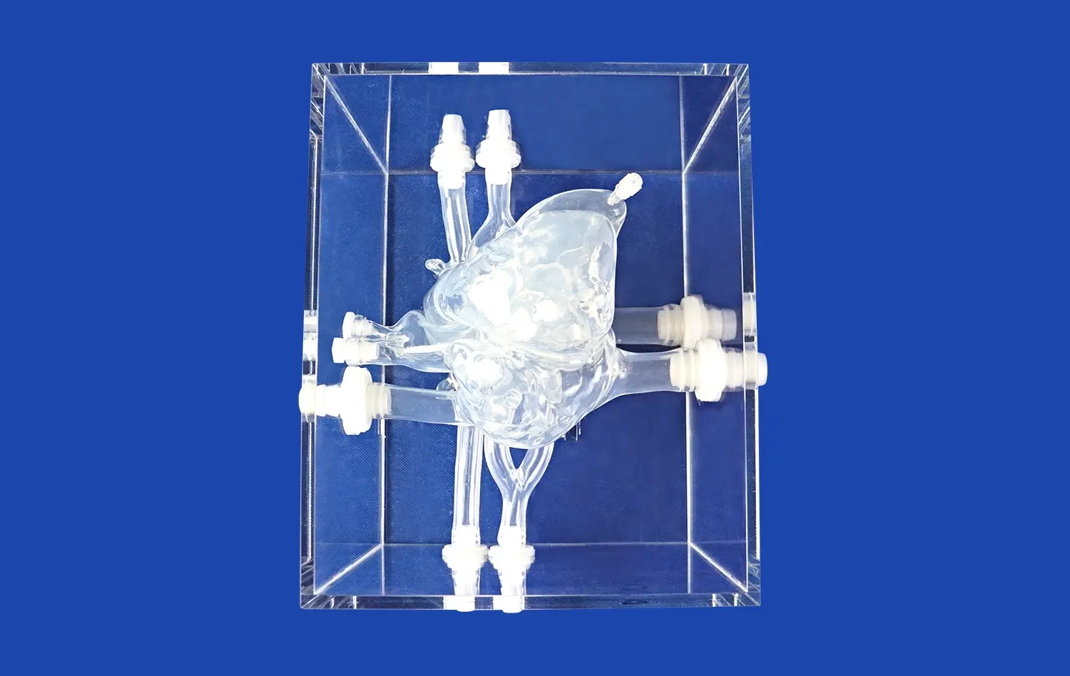
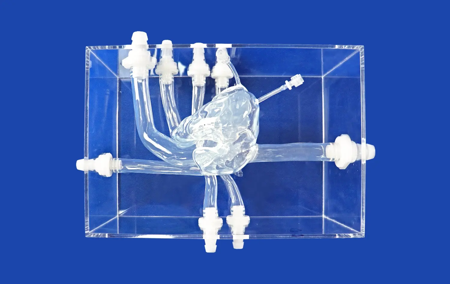
The creation of tailored neurovascular models has changed the way doctors are trained and how procedures are planned. Here at Trandomed, our No. The SJX005, which is also called the Middle Cerebral Artery model, is a great example of this new technology because of its complex design and accurate anatomy.
The Technology Behind High-Fidelity Vascular Simulation
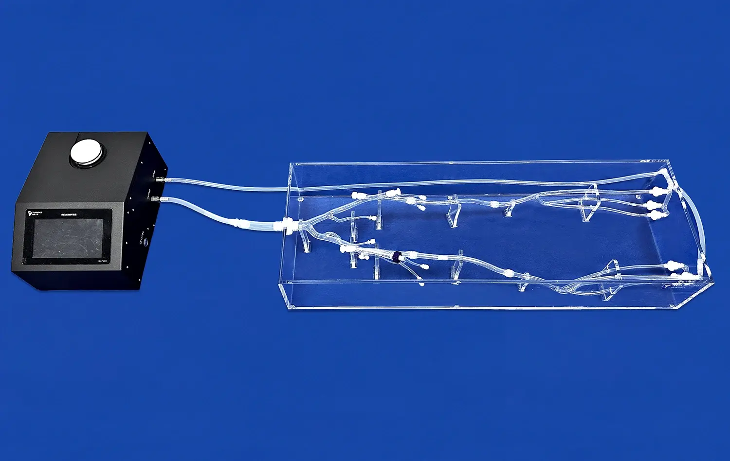
Modern arterial simulations use cutting edge 3D printing to make models that are exact copies of human bodies, down to the smallest detail. The process starts with medical imaging data from CT scans or CAD files. Our team then uses advanced additive manufacturing methods to turn this data into real models. With this technology, we can very accurately copy the natural curvature of blood vessels, the exact sites of bifurcations, and the complicated shape of aneurysms.
Silicone Shore 40A, a material carefully chosen for its tissue-like qualities, is used to make the SJX005 model. Working with this medical-grade plastic feels a lot like working with real blood vessels, which helps doctors build muscle memory and confidence in their procedures. The material is flexible enough to make realistic simulations of catheter travel through complicated venous paths, but strong enough to last through multiple training sessions.
Anatomical Accuracy and Customization Capabilities
The unique thing about advanced neurovascular training tools is that they can be changed to fit specific clinical or teaching needs. Our customization service lets you make changes without asking extra for the design, so institutions with different needs can get specialized training. The model can be changed in a number of ways. For example, the number, size, and location of aneurysms can be changed to represent different medical conditions. The degree of curvature on the internal carotid artery segment can be changed to reflect the anatomy of a specific patient. Finally, the training goals can be used to finetune the tortuosity radius of the middle cerebral artery (MCA) and anterior cerebral artery (ACA).
This level of tailoring is very helpful for surgery teams getting ready for difficult cases. Surgeons can practice their method, find possible problems, and make backup plans before the actual surgery by duplicating a patient's exact venous anatomy from image data. Leading medical schools have done research that shows this kind of advance planning can cut the time needed for surgery by up to 25% and the number of complications by a large amount.
Benefits for Medical Education and Procedural Rehearsal
Neurovascular treatments are easier to learn when you use high-quality anatomy models like the 3D artery model for training. Medical students and trainees can practice moving catheters, navigating guidewires, and coiling aneurysms over and over again without putting patients at risk. In a way that computer models or textbooks can't match, getting hands-on experience boosts confidence and skill.
The model is useful for medical teaching in many ways. It can be used to test how trackable different catheter systems are, to simulate aneurysm tamponade procedures, and to practice using embolic coils and flow diversion devices. Manufacturers of medical devices also use these models to try new tools in real-life settings before putting them through clinical studies.
Comparing 3D Artery Models with Traditional and Alternative Solutions
The field of medical education has changed a lot over the years, but many schools still use old methods that make learning less effective. Figuring out how modern neurovascular models stack up against older methods helps people in charge of buying things justify spending money on new training tools.
Advantages Over Cadaver-Based Training
Cadaver studies have been thought of as the best way to learn about anatomy for a long time, but they have a lot of problems. It is hard to correctly model catheter guidance or device placement because preserved tissue loses a lot of its natural flexibility and structure. Another problem is that it's hard to get new cadaveric examples because they are expensive, limited, and subject to ethical and legal limits.
These problems can be solved by silicone-based arterial models that offer uniform, repeated training experiences. Instead of breaking down over time like cadavers do, our models stay strong through hundreds of practice sessions. The Silicone Shore 40A material used in the SJX005 model gives realistic physical feedback that feels a lot like real tissue, giving students real procedural feelings. Because anatomy models can be changed, teachers can show students a wider range of disease states than they could with cadaveric objects alone.
Material Properties: Flexible Versus Rigid Models
Which one to use - flexible or hard anatomy models - depends on the training goals and uses. Rigid models, which are usually made from photopolymer resins, are great for showing how bodies are connected and where things are in space. They are good for teaching the basics of anatomy and showing how a gadget works when movement isn't needed.
For routine training, it's important to have flexible plastic models like our brain simulator that let you work with them in a dynamic way. The material's flexibility lets the vessel bend realistically as the tube moves through it, mimicking the push-and-pull movements that doctors see during real treatments. This trait is especially helpful when practicing for sensitive treatments where too much force could break or perforate a blood vessel. The feedback from the touch helps practitioners learn how to use a gentle touch to safely move through fragile neurovascular systems.
Over twenty years of making medical training gadgets, we've learned that the choice of material has a huge effect on how well training works. The Shore 40A hardness grade is the best compromise between being soft enough to feel like real tissue and being firm enough to keep its shape after repeated use.
Cost-Efficiency and Return on Investment
When buying managers look at training options for a 3D artery model, they need to think about both the short-term and long-term costs and benefits. Even though good arterial models cost a lot at first, they pay for themselves over time because they last a long time and can be used again and again. A single corpse purchase has costs for getting the body, storing it, getting rid of it, and limited times when it can be used. Hardware, software rights, and ongoing professional assistance for virtual reality systems are all very expensive.
Silicone skeletal models don't need much upkeep and don't come with ongoing license fees. With the right care, they can give thousands of hours of training to many groups of students. Our unique models have a lead time of 7–10 days, which means that institutions can quickly start training programs without having to wait a long time. We offer fast shipping around the world through well-known carriers like FedEx, DHL, EMS, UPS, and TNT, so your package will arrive on time no matter where it is.
More than just straight cost savings are part of the return on investment. Better technical skill leads to better results for patients, fewer complications, and shorter operating times, all of which improve the institution's image and bottom line. When medical device companies use these models to test their products, they can shorten the time it takes to build new products and avoid having to make expensive changes to the designs later on.
Applications of Custom 3D Artery Models in Surgery and Cardiovascular Research
Precision neurovascular models can be used in a lot of different areas of modern medicine. From getting ready for surgery to doing study in the lab, these tools are becoming more and more important for improving patient care and medical progress.
Pre-Surgical Planning and Risk Reduction
Neurovascular surgeries are some of the most highly difficult treatments in medicine. Outcomes for patients can depend on decisions made at the millimeter level. Surgeons who have to deal with complicated aneurysm cases are using patient-specific models for practice more and more. By using a real copy of the patient's body, the medical team can try out different ways to treat them, choose the best sizes for the devices, and plan for any technology problems that might come up before they make the first cut.
The Journal of NeuroInterventional Surgery released a study that showed how surgery teams used anatomy models to plan how to treat big brain aneurysms. The practice run before the surgery revealed unexpected problems with catheter entry in 30% of cases. This led to changes in the procedure that eventually led to higher success rates. Being able to directly move devices through the replicant's body gave researchers information that image analysis alone could not.
Training Benefits for ICA and Aneurysm Procedures
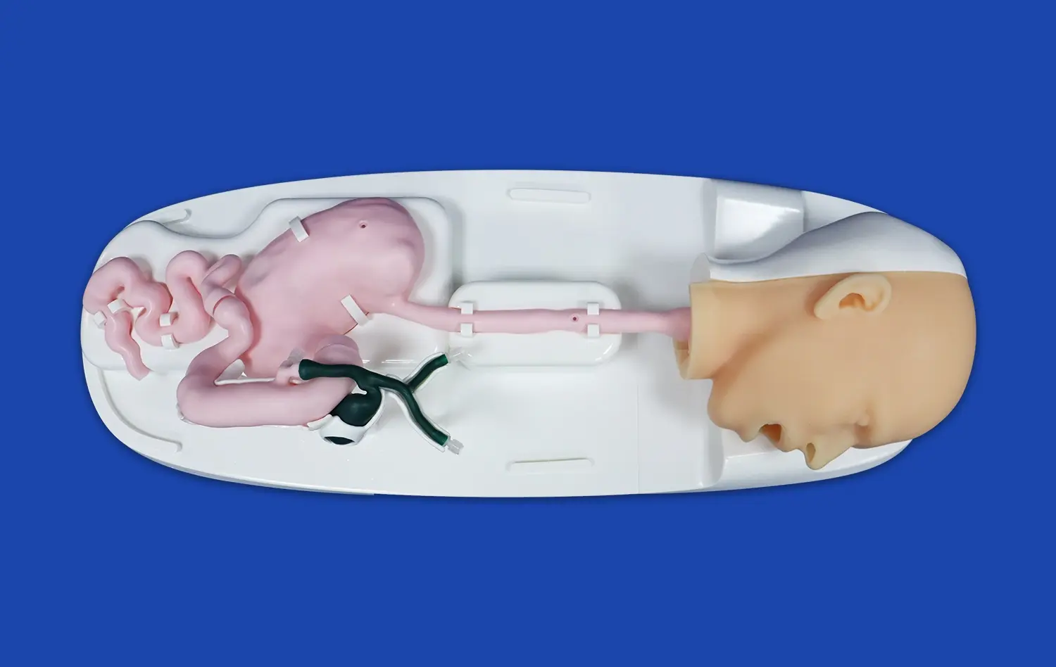
Because it is so important in patients and has a complicated anatomy, the internal carotid artery in a 3D artery model is especially hard to learn how to work on. The vessel's winding path from the neck into the brain, along with important branch vessels, makes it necessary to have excellent piloting skills. Our model of the middle cerebral artery includes accurate ICA shape, which helps trainees get better at handling this difficult arterial area.
Another high-stakes clinical situation is covered by aneurysm treatment exercise. The model's design lets you practice using different types of treatment, such as coil occlusion and flow diversion. Through physical input, trainees can feel where the microcatheter is in an aneurysm sac, learn to tell when the coils are packed densely enough, and come up with ways to deal with problems like coil leakage. Before trying these moves on live patients, this hands-on practice is very helpful.
Device Testing and Research Applications
In addition to being used for practical training, our arterial models are very important for developing medical devices and doing study on the heart. Before putting their products through human trials, companies that are making new tubes, guidewires, stents, and embolic devices need to try them on systems that are like real life. Our models' physical accuracy and material qualities make them reliable testing grounds for checking how well devices can be tracked, delivered, and put into use.
Biomechanical studies at research institutions use custom models to look into things like how blood flows in arteries, how blood pressure affects tissues, and how devices affect tissues. By changing structural features like aneurysm size, vessel tortuosity, or branching patterns, researchers can look into factors that would be hard to control in clinical studies. We can make models fit your needs using data files in forms like CT, CAD, STL, STP, and STEP. This lets experts have full control over the features of the specimens they are studying.
Translational medicine labs have used our models to create methods that connect what scientists find in the lab with what they do in the clinic. Because the structure is constant and can be repeated, researchers can make sure that trial methods work and train doctors on new treatments in controlled settings before putting them on real patients.
Conclusion
Custom neurovascular models like the 3D artery model are important tools for medical teaching, practicing for surgery, and making new medical devices. Models like Trandomed's SJX005 let medical workers practice and improve their skills without putting real patients at risk because they are accurate in terms of anatomy and materials and can be customized. These silicone-based models are more cost-effective, always available, and can be used in a wider range of situations than traditional training methods. They can be used for biological research, pre-surgical planning, and clinical training. Healthcare systems are putting more emphasis on patient safety and excellent procedures. Investing in high-fidelity modeling technology pays off in the form of better results, fewer problems, and faster skill development for practitioners.
FAQ
What amount of realism in anatomy can I expect from my tailored neurovascular models?
High-quality makers get physical accuracy to within a few millimeters by getting model shapes from trusted medical imaging sources and CT or MRI pictures of the patient. To make sure that Trandomed's models accurately reflect clinical anatomy, they go through strict measurement testing and are approved by neurosurgeons. The accuracy of the recreation includes vessel lengths, branching angles, aneurysm shapes, and surface texture. This makes training situations that are very similar to real-life procedures.
How do these models stack up against systems that simulate virtual reality?
Virtual reality tools and physical plastic models work together, not against each other. Virtual platforms are great at giving visual feedback, keeping track of performance data, and modeling rare problems that can't be seen or touched. Physical models provide unmatched physical input, accurate gadget contact, and real-life procedural feel that helps build muscle memory. Virtual systems are used for cognitive learning, and real models are used for manual skill development. This is common in advanced training programs. Practitioners are better prepared for the resistance, friction, and slight feedback they will feel during real treatments when they move a catheter through silicone channels.
How long does it usually take to get a custom model?
Standard requests for customization are finished within 7–10 days of the order being confirmed, which includes production and quality checks. This schedule suggests that clear details about the anatomy are given and that design ideas are approved. Custom projects that are more complicated and need to change a lot of anatomy or add new features may need more development time. We work closely with clients to set realistic dates that meet both production needs and project deadlines. If training needs or medical case preparation are urgent, we can also look into expedited production options.
Can models be changed to fit the body of a specific patient so that surgery can be planned?
Yes, modeling that is special to a patient is one of the best ways to use our customization tools. Surgical teams can send us CT or MR angiography data in the standard DICOM format, and our team will turn it into printed models that show how the patient's blood vessels are structured. This service is especially helpful for cases of complicated aneurysms, odd body variations, or treatments that might go wrong with normal methods. Being able to practice on a copy of the exact body that doctors will be working with can greatly improve planning procedures, choosing the right tools, and working together as a team.
Partner with a Trusted 3D Artery Model Manufacturer
Trandomed has over 20 years of experience making anatomy models that are very accurate and ready to help you with your neurovascular training and study projects using a 3D artery model. Our middle cerebral artery model (Product No. SJX005) can be changed to fit your needs and is accurate in terms of anatomy, materials, and functions. It is perfect for routine training and getting ready for surgery. You are welcome to feel the difference in quality that has made us the source of choice for medical facilities all over the world.You can email our expert team at jackson.chen@trandomed.com to talk about your unique needs, get full product specs, or set up a trial copy. We're committed to providing solutions that are tailored to your specific needs with fast delivery and dedicated support throughout our relationship, whether you're starting a new training program, getting ready for complicated surgery cases, or making new medical products.
References
Mashiko, T., Otani, K., Kawano, R., et al. (2015). Development of three-dimensional hollow elastic model for cerebral aneurysm clipping simulation enabling rapid and low cost prototyping. World Neurosurgery, 83(3), 351-361.
Ryan, J. R., Almefty, K. K., Nakaji, P., & Frakes, D. H. (2016). Cerebral aneurysm clipping surgery simulation using patient-specific 3D printing and silicone casting. World Neurosurgery, 88, 175-181.
Anderson, J. R., Thompson, W. L., Alkattan, A. K., et al. (2017). Three-dimensional printing of anatomically accurate, patient specific intracranial aneurysm models. Journal of NeuroInterventional Surgery, 9(6), 531-536.
Kimura, T., Morita, A., Nishimura, K., et al. (2009). Simulation of and training for cerebral aneurysm clipping with 3-dimensional models. Neurosurgery, 65(4), 719-726.
Waran, V., Narayanan, V., Karuppiah, R., et al. (2014). Utility of multimaterial 3D printers in creating models with pathological entities to enhance the training experience of neurosurgeons. Journal of Neurosurgery, 120(2), 489-492.
Torres, I., & De Luccia, N. (2017). A simulator for training in endovascular aneurysm repair: The use of three dimensional printers. European Journal of Vascular and Endovascular Surgery, 54(2), 247-253.
_1736215128474.webp)
_1734504197376.webp)