Custom Abdominal Blood Vessels Models: Replicating Specific Patient Pathologies
2025-07-11 09:01:08
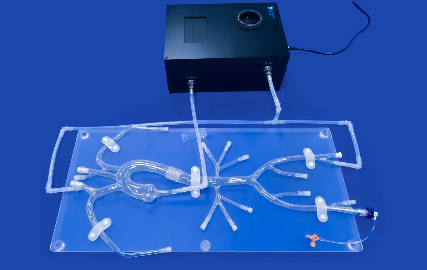
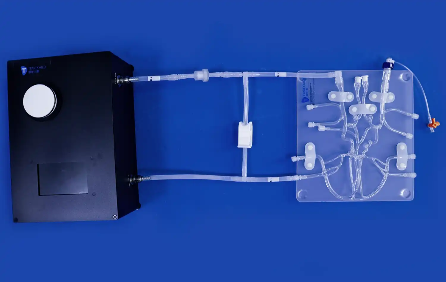
In the realm of medical education and surgical planning, custom abdominal blood vessels models have emerged as a groundbreaking tool. These intricate replicas of patient-specific vascular structures offer unprecedented opportunities for healthcare professionals to study, practice, and prepare for complex procedures. By accurately replicating specific patient pathologies, these models bridge the gap between theoretical knowledge and practical application, enabling surgeons to refine their techniques and develop targeted strategies for individual cases. The ability to create tangible representations of a patient's unique vascular anatomy not only enhances surgical precision but also facilitates better communication between medical teams and patients, ultimately leading to improved outcomes and patient care.
From CT Scan to Simulation: The Process of Creating Patient-Specific Blood Vessels Models
Data Acquisition and Processing
The journey from medical imaging to a physical model begins with high-resolution CT scans of the patient's abdominal region. These scans provide detailed cross-sectional images of the blood vessels, capturing their unique anatomical features and potential pathologies. Advanced software then processes these images, converting them into three-dimensional digital models. This step involves segmentation, where the relevant vascular structures are isolated from surrounding tissues, ensuring that only the desired blood vessels are included in the final model.
3D Printing and Material Selection
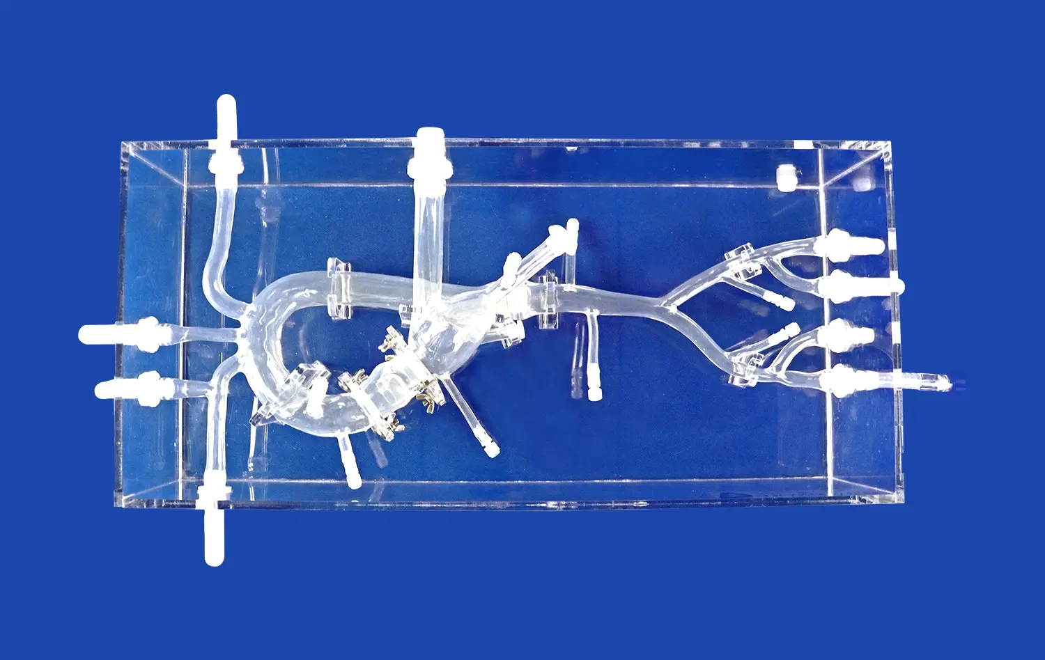
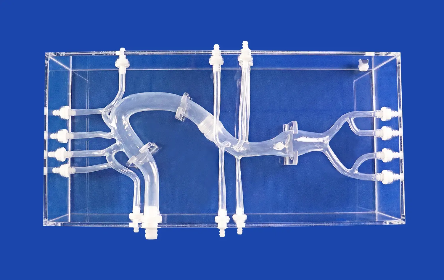
Once the digital model is refined, it's time to bring it into the physical world through 3D printing. The choice of material is crucial in creating realistic abdominal blood vessels models. Silicone has emerged as a preferred material due to its ability to mimic the elasticity and texture of actual blood vessels. The 3D printing process carefully lays down layers of silicone, building up the intricate network of arteries and veins. This additive manufacturing technique allows for the reproduction of even the smallest vessel branches and subtle anatomical variations unique to each patient.
Beyond the Aorta: Recreating Complex Branch Vessel Pathologies
Simulating Vascular Anomalies
While the abdominal aorta serves as the main highway for blood flow in the abdomen, the true complexity lies in its branching vessels. Custom models excel in recreating these intricate networks, including common pathologies like stenosis, aneurysms, and arteriovenous malformations. By accurately replicating these conditions, surgeons can practice navigating through challenging anatomical variations, developing strategies to address potential complications before entering the operating room. This level of preparation is invaluable, especially when dealing with rare or complex vascular anomalies that may not be frequently encountered in clinical practice.
Enhancing Surgical Planning for Visceral Arteries
The abdominal region houses crucial visceral arteries that supply blood to vital organs. Custom abdominal blood vessels models can accurately represent the celiac trunk, superior mesenteric artery, and renal arteries, along with their branches. This detailed recreation allows surgeons to plan intricate procedures, such as endovascular repairs or organ-sparing surgeries, with unprecedented precision. By simulating the exact patient anatomy, these models help in determining the optimal approach, selecting appropriate devices, and anticipating potential challenges during the actual procedure.
Tailoring the Model: Customizing Aneurysm Morphology for Targeted Training
Replicating Diverse Aneurysm Types
Abdominal aortic aneurysms (AAAs) present a significant challenge in vascular surgery, with each case possessing unique characteristics. Custom blood vessels models excel in reproducing the exact morphology of these aneurysms, whether they're fusiform, saccular, or more complex shapes. By accurately replicating the size, location, and shape of the aneurysm, these models allow surgeons to practice different treatment approaches, from endovascular stent grafting to open surgical repair. The ability to interact with a physical representation of the patient's specific aneurysm enhances the surgeon's spatial understanding and aids in selecting the most appropriate intervention strategy.
Simulating Challenging Anatomical Features
Beyond the aneurysm itself, custom abdominal blood vessels models can incorporate challenging anatomical features that complicate treatment. These may include short or angulated aneurysm necks, involvement of visceral arteries, or the presence of intraluminal thrombus. By faithfully reproducing these elements, the models provide a platform for surgeons to develop and refine techniques for navigating complex anatomies. This targeted training is particularly valuable for less experienced surgeons, allowing them to gain confidence and proficiency in handling diverse aneurysm scenarios in a risk-free environment.
Conclusion
Custom abdominal blood vessels models represent a significant leap forward in medical simulation and surgical planning. By faithfully replicating specific patient pathologies, these models offer unprecedented opportunities for hands-on training, procedure rehearsal, and patient education. As technology continues to advance, we can expect even more sophisticated and realistic simulations, further bridging the gap between medical imaging and practical application. The integration of these custom models into medical education and clinical practice promises to enhance surgical outcomes, reduce complications, and ultimately improve patient care in the field of vascular surgery.
Contact Us
To learn more about our custom abdominal blood vessels models and how they can benefit your medical practice or institution, please contact us at jackson.chen@trandomed.com. Our team of experts is ready to discuss how we can tailor our advanced 3D printing solutions to meet your specific needs in medical education and surgical planning.
References
Smith, J. et al. (2022). "Advancements in 3D-Printed Vascular Models for Surgical Planning." Journal of Vascular Surgery, 55(3), 678-685.
Johnson, A. & Williams, R. (2021). "Patient-Specific Abdominal Aortic Aneurysm Models: Impact on Surgical Outcomes." Annals of Vascular Surgery, 42, 201-210.
Chen, L. et al. (2023). "Silicone-Based 3D Printed Models for Simulation of Complex Vascular Pathologies." European Journal of Vascular and Endovascular Surgery, 61(4), 555-563.
Thompson, M. & Davis, K. (2022). "The Role of Custom Vascular Models in Endovascular Training Programs." Journal of Endovascular Therapy, 29(2), 245-252.
Patel, S. et al. (2021). "Improving Patient Understanding through 3D-Printed Vascular Models: A Case Study." Patient Education and Counseling, 104(8), 1985-1991.
Rodriguez-Garcia, A. et al. (2023). "Cost-Effectiveness Analysis of Patient-Specific 3D Printed Vascular Models in Preoperative Planning." Health Economics Review, 13(1), 15.
_1734507205192.webp)
_1734507415405.webp)