How Can Mitral Valve Models Aid in Regurgitation Diagnosis?
Enhancing Visualization of Valve Anatomy
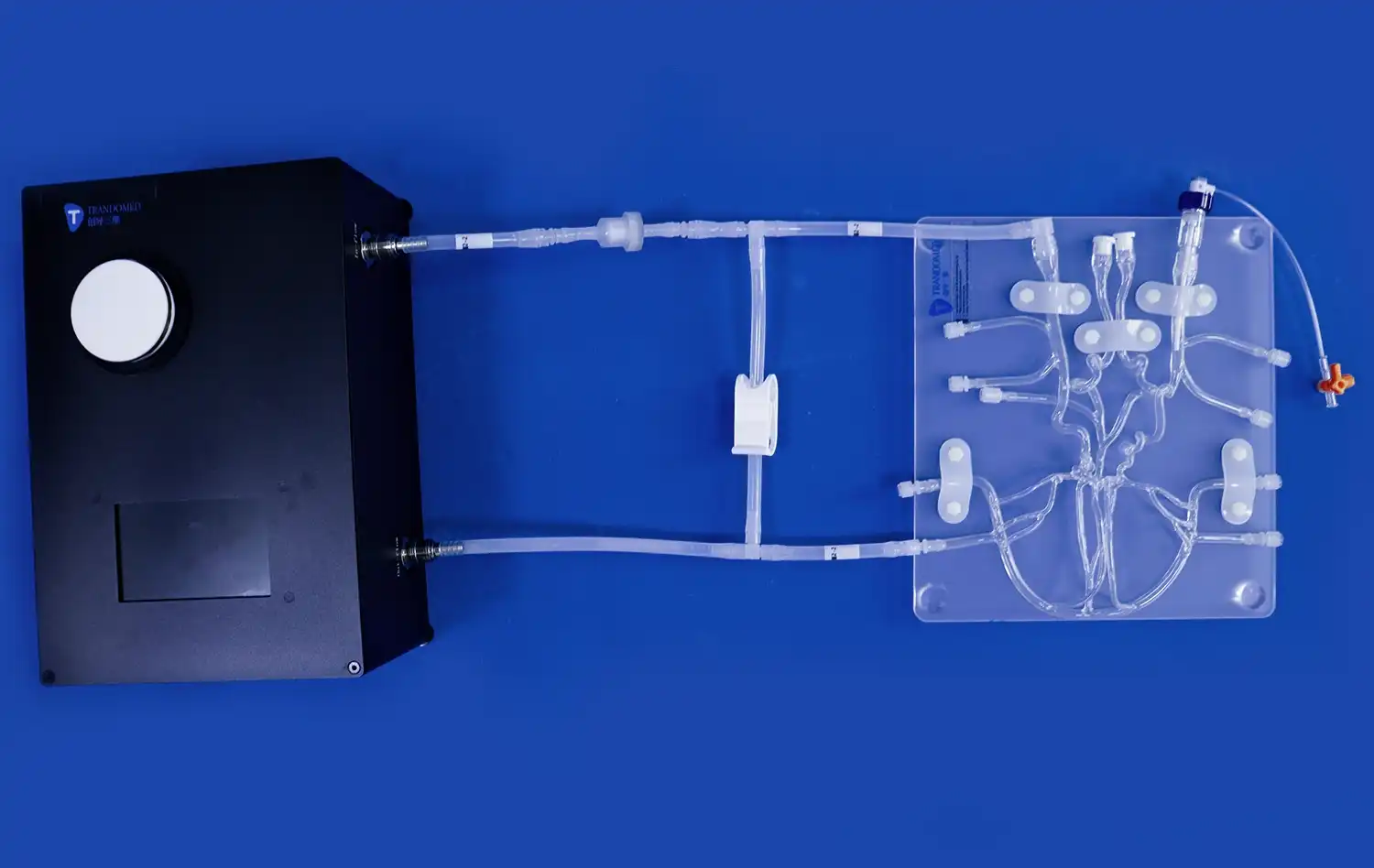
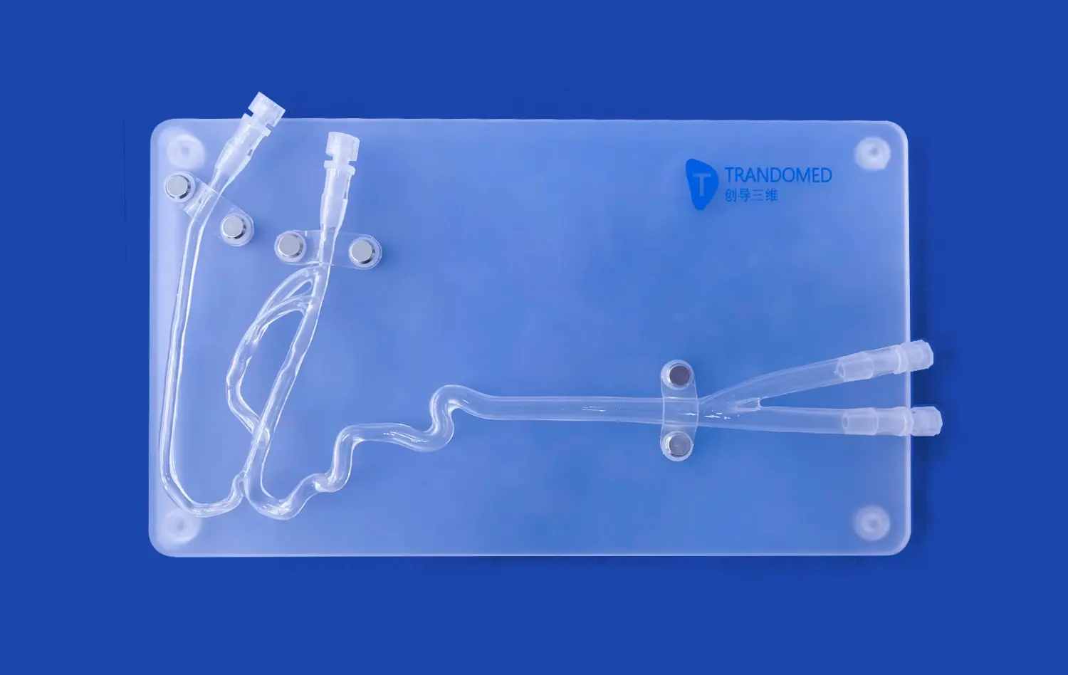
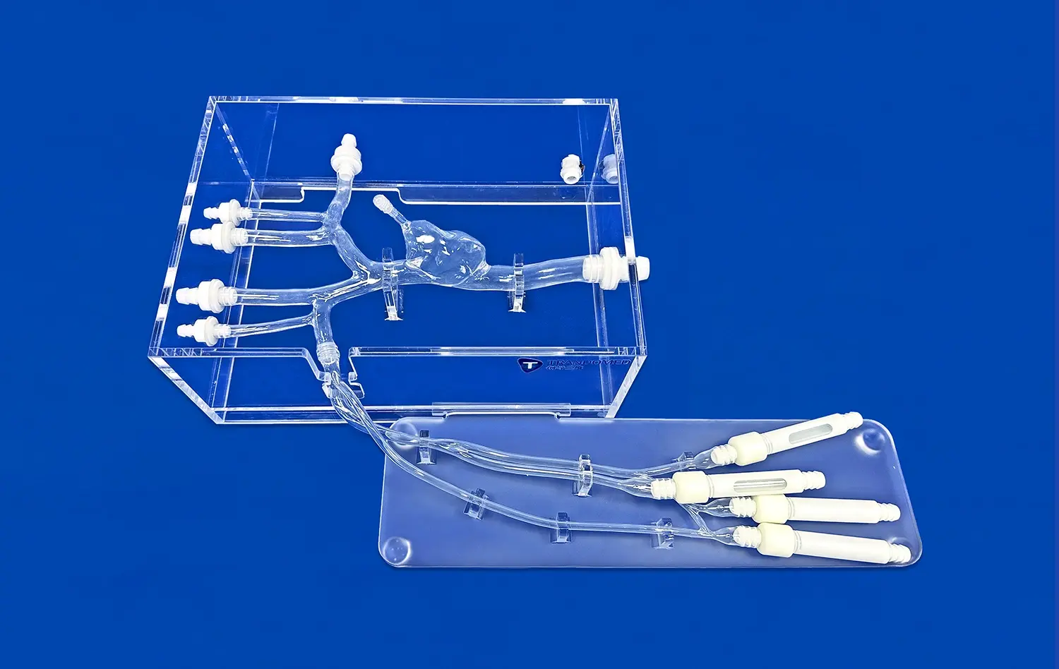
Mitral valve models provide an exceptional level of detail, allowing clinicians to visualize the intricate anatomy of the valve complex. The Mitral Valve Model (XXD006) from Trandomed, for instance, replicates the detailed structure of the mitral valve, including the annulus, leaflets, chordae tendineae, and papillary muscles. This comprehensive representation enables healthcare professionals to gain a deeper understanding of normal valve function and identify abnormalities associated with regurgitation.
Simulating Pathological Conditions
Advanced mitral valve models can be customized to simulate various pathological conditions, including posterior leaflet prolapse, which is a common cause of mitral regurgitation. These models allow clinicians to observe how structural abnormalities affect valve function, helping them correlate clinical findings with anatomical changes. By manipulating the model to recreate specific patient scenarios, doctors can refine their diagnostic skills and develop more targeted treatment plans.
Facilitating Echocardiographic Interpretation
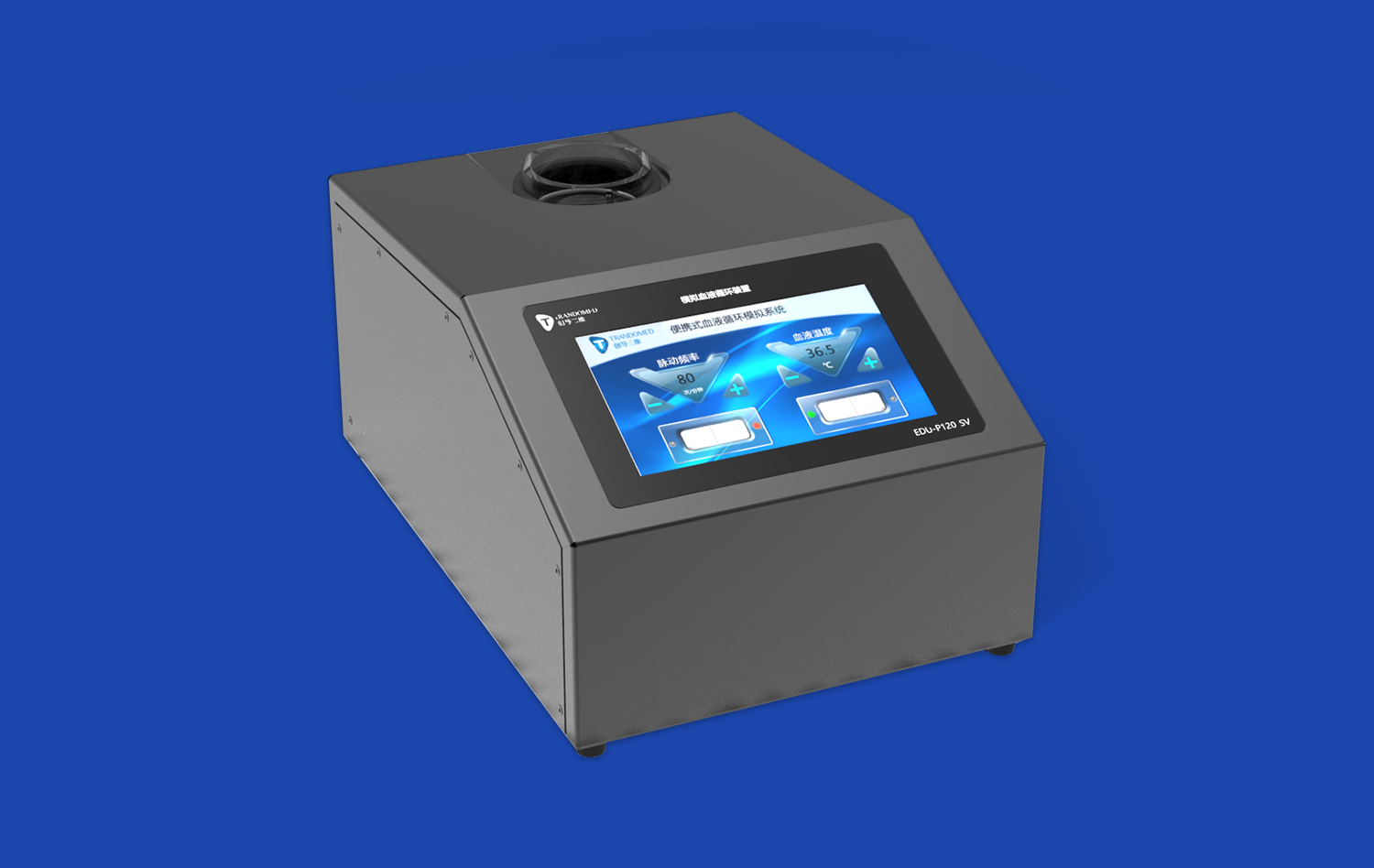
Echocardiography is a primary tool for diagnosing mitral valve regurgitation. Mitral valve models can be used in conjunction with echocardiographic imaging to improve interpretation skills. By comparing the three-dimensional model with two-dimensional echocardiographic images, clinicians can better understand the spatial relationships within the heart and more accurately assess the severity and mechanism of regurgitation.
Simulation Techniques for Mitral Valve Regurgitation Training
Hands-on Surgical Skill Development
Mitral valve models serve as invaluable tools for surgical training. The LifeLike Mitral Valve Model, for example, allows surgeons to practice techniques for mitral valve repair and replacement in a risk-free environment. Trainees can perform procedures such as leaflet resection, chordal replacement, and annuloplasty, honing their skills before operating on actual patients. This hands-on experience is crucial for developing the dexterity and confidence needed for complex mitral valve surgeries.
Catheter-Based Intervention Practice
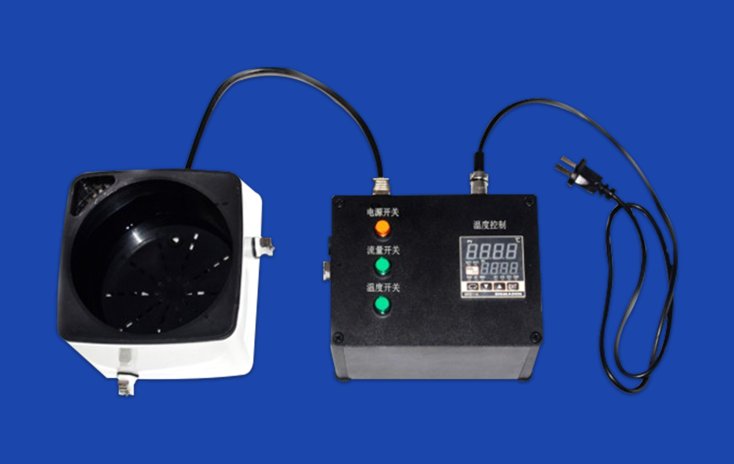
With the rise of transcatheter mitral valve repair and replacement procedures, simulation models have become essential for training interventional cardiologists. Models like the Mitral Valve Model (XXD006) can be integrated with simulated catheterization systems, allowing practitioners to navigate the complex anatomy from the femoral vein to the left heart. This training enhances the ability to accurately position and deploy devices used in minimally invasive mitral valve interventions.
Team-Based Scenario Training
Mitral valve models facilitate team-based training scenarios that mimic real-life clinical situations. These simulations can involve multiple healthcare professionals, including surgeons, cardiologists, anesthesiologists, and nurses. By working together on a realistic model, teams can improve their communication, decision-making, and crisis management skills in a controlled environment, ultimately leading to better patient outcomes in actual procedures.
Clinical Application of Mitral Valve Models in Diagnostic Accuracy
Improving Pre-operative Planning
The use of mitral valve models in pre-operative planning has significantly enhanced surgical outcomes. By creating patient-specific models based on imaging data, surgeons can meticulously plan their approach before entering the operating room. This level of preparation allows for more precise decision-making regarding repair techniques, valve replacement options, and potential complications, ultimately leading to more successful surgeries and reduced operative times.
Enhancing Patient Education and Consent
Mitral valve models serve as powerful educational tools for patients and their families. By using these visual aids, healthcare providers can clearly explain the nature of mitral valve regurgitation, the proposed treatment plan, and expected outcomes. This improved understanding leads to more informed consent and can alleviate patient anxiety by demystifying the surgical process.
Validating New Diagnostic Techniques
As new diagnostic technologies emerge, mitral valve models play a crucial role in their validation and refinement. Researchers can use these models to test and calibrate novel imaging modalities or diagnostic algorithms, ensuring their accuracy before implementation in clinical practice. This process accelerates the development and adoption of innovative techniques for detecting and assessing mitral valve regurgitation.
Conclusion
The integration of mitral valve models into medical education, training, and clinical practice has markedly enhanced the detection and management of mitral valve regurgitation. These sophisticated tools offer unparalleled opportunities for hands-on learning, surgical skill development, and improved diagnostic accuracy. As technology continues to advance, we can expect even more realistic and customizable models that will further revolutionize cardiovascular care. The ongoing collaboration between medical professionals, researchers, and model manufacturers will undoubtedly lead to continued improvements in patient outcomes and quality of life for those affected by mitral valve disorders.
Contact Us
To learn more about how Trandomed's cutting-edge mitral valve models can enhance your institution's cardiovascular training and diagnostic capabilities, please contact us at jackson.chen@trandomed.com. Our team of experts is ready to help you explore the benefits of integrating these advanced simulation tools into your practice.