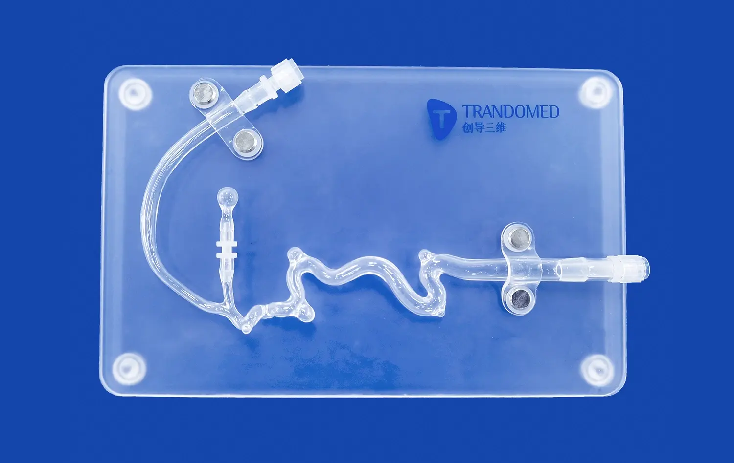
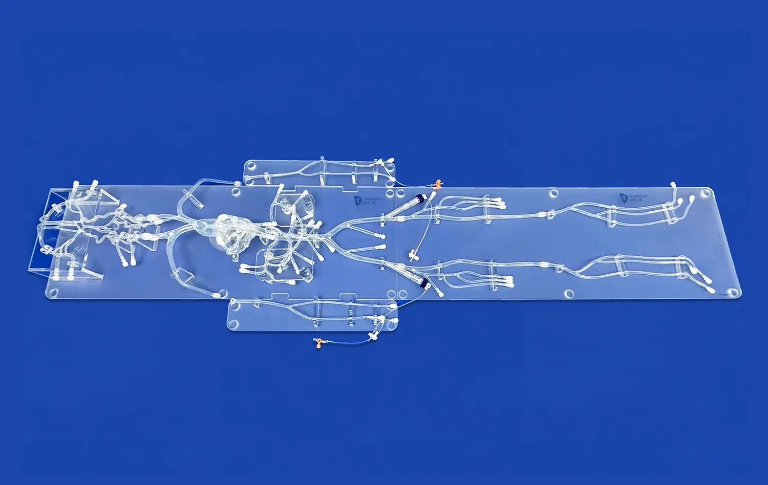
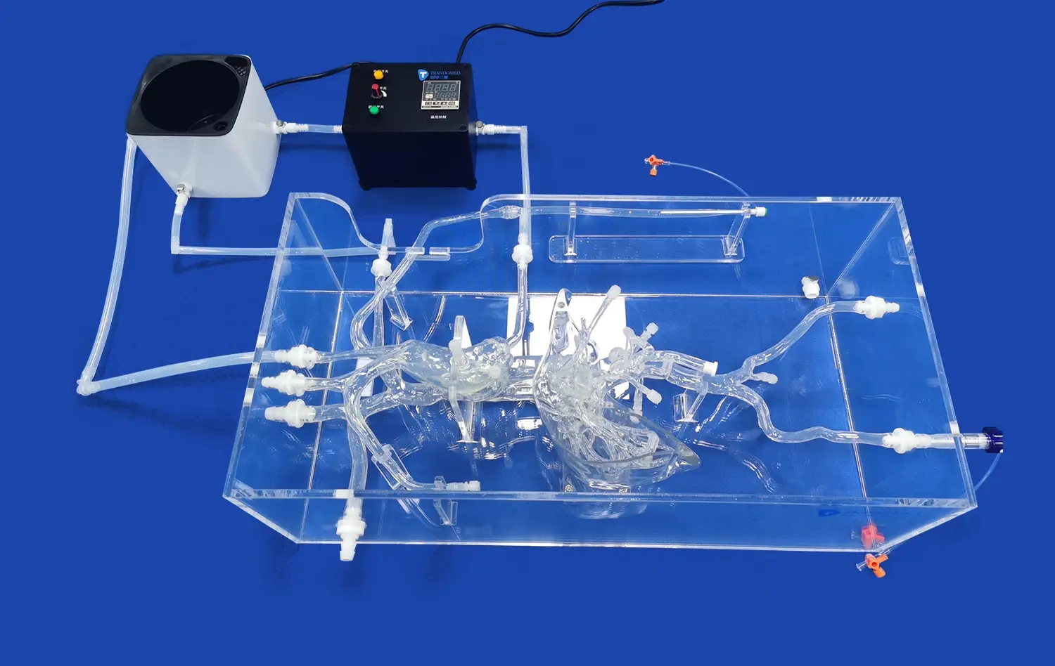
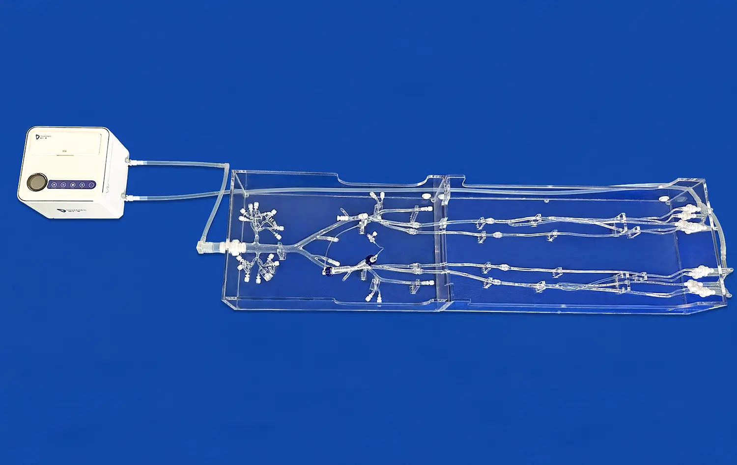
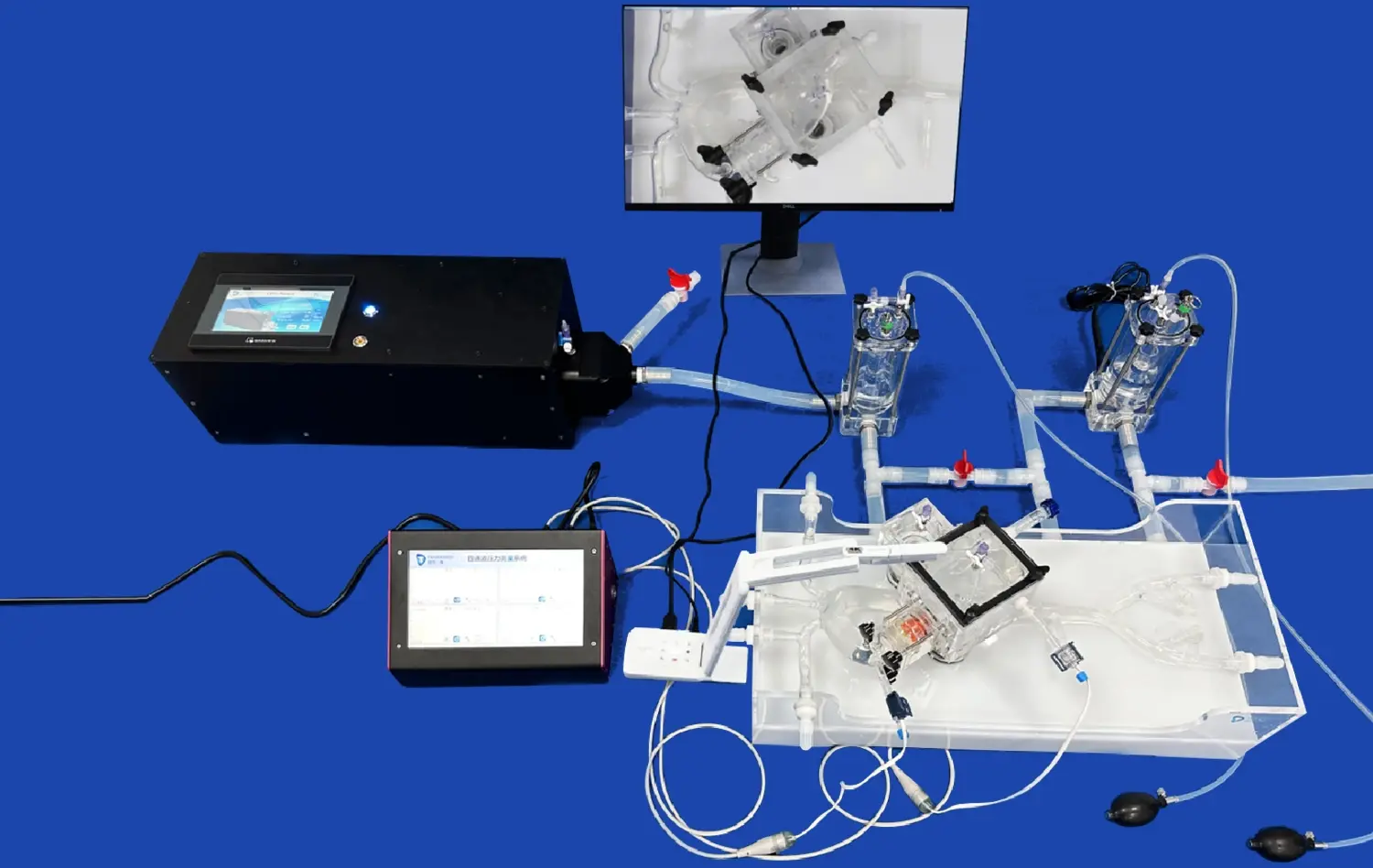
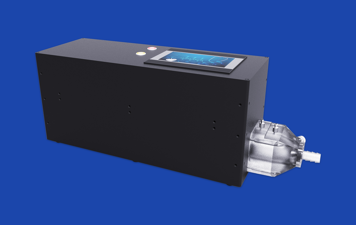
Training in electrophysiology (EP) catheter navigation is a key part of getting heart specialists ready to identify and treat arrhythmias with confidence and accuracy. The venous cardiac electrophysiology model is a revolutionary method that replicates the heart's complex venous routes and electrical activity. In contrast to traditional training methods that use flat images or dead bodies, these advanced simulation tools give clinicians hands-on experience with realistic anatomical structures, such as the superior vena cava, inferior vena cava, right atrium, and right ventricle. This lets them practice inserting catheters, mapping, and ablation techniques without any risk.
Understanding Venous Cardiac Electrophysiology Models in EP Catheter Navigation
Core Concepts and Operational Mechanisms
Cardiac arrhythmia training requires accuracy that can mean the difference between a patient getting better or having problems. Venous electrophysiology models mimic both the structural and functional parts of the cardiac venous system. They give trainees a complete way to see and understand the complicated pathways involved in managing arrhythmias.
These modeling tools have accurate copies of vein structures' anatomical parts and the adaptability needed for repeated practice. The models have important anatomical landmarks that help doctors place catheters during real treatments. This helps them improve their muscle memory and sense of space before they work on real patients. What makes these models unique is that they can imitate the physical feedback that is felt when a real catheter is being navigated, including the resistance that is felt when the catheter is being moved through vessels and chambers.
Distinct Advantages for Training Institutions
There is more and more pressure on medical schools and clinical skills centers to turn out skilled experts while keeping patient safety at the highest level during the learning curve. This problem can be solved with simulation-based training, which lets you practice as much as you want without worrying about ethics. The venous cardiac electrophysiology model lets schools make sure that all of their students get the same training, so that students always get better at their skills, even if the teacher isn't available or there are a lot of patients.
Research shows that simulation training makes it much easier for doctors to handle problems when they move on to real patient care. These models boost confidence by letting trainees experience and deal with uncommon body differences and tough situations in a safe environment. This directly leads to better clinical results. Being able to do things over and over again until they are perfected makes practitioners skilled, and when they go into a clinical setting, their technical skills are much better.
Challenges and Opportunities in Using Venous Heart Models for Catheter Navigation
Technical Limitations and Validation Concerns
Traditional ways of teaching catheter skills had a lot of problems that made it harder to learn. Access to cadaveric specimens was limited, patient-based training was fraught with ethical issues, and it wasn't possible to model certain pathological conditions. These problems led to big gaps in practitioner preparation. Because of these limits, learning took longer and there was more risk during the first procedures on patients.
Many of these problems are fixed by venous heart models, but they also have their own things to think about. Making sure the anatomy is correct needs complex manufacturing methods based on imaging data from real patients. The choice of material is very important because the model needs to give true tactile feedback and last through thousands of training sessions. Early computer models had trouble meeting these needs, which made people doubt their usefulness for training.
Technological Advances Overcoming Barriers
Heart simulation training has become much more realistic and useful thanks to new technologies. Modern 3D printing technologies now make it possible to make models from CT and MRI pictures of real patients. These models can show the different body parts that doctors will see in real life. Materials like medical-grade silicone that is 40A hardness can very accurately mimic the shape and feel of biological flesh.
Another big step forward is software integration, which lets models talk to imaging systems and tracking gear that are used in real EP labs. As these technologies come together, they make realistic training environments where operators can practice navigating catheters while looking at fluoroscopic images and electrical mapping data, which is similar to how they have to do multiple things during real procedures. With these improvements, simulation goes from being a rough estimate to a high-fidelity practice that gets doctors ready for the complicated work they will do in real life.
Choosing the Right Venous Cardiac Electrophysiology Model and Software
Comparative Analysis of Model Options
Before making a purchase choice, it's important to carefully consider how each model fits with the training goals and clinical focus areas of the institution. For interventional cardiology training, traditional cardiac models tend to focus on arterial anatomy. On the other hand, venous-focused electrophysiology models focus on the pathways that are important for diagnosing arrhythmias and performing catheter ablation treatments.
The Venous Cardiac Electrophysiology Model (Product No. XXS004) is an example of a model that was specially made to meet the needs of EP training. This model is made of silicone Shore 40A and has the inferior vena cava, right atrium, right ventricle, superior vena cava, and subclavian vein—exactly the structures that doctors move around in during EP tests. The properties of the material make the resistance during catheter advancement realistic. This helps trainees develop the tactile awareness that is needed for safe procedures.
Being able to customize training makes it much more useful. Institutions can get ready for difficult cases or rare anatomical versions by changing anatomical features based on imaging data that is unique to each patient. Some models can read data files in CT, CAD, STL, STP, and STEP forms, which lets doctors reconstruct a patient's body with great accuracy. This adaptability is very helpful for planning surgeries and testing devices in situations other than normal training.
Software and Vendor Support Considerations
The ecosystem of simulations includes more than just physical models. It also includes software systems that make training more realistic and improve assessment tools. The best providers have tools that keep track of where the catheter is, record procedural metrics, and give objective comments on performance. These analytics help teachers see where their students need more practice and keep track of their progress so they can give them credentials.
Relationships with vendors are very important for making training programs work well. Partners who offer full support, such as help with installation, staff training, upkeep services, and model updates as clinical techniques change, are good for organizations. Manufacturers with a lot of experience in medical simulation know what is needed in healthcare training settings and can give advice that speeds up the implementation of programs.
Institutions should look at a supplier's ability to make things, how flexible they are with customization, and how they handle help after the sale. Companies that use their own 3D printing methods and large collections of real human imaging data usually get better results when it comes to anatomy. Short lead times, like seven to ten days, let programs respond quickly to new training needs without having to wait for long buying delays.
Implementing EP Catheter Navigation Training Using Venous Heart Models
Procedural Workflow Design and Technology Integration
Successful implementation requires thoughtful integration of simulation training into broader educational curricula. Effective programs typically follow a progressive structure, beginning with familiarization with cardiac anatomy and catheter equipment, advancing through guided practice sessions, and culminating in independent performance of complete procedures under observation.
Structured protocols enhance learning effectiveness by establishing clear competency milestones and assessment criteria. Training sessions benefit from scenarios that progress from straightforward anatomies to challenging variations, building confidence incrementally. Integration with imaging modalities and monitoring equipment used in clinical practice ensures trainees develop proficiency with the complete procedural environment rather than just catheter manipulation skills.
Measuring training outcomes provides essential data for program refinement and demonstrates return on investment. Performance metrics might include procedural completion time, catheter positioning accuracy, fluoroscopy time, and complication rates. Studies comparing simulation-trained practitioners to those receiving traditional instruction consistently show improved metrics across these dimensions, validating the investment in high-fidelity training tools.
Case Evidence and Future Directions
Healthcare institutions implementing venous electrophysiology model training report tangible improvements in operator confidence and procedural outcomes. Trainees who complete structured simulation curricula demonstrate reduced learning curves when transitioning to supervised patient care, requiring fewer cases to achieve competency benchmarks. Program directors note decreased anxiety among novice operators and improved patient safety metrics during the critical early phase of independent practice.
Looking ahead, enhanced model fidelity promises even greater training effectiveness. Developments in materials science may soon enable models that replicate patient-specific tissue properties, including variations in vessel stiffness associated with disease states. Integration of haptic feedback systems could provide quantitative measures of catheter manipulation forces, helping trainees develop optimal technique. Collaborative training platforms connecting multiple institutions would enable shared learning experiences and exposure to diverse case scenarios beyond what individual centers encounter.
Procurement Guide: How to Acquire and Utilize Venous Cardiac Electrophysiology Models?
Vendor Selection and Negotiation Strategies
Identifying the appropriate supplier begins with clarifying institutional requirements, including training volume, clinical specialties, customization needs, and budget parameters. Request detailed specifications that address anatomical accuracy, material properties, durability expectations, and available customization options. Reputable manufacturers should provide evidence of their models' anatomical fidelity, typically through validation against medical imaging databases and input from clinical experts.
Trandomed, as a specialized venous cardiac electrophysiology model manufacturer, offers particular advantages for institutions seeking reliable simulation solutions. With over two decades of experience in medical 3D printing technology, the company has developed proprietary manufacturing processes that deliver exceptional anatomical accuracy. Their models incorporate data from extensive CT and MRI databases, ensuring representations that reflect actual patient anatomy rather than idealized textbook illustrations.
The customization capabilities available from experienced manufacturers prove invaluable for specialized applications. Institutions can request modifications to anatomical features, such as adjusting the dimensions and placement of structures like the foramen ovale or reconstructing specific chambers based on actual patient imaging data. Significantly, leading suppliers like Trandomed provide these design services without additional charges, recognizing that tailored solutions enhance training effectiveness and customer satisfaction.
Building Sustainable Vendor Partnerships
Procurement extends beyond the initial purchase transaction to encompass the ongoing relationship that supports long-term program success. Evaluate potential suppliers based on their commitment to post-sale support, including technical assistance, model maintenance, and access to updated designs as clinical practice evolves. Reliable communication channels prove essential—responsive support teams who understand healthcare training requirements can quickly resolve issues and minimize program disruptions.
Payment terms and shipping logistics deserve careful consideration, particularly for international procurement. Standard business-to-business payment methods like telegraphic transfer provide security for both parties. Shipping arrangements through established carriers such as FedEx, DHL, EMS, UPS, and TNT ensure timely delivery with tracking capabilities and insurance protection for valuable simulation equipment.
The procurement process offers opportunities to establish partnerships that deliver value beyond individual product purchases. Suppliers with comprehensive product portfolios can support evolving training needs across multiple specialties, simplifying vendor management and potentially offering volume considerations for broader institutional relationships. Companies investing in research and development continuously improve their offerings, ensuring customers benefit from advances in materials, manufacturing techniques, and simulation technology.
Conclusion
EP catheter navigation training has evolved dramatically through the adoption of advanced venous cardiac electrophysiology models. These sophisticated simulation tools address long-standing training challenges by providing realistic, repeatable practice opportunities that build competence before clinicians treat patients. The anatomical accuracy, tactile fidelity, and customization capabilities of modern models create training experiences that translate directly to improved clinical performance. As healthcare institutions navigate procurement decisions, partnering with experienced manufacturers who understand both simulation technology and clinical training requirements maximizes program effectiveness and return on investment.
FAQ
What distinguishes venous cardiac models from standard heart simulators?
Venous cardiac electrophysiology models specifically emphasize the venous pathways—superior vena cava, inferior vena cava, right atrium, and associated structures—critical to EP catheter navigation. While standard heart simulators may focus on arterial anatomy for interventional procedures or provide generalized cardiac representations, venous models deliver the anatomical detail and tactile realism essential for arrhythmia diagnosis and ablation training. This specialized focus ensures practitioners develop skills directly applicable to electrophysiology procedures.
How do institutions measure training effectiveness with simulation models?
Effective assessment combines objective performance metrics with competency evaluations. Programs track procedural completion times, catheter positioning accuracy, fluoroscopy duration, and error rates during simulation sessions. Many institutions establish competency thresholds that trainees must achieve before advancing to supervised patient care. Comparing simulation-trained practitioners' clinical outcomes to historical data provides compelling evidence of training program impact on patient safety and procedural success rates.
Can these models accommodate rare anatomical variations?
High-quality venous electrophysiology models offer substantial customization capabilities. Manufacturers accepting patient-specific imaging data in formats like CT, STL, and STEP can create models replicating unusual anatomies or pathological conditions. This capability proves invaluable for surgical planning, allowing teams to rehearse complex procedures before entering the operating room, and for training programs preparing practitioners to recognize and navigate anatomical variants they'll eventually encounter in clinical practice.
Partner with Trandomed for Superior EP Training Solutions
Trandomed stands ready to support your institution's electrophysiology training objectives with industry-leading simulation models designed for excellence. Our Venous Cardiac Electrophysiology Model (XXS004) combines anatomical precision with practical durability, manufactured using proprietary 3D printing techniques refined over two decades. As a trusted venous cardiac electrophysiology model supplier, we offer customization without design fees, rapid seven-to-ten-day production timelines, and comprehensive support throughout your procurement journey. Contact our team at jackson.chen@trandomed.com to discuss your specific training requirements and discover how our solutions can elevate practitioner competence and patient outcomes.
References
Josephson, M.E. (2020). Clinical Cardiac Electrophysiology: Techniques and Interpretations (5th ed.). Philadelphia: Wolters Kluwer.
Zipes, D.P., Jalife, J., & Stevenson, W.G. (2018). Cardiac Electrophysiology: From Cell to Bedside (7th ed.). Philadelphia: Elsevier.
Huang, S.K.S., Wood, M.A., & Miller, J.M. (2019). Catheter Ablation of Cardiac Arrhythmias (3rd ed.). Philadelphia: Elsevier Saunders.
Calkins, H., Hindricks, G., Cappato, R., et al. (2017). 2017 HRS/EHRA/ECAS/APHRS/SOLAECE expert consensus statement on catheter and surgical ablation of atrial fibrillation. Heart Rhythm, 14(10), e275-e444.
McGaghie, W.C., Issenberg, S.B., Cohen, E.R., et al. (2011). Does simulation-based medical education with deliberate practice yield better results than traditional clinical education? A meta-analytic comparative review. Academic Medicine, 86(6), 706-711.
Kneebone, R.L., Nestel, D., Wetzel, C., et al. (2006). The human face of simulation: patient-focused simulation training. Academic Medicine, 81(10), 919-924.