Exploring Aneurysm Morphology with Neuro Vascular System with Aneurysm
2025-07-01 09:00:00
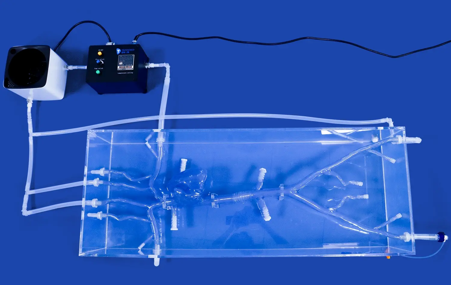
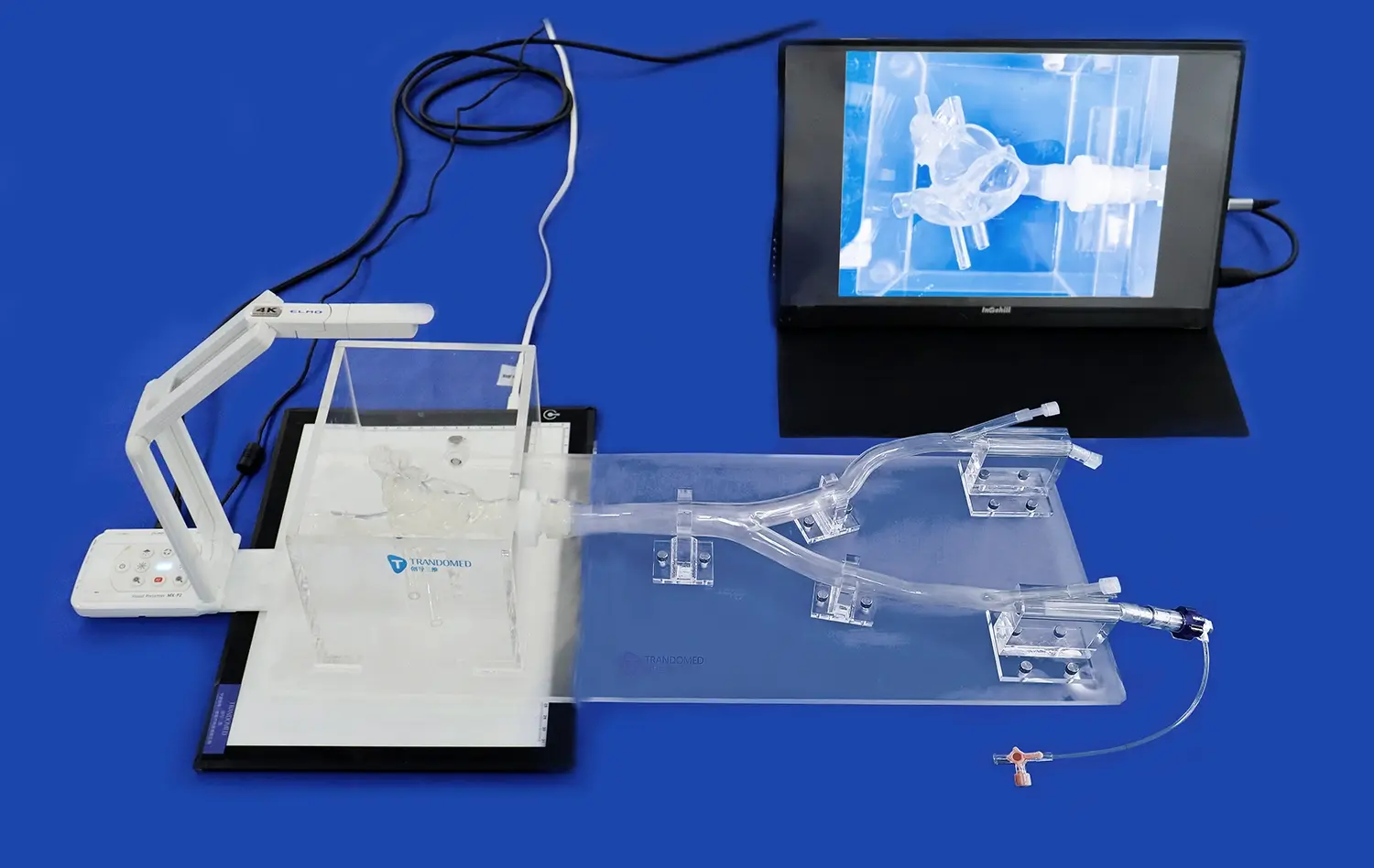
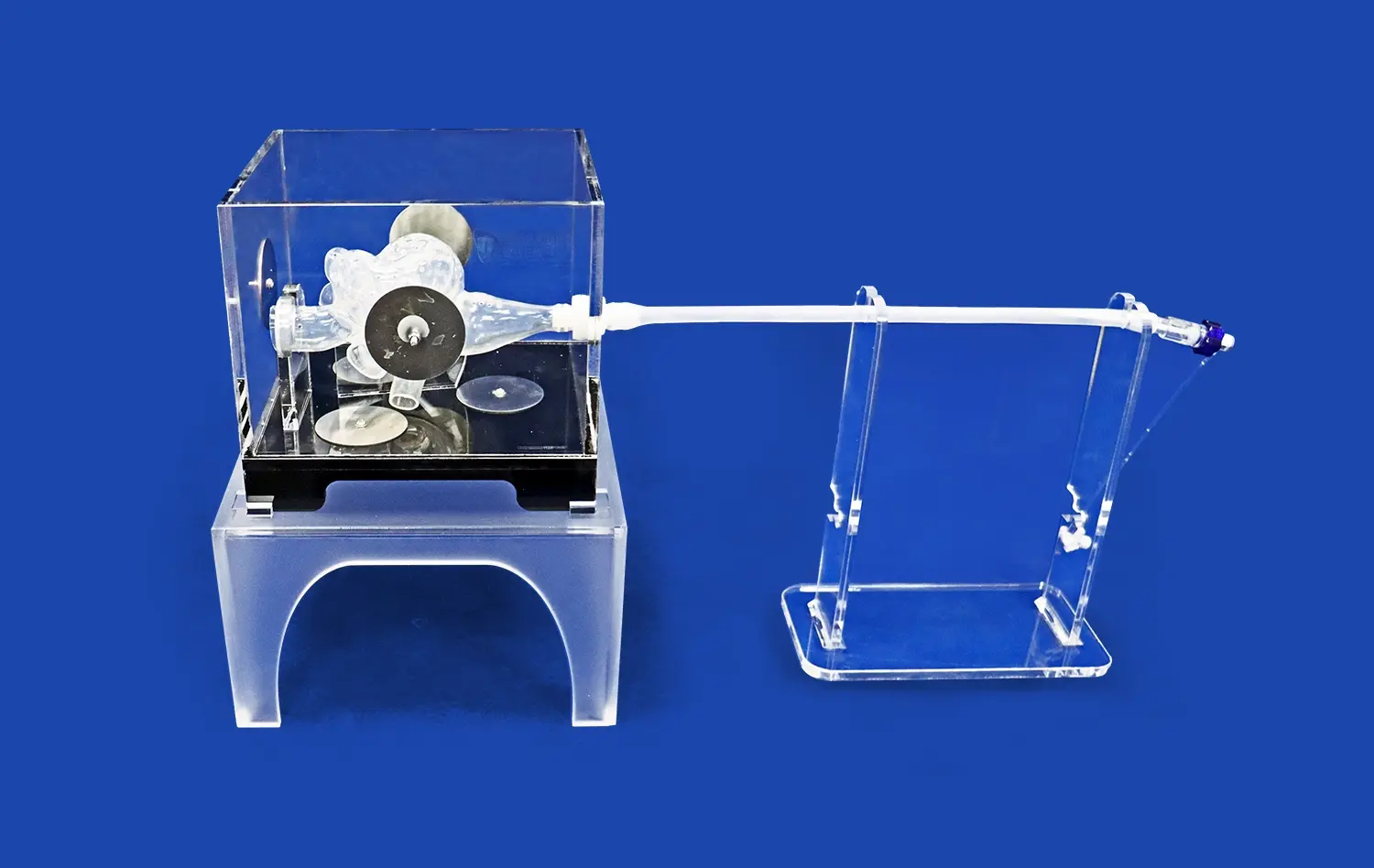
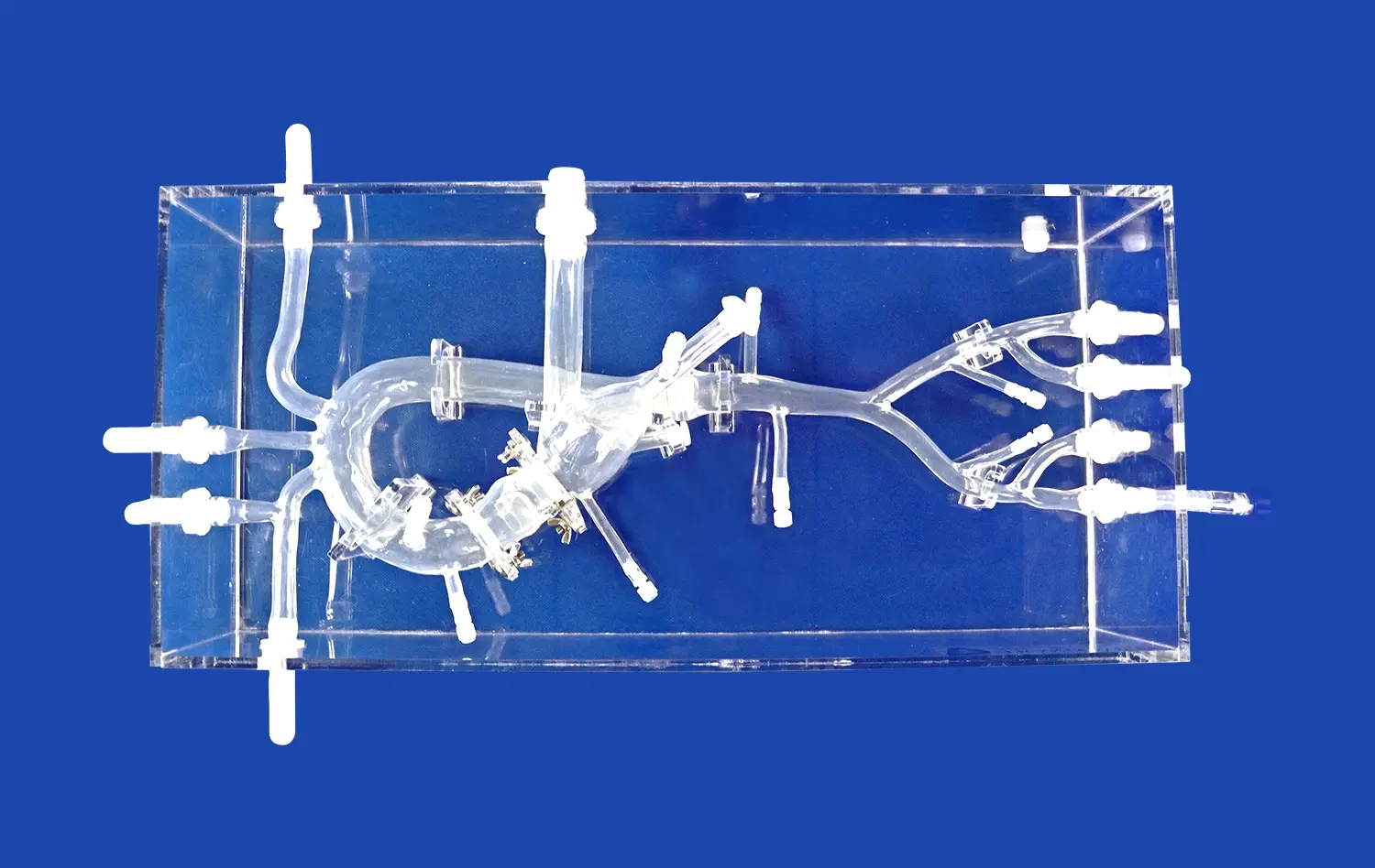
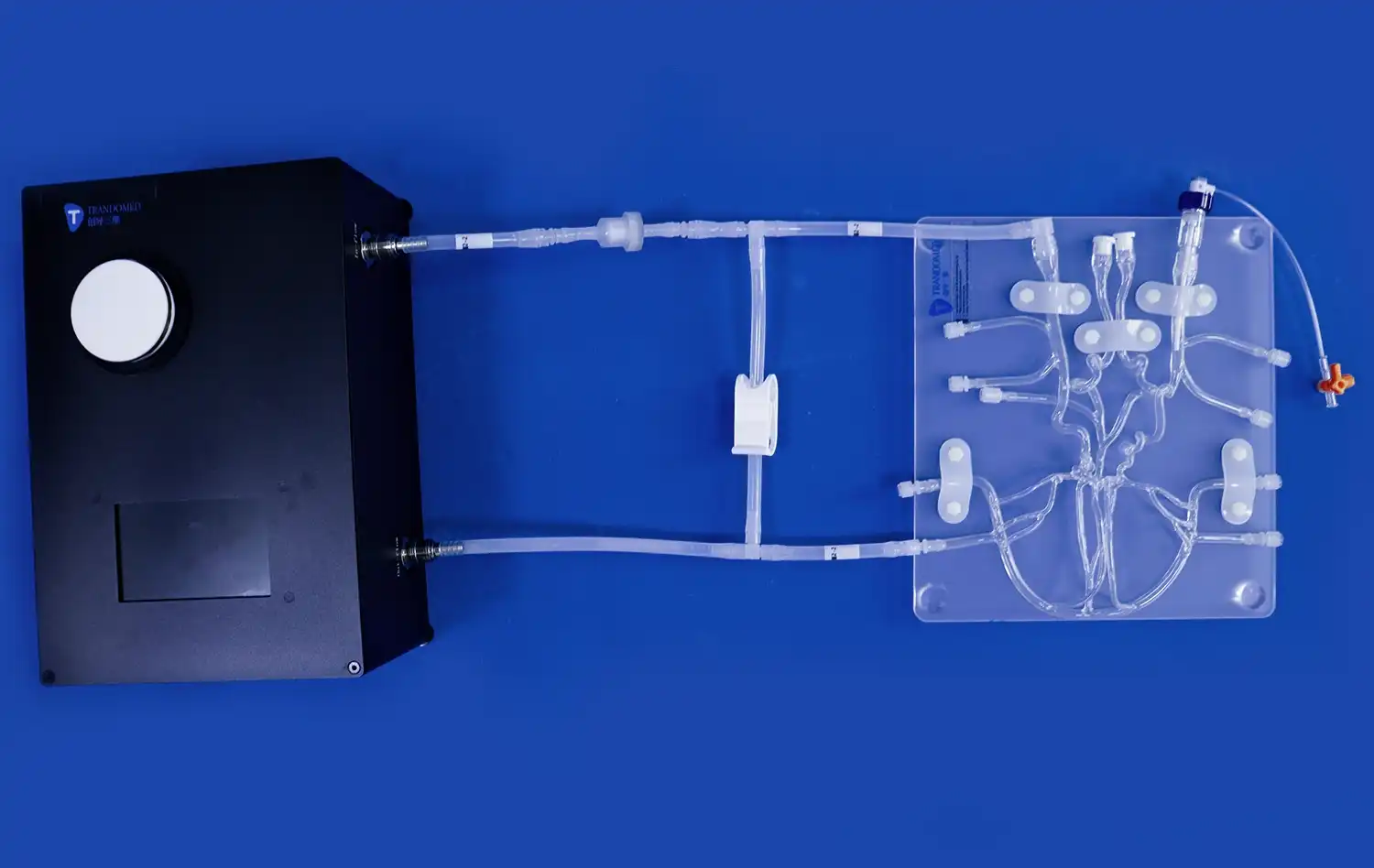
Delving into the complex world of cerebrovascular disorders, the study of aneurysm morphology using a neuro vascular system with aneurysm models has become an indispensable tool for medical professionals and researchers alike. These advanced 3D-printed silicone simulators offer unprecedented insights into the varied shapes, sizes, and types of aneurysms, revolutionizing our understanding of these potentially life-threatening conditions. By providing a tangible, highly detailed representation of the intricate blood vessel structures and aneurysm formations, these models enable healthcare providers to enhance their diagnostic accuracy, refine treatment strategies, and improve patient outcomes. The ability to explore aneurysm morphology in a hands-on, risk-free environment not only facilitates better clinical decision-making but also serves as an invaluable educational resource for medical students and practicing clinicians. As we navigate the nuances of aneurysm characteristics, we uncover the critical role that shape and size play in determining the most effective management approaches for each unique case.
Why Shape Matters in Aneurysm Management?
The shape of an aneurysm is far more than a mere anatomical curiosity; it's a crucial factor that significantly influences treatment decisions and patient prognosis. Understanding the intricate relationship between aneurysm morphology and clinical outcomes is essential for developing effective management strategies.
Impact on Rupture Risk
Aneurysm shape plays a pivotal role in determining the risk of rupture. Irregular or multilobular aneurysms often exhibit higher rupture rates compared to their smooth, regular counterparts. This is due to the uneven distribution of hemodynamic stress across the aneurysm wall. Areas of increased stress, particularly at points of inflection or protrusion, are more susceptible to weakening and eventual rupture. By studying these variations using neuro vascular system models with aneurysms, clinicians can better assess the urgency of intervention and tailor treatment plans accordingly.
Influence on Treatment Selection
The morphology of an aneurysm significantly impacts the choice of treatment modality. For instance, wide-necked aneurysms may pose challenges for traditional endovascular coiling, necessitating advanced techniques such as stent-assisted coiling or flow diversion. Conversely, narrow-necked aneurysms might be more amenable to standard coiling procedures. The ability to visualize and manipulate 3D models of various aneurysm shapes allows neurosurgeons and interventional neuroradiologists to anticipate potential difficulties and select the most appropriate treatment approach. This preoperative planning can lead to improved procedural success rates and reduced complications.
Visualizing the Variations: A Detailed Look at Different Aneurysm Shapes and Sizes
The diversity of aneurysm morphology is vast, ranging from simple saccular formations to complex, multi-lobed structures. Utilizing advanced neuro vascular system with aneurysms enables healthcare professionals to gain a comprehensive understanding of these variations and their clinical implications.
Saccular Aneurysms: The Most Common Type
Saccular aneurysms, often described as "berry aneurysms" due to their rounded appearance, are the most frequently encountered type. These balloon-like outpouchings typically form at arterial bifurcations or along the outer curve of a blood vessel. The size of saccular aneurysms can vary dramatically, from tiny 2mm blebs to giant aneurysms exceeding 25mm in diameter. Each size category presents unique challenges and considerations for treatment. Small aneurysms may be candidates for conservative management with regular monitoring, while larger ones often require more aggressive intervention due to their higher rupture risk. The neck-to-dome ratio of saccular aneurysms is another critical factor that influences treatment decisions, particularly when considering endovascular approaches.
Complex Morphologies: Multilobular and Irregular Aneurysms
Not all aneurysms conform to the classic saccular shape. Complex morphologies, including multilobular and irregular aneurysms, present additional challenges in both diagnosis and treatment. Multilobular aneurysms feature multiple sacs or lobes, often with a shared neck. These formations can complicate endovascular treatments, as ensuring complete occlusion of all lobes is crucial to prevent recurrence. Irregular aneurysms, characterized by asymmetrical or non-uniform shapes, may have areas of focal weakness that are prone to rupture. The intricate geometry of these complex aneurysms necessitates careful study and planning. Neuro vascular system with aneurysms that accurately replicate these complex morphologies provide invaluable insights, allowing clinicians to develop tailored treatment strategies that address the unique challenges posed by each aneurysm's shape and size.
Beyond Saccular: Exploring Fusiform, Dissecting, and Other Aneurysm Types
While saccular aneurysms are the most commonly encountered, a comprehensive understanding of aneurysm morphology must extend to less frequent but equally important types. Fusiform and dissecting aneurysms, along with other rare variants, present unique challenges that require specialized approaches to management and treatment.
Fusiform Aneurysms: Elongated Challenges
Fusiform aneurysms are characterized by a gradual, circumferential dilation of the entire vessel wall, resulting in an elongated, spindle-shaped enlargement. Unlike saccular aneurysms, fusiform aneurysms lack a distinct neck, which can complicate treatment options. These aneurysms often occur in the vertebrobasilar system and can reach substantial sizes, potentially causing mass effect on surrounding neural structures. The management of fusiform aneurysms frequently requires innovative approaches, such as flow diversion or bypass procedures, to reconstruct the affected vessel segment. Neuro vascular system with aneurysms that accurately depict fusiform morphology are invaluable for surgical planning and technique refinement, allowing surgeons to visualize the complex geometry and practice various reconstruction strategies in a risk-free environment.
Dissecting Aneurysms: A Unique Pathology
Dissecting aneurysms represent a distinct pathological entity, arising from a tear in the vessel's inner lining that allows blood to enter the wall itself. This process can lead to the formation of a false lumen within the vessel wall, potentially compromising blood flow and weakening the overall structure. Dissecting aneurysms are particularly challenging to manage due to their dynamic nature and the risk of progression. They often require a multifaceted approach that may include medical management, endovascular techniques, or open surgical repair, depending on the location and extent of the dissection. Advanced neuro vascular system models that incorporate dissecting aneurysm features provide an unparalleled opportunity for clinicians to study the complex interplay between true and false lumens, assess the impact on branch vessels, and develop targeted treatment strategies. These models serve as powerful educational tools, enhancing the understanding of this complex pathology among medical trainees and experienced practitioners alike.
Conclusion
The exploration of aneurysm morphology through advanced neuro vascular system with aneurysms has ushered in a new era of understanding and treating these complex cerebrovascular conditions. By providing tangible, highly detailed representations of various aneurysm shapes, sizes, and types, these models enable healthcare professionals to refine their diagnostic skills, optimize treatment planning, and improve patient outcomes. As we continue to unravel the intricacies of aneurysm morphology, the integration of 3D-printed silicone simulators in clinical practice and medical education promises to drive further advancements in the field of neurovascular surgery and interventional neuroradiology.
Contact Us
To learn more about our advanced neuro vascular system models with aneurysms and how they can enhance your clinical practice or research, please contact us at jackson.chen@trandomed.com. Our team is dedicated to providing cutting-edge 3D-printed medical simulators that push the boundaries of medical education and surgical planning.
References
Morita, A., Kirino, T., Hashi, K., et al. (2012). The natural course of unruptured cerebral aneurysms in a Japanese cohort. New England Journal of Medicine, 366(26), 2474-2482.
Cebral, J. R., Mut, F., Weir, J., & Putman, C. M. (2011). Association of hemodynamic characteristics and cerebral aneurysm rupture. American Journal of Neuroradiology, 32(2), 264-270.
Wiebers, D. O., Whisnant, J. P., Huston, J., et al. (2003). Unruptured intracranial aneurysms: natural history, clinical outcome, and risks of surgical and endovascular treatment. The Lancet, 362(9378), 103-110.
Brinjikji, W., Zhu, Y. Q., Lanzino, G., et al. (2016). Risk factors for growth of intracranial aneurysms: a systematic review and meta-analysis. American Journal of Neuroradiology, 37(4), 615-620.
Etminan, N., Rinkel, G. J., & International Study of Unruptured Intracranial Aneurysms Investigators. (2016). Unruptured intracranial aneurysms: development, rupture and preventive management. Nature Reviews Neurology, 12(12), 699-713.
Spetzler, R. F., McDougall, C. G., Albuquerque, F. C., et al. (2019). The Barrow Ruptured Aneurysm Trial: 10-year results. Journal of Neurosurgery, 130(6), 1790-1798.
_1736216292718.webp)
