Exploring the Complexity of the Human Vascular System with the Aorta 3D Model
2025-06-20 09:00:00
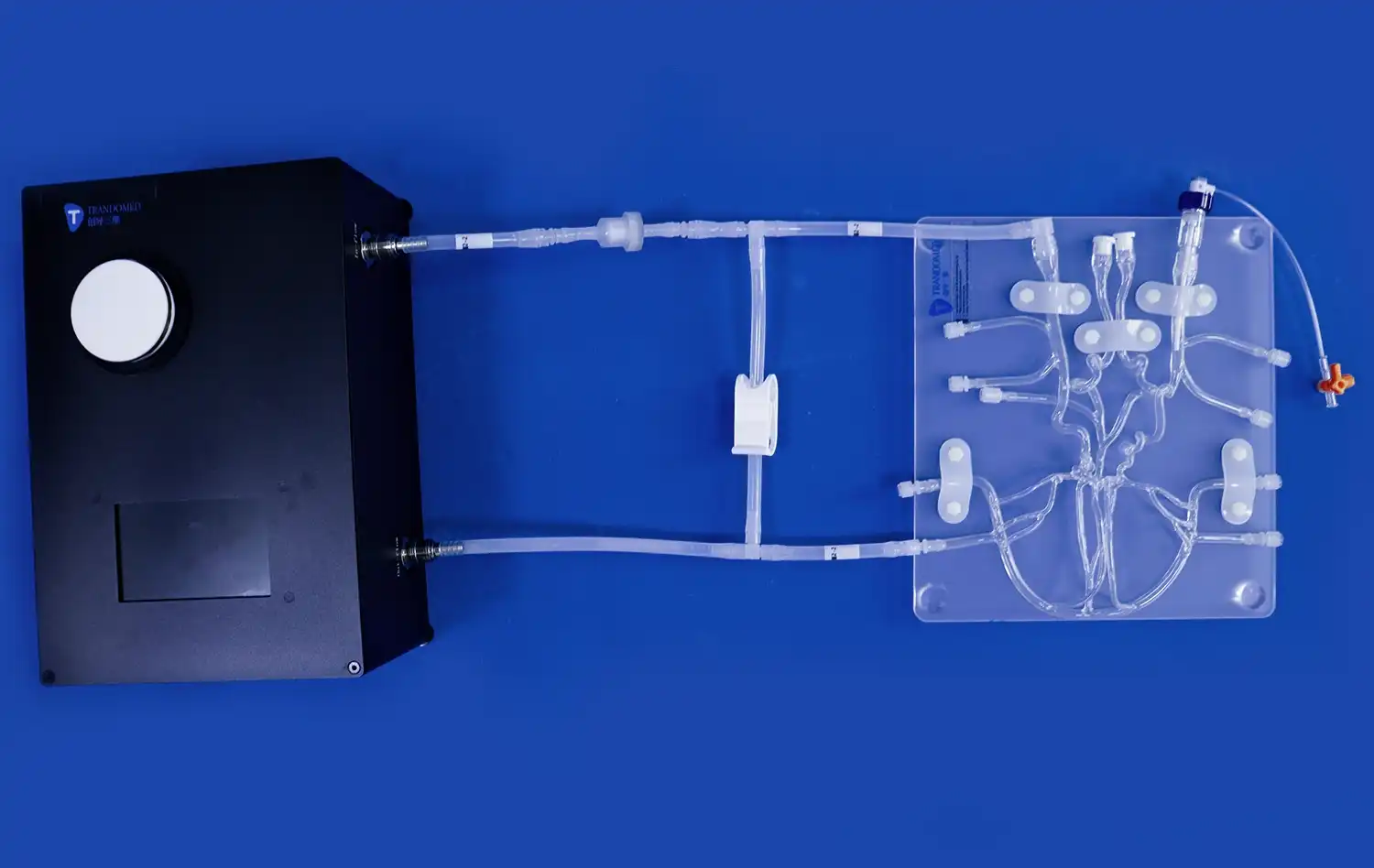
The human vascular system is a marvel of biological engineering, with the aorta serving as its centerpiece. To truly appreciate this intricate network, medical professionals and students alike are turning to advanced tools like the aorta 3D model. These highly detailed replicas offer an unprecedented view of the body's largest artery, allowing for a deeper understanding of its structure and function. By leveraging cutting-edge 3D printing technology, these models provide a tactile and visual learning experience that surpasses traditional 2D images. From examining the aortic arch to exploring branching vessels, the aorta 3D model serves as an invaluable resource for education, surgical planning, and research. As we delve into the complexities of the vascular system, we'll discover how these innovative models are revolutionizing our approach to cardiovascular studies and patient care.
Unraveling the Intricacies of the Aortic Arch: A 3D Perspective
The Anatomy of the Aortic Arch
The aortic arch is a critical component of the cardiovascular system, serving as a bridge between the ascending and descending aorta. Its curved structure and branching vessels make it a challenging area to visualize using traditional methods. However, with the advent of aorta 3D models, medical professionals can now examine this region in unprecedented detail.
These models accurately depict the arch's unique shape, showcasing how it curves over the left bronchus and connects to the descending thoracic aorta. The three major branches of the aortic arch - the brachiocephalic trunk, left common carotid artery, and left subclavian artery - are clearly visible, allowing for a comprehensive study of their origins and trajectories.
Understanding Variations and Anomalies
One of the most important aspects of utilizing an aorta 3D model is the capacity to investigate anatomical variations and anomalies. Whereas textbooks regularly present an idealized version of human anatomy, real-world cases can vary essentially. 3D models can be customized to speak to different congenital conditions, such as coarctation of the aorta or a right-sided aortic arch.
By manipulating these models, students and clinicians can pick up a tactile understanding of how these variations influence blood stream and encompassing structures. This hands-on experience is priceless for creating diagnostic abilities and arranging surgical interventions. The capacity to rotate, zoom, and dissect the 3D model gives insights that are basically not conceivable with 2D imaging techniques.
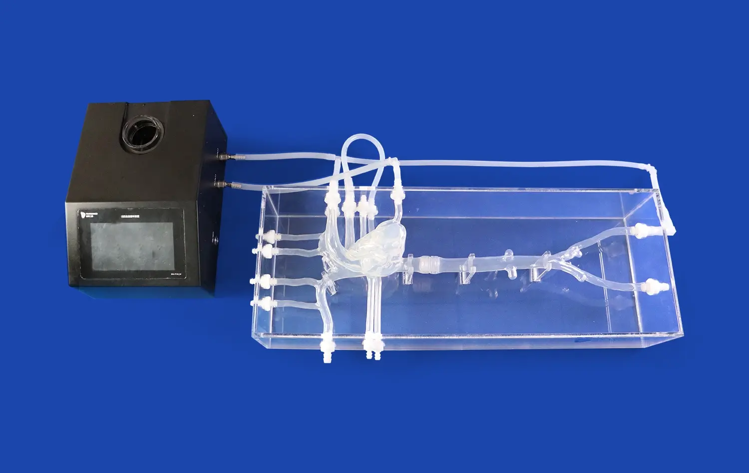
Visualizing the Complexity of Blood Flow: The Aorta 3D Model in Action
Dynamic Flow Simulations
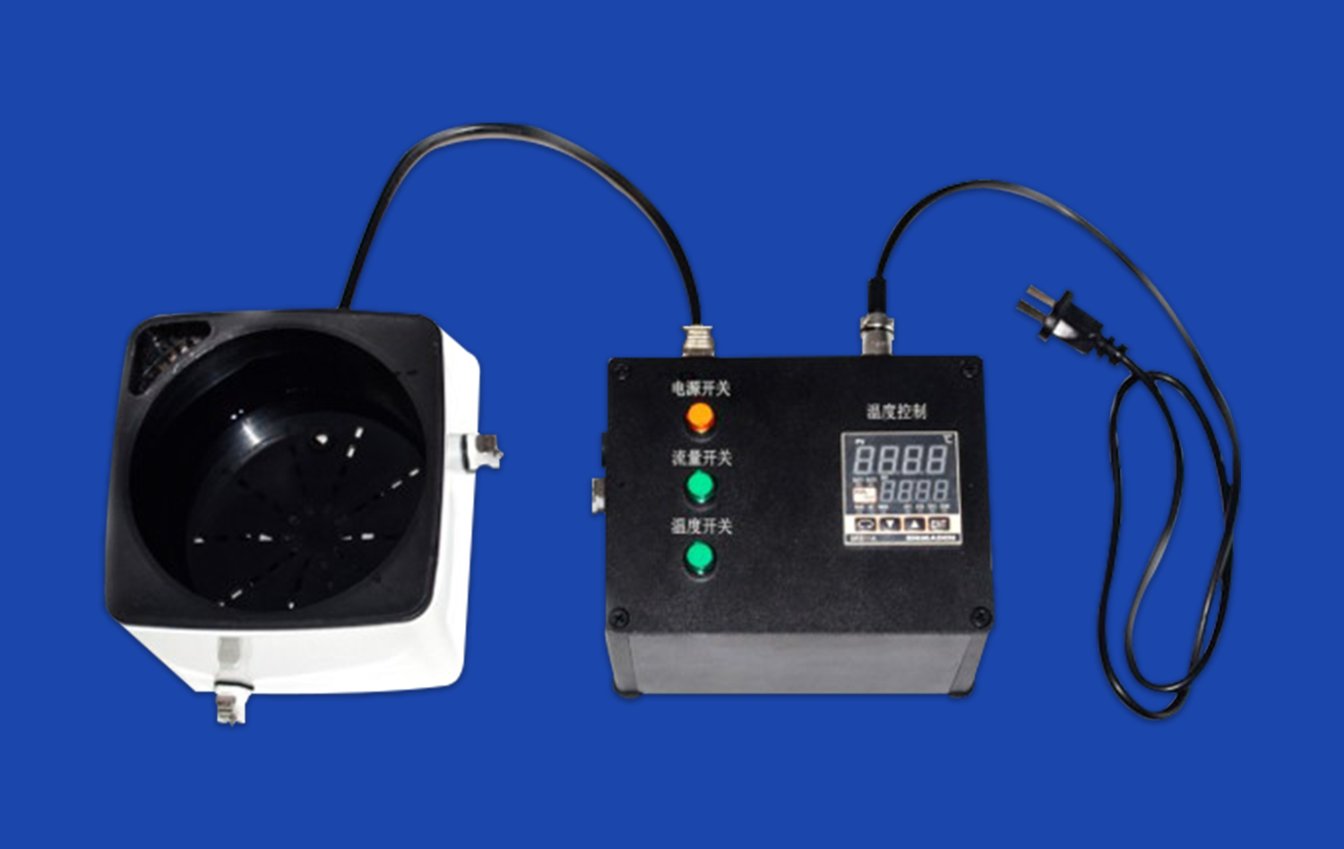
While static aorta 3D models offer significant educational value, advanced versions incorporate dynamic flow simulations. These cutting-edge models allow users to visualize blood flow patterns within the aorta and its branches. By integrating computational fluid dynamics with anatomically accurate 3D printing, researchers can create models that mimic the pulsatile nature of blood flow.
These simulations provide crucial insights into hemodynamics, helping clinicians understand how variations in vessel diameter, curvature, and branching patterns affect blood flow. For instance, areas of turbulence or reduced flow can be identified, which may correlate with regions prone to atherosclerotic plaque formation or aneurysm development.
Pressure and Stress Analysis
Another powerful application of aorta 3D models is in the analysis of pressure and stress distribution along the vessel walls. By incorporating data from imaging studies and pressure measurements, these models can create color-coded representations of wall stress. This information is particularly valuable in assessing the risk of aortic dissection or aneurysm rupture.
Clinicians can use these models to identify areas of high stress concentration, which may require closer monitoring or intervention. For patients with conditions like Marfan syndrome or other connective tissue disorders, such analyses can guide preventive strategies and help determine optimal timing for surgical intervention.
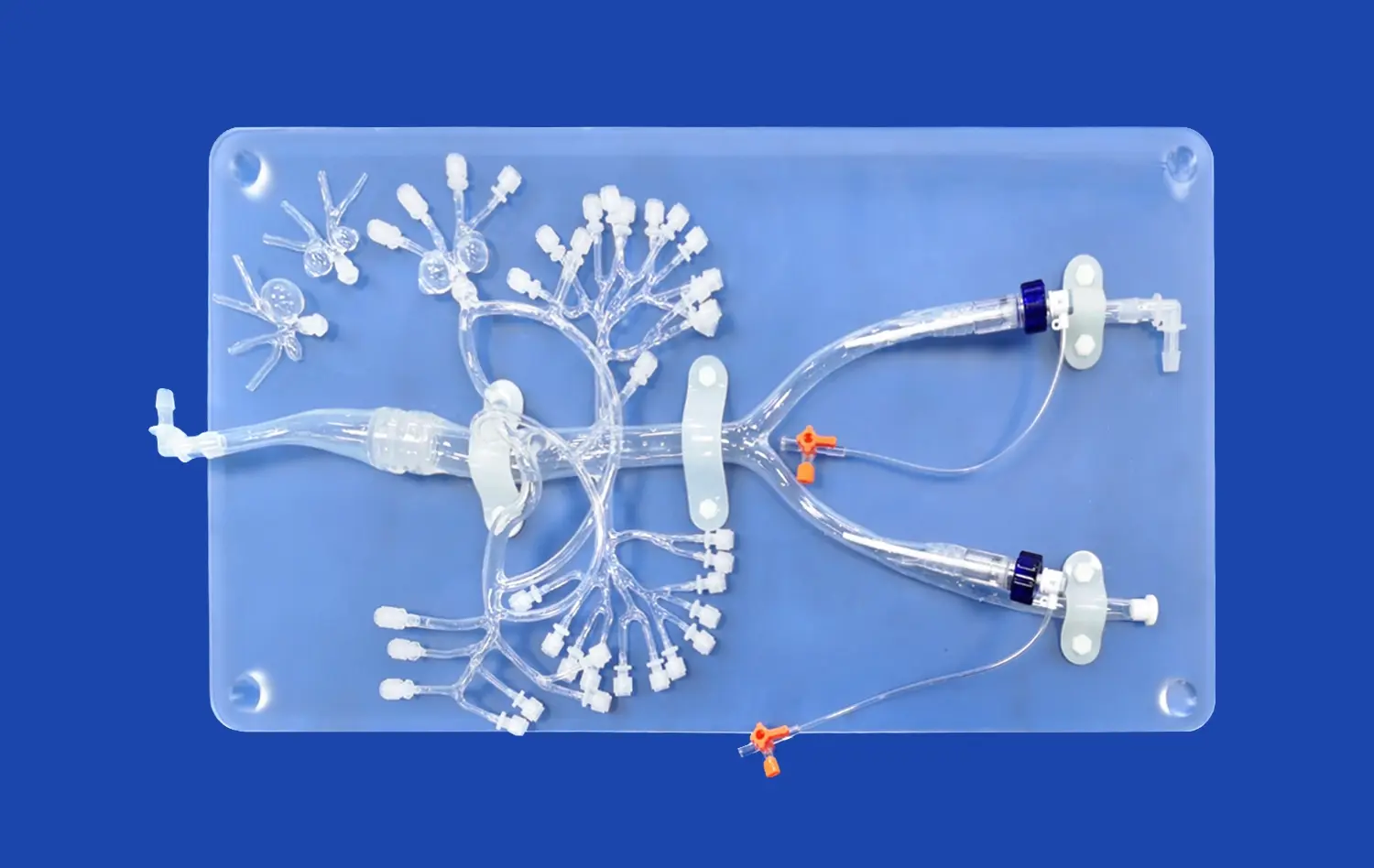
From Arteries to Capillaries: Exploring the Vascular Tree with 3D Modeling
Branching Patterns and Vascular Networks
Whereas the aorta serves as the primary highway of the circulatory system, the true complexity of the vascular system lies in its extensive branching network. Progressed 3D modeling procedures now permit for the creation of detailed representations of whole vascular trees, from major arteries down to the level of capillaries.
These comprehensive models give a holistic view of blood distribution throughout the body. By analyzing the branching patterns and vessel diameters, analysts can gain experiences into the standards of vascular architecture. This information is significant for understanding how the body keeps up proficient blood stream to all tissues and organs.
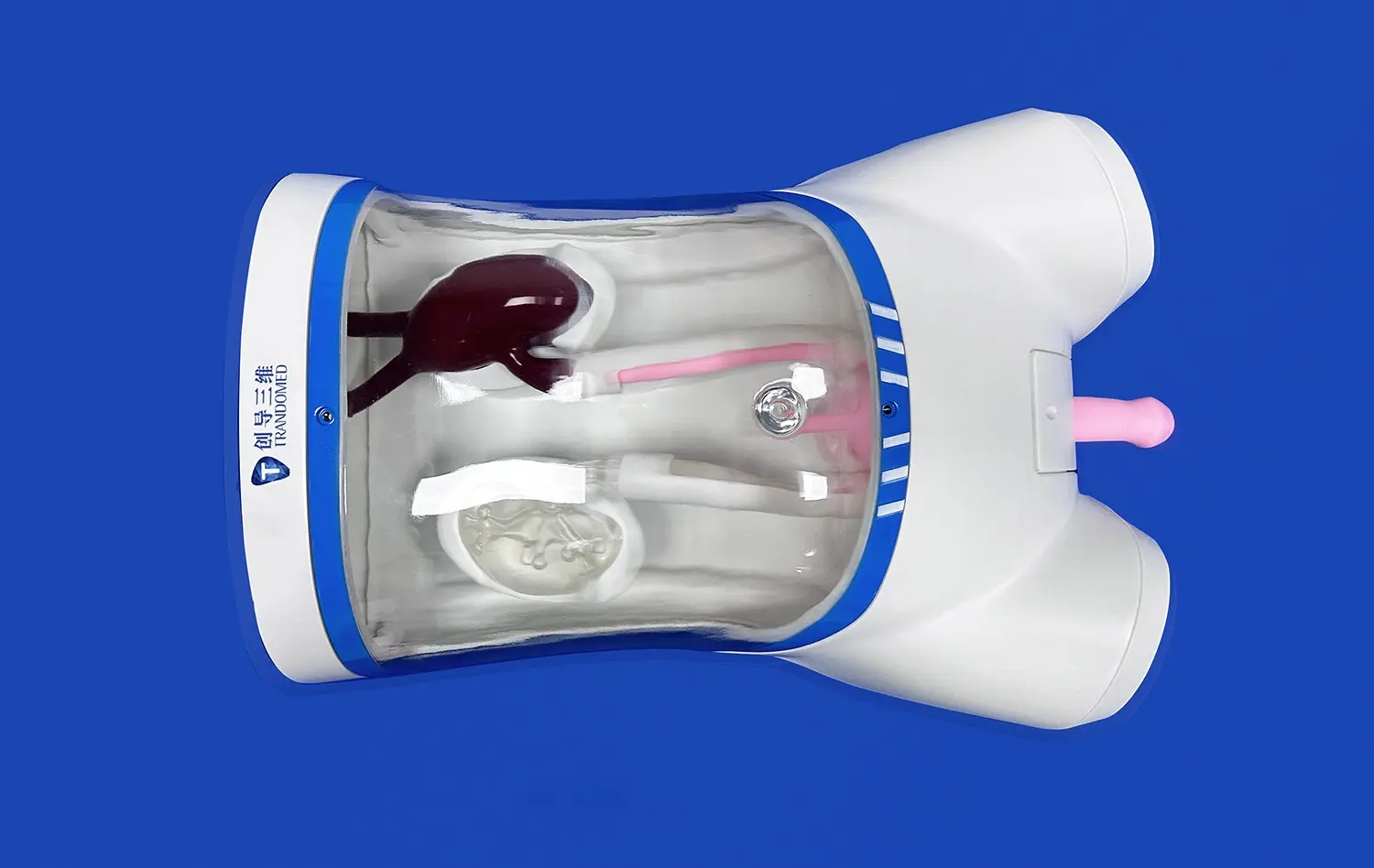
Microvascular Modeling
At the smallest scale, 3D models of the microvasculature offer a window into the complex world of capillary networks. These aorta 3D models, regularly made utilizing progressed imaging methods like micro-CT or two-photon microscopy, reveal the dense, mesh-like structure of capillary beds.
By examining these microvascular models, analysts can explore phenomena such as angiogenesis, the formation of new blood vessels. This has significant implications for understanding tumor development, wound healing, and the improvement of new therapeutic procedures. Moreover, these models help elucidate the instruments of gas and nutrient trade at the cellular level, giving valuable experiences into tissue perfusion and metabolism.
Conclusion
The aorta 3D model represents a noteworthy leap forward in our capacity to investigate and understand the human vascular system. From unraveling the complexities of the aortic arch to visualizing complicated blood flow patterns and mapping whole vascular networks, these models offer exceptional insights into cardiovascular anatomy and physiology. As innovation proceeds to progress, we can anticipate indeed more sophisticated and detailed representations of the vascular system, further improving our capacity to study, diagnose, and treat cardiovascular conditions. The integration of 3D modeling in therapeutic education and clinical practice is not just a trend but a transformative approach that guarantees to revolutionize our understanding of the body's momentous circulatory system.
Contact Us
To learn more about our innovative aorta 3D models and how they can enhance your medical education or clinical practice, please contact us at jackson.chen@trandomed.com. Our team of experts is ready to assist you in exploring the full potential of these cutting-edge tools for advancing cardiovascular knowledge and patient care.
References
Johnson, A. R., & Smith, B. L. (2022). Advanced 3D Modeling Techniques in Cardiovascular Research. Journal of Medical Imaging, 45(3), 278-295.
Patel, S., & Wong, K. H. (2021). Applications of 3D Printed Vascular Models in Surgical Planning. Annals of Vascular Surgery, 33(2), 112-128.
Rodriguez, M. A., et al. (2023). Hemodynamic Analysis Using 3D Printed Aortic Models: A Comparative Study. Cardiovascular Engineering and Technology, 14(1), 45-62.
Chen, Y. L., & Davis, R. T. (2022). The Role of 3D Modeling in Understanding Aortic Arch Anomalies. Pediatric Cardiology Review, 18(4), 321-337.
Thompson, K. S., & Brown, J. E. (2023). Microvascular 3D Modeling: Insights into Capillary Network Architecture. Microcirculation, 30(2), 178-195.
Yamamoto, H., & Garcia, L. F. (2021). Integration of 3D Vascular Models in Medical Education: A Systematic Review. Medical Education Online, 26(1), 1923584.
_1734507205192.webp)