From Femoral Vein to Superior Vena Cava: Exploring the Comprehensive Anatomy of the Mitral Valve Model
2025-06-27 09:00:00
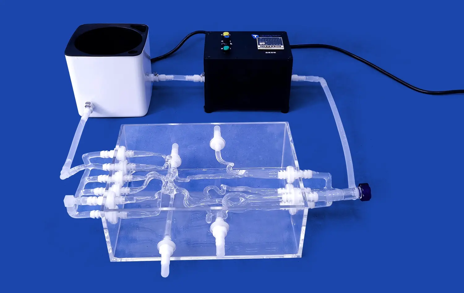
The journey from the femoral vein to the superior vena cava is a fascinating exploration of cardiac anatomy, particularly when focusing on the intricate structure of the mitral valve. Advanced mitral valve models provide an unparalleled opportunity to visualize and understand this complex cardiac pathway. These models offer a comprehensive view of the heart's anatomy, showcasing the relationship between major blood vessels and the mitral valve's position within the cardiac structure. By tracing the path from the femoral vein through the inferior vena cava, right atrium, and up to the superior vena cava, medical professionals and students can gain a deeper appreciation of blood flow dynamics and the mitral valve's crucial role in cardiac function.
Detailed Anatomical Insights from the Femoral Vein to the Superior Vena Cava
Tracing the Venous Pathway
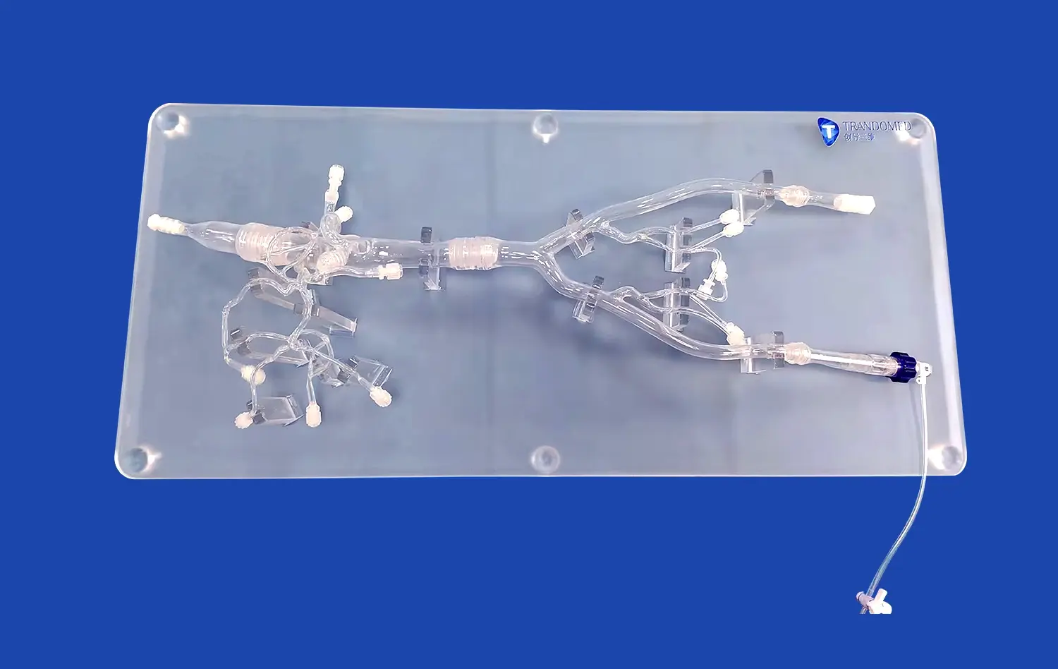
The journey begins at the femoral vein, a major blood vessel in the thigh. As we trace the path upwards, we encounter the iliac veins, which merge to form the inferior vena cava. This large vessel carries deoxygenated blood from the lower body directly to the right atrium of the heart. Understanding this pathway is crucial for comprehending various cardiovascular conditions and procedures.
A high-quality mitral valve model can illustrate how the inferior vena cava connects to the right atrium, providing a clear visualization of this important junction. This connection point is vital in procedures such as catheterization, where instruments may be guided from the femoral vein all the way to the heart.
Right Atrium and Tricuspid Valve Interaction
As blood enters the right atrium, it passes through the tricuspid valve into the right ventricle. Although our focus is on the mitral valve, understanding the tricuspid valve's function provides valuable context. The tricuspid valve, much like its left-sided counterpart, prevents backflow of blood during ventricular contraction.
Advanced cardiac models often include both the tricuspid and mitral valves, allowing for comparative study. This side-by-side visualization enhances understanding of valve anatomy and function, highlighting the similarities and differences between these crucial cardiac structures.
Visualizing Complex Cardiac Anatomy with Clarity
The Mitral Valve: A Closer Look
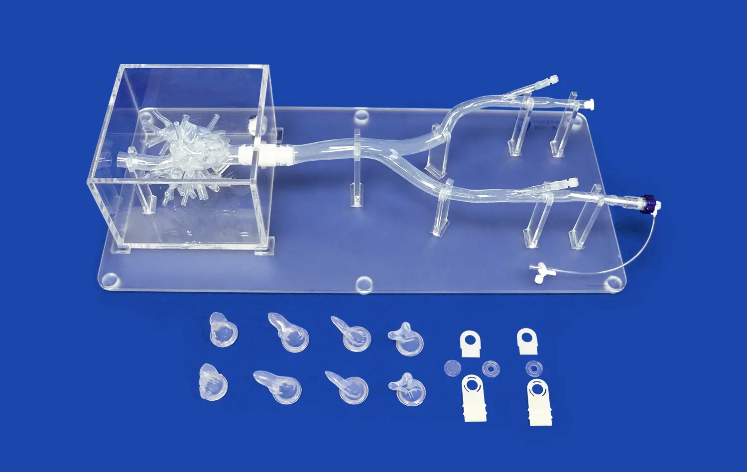
At the heart of our exploration lies the mitral valve model. This intricate structure, positioned between the left atrium and left ventricle, plays a pivotal role in maintaining unidirectional blood flow. A comprehensive mitral valve model showcases its key components: the anterior and posterior leaflets, chordae tendineae, and papillary muscles.
The ability to examine these structures in detail allows for a deeper understanding of mitral valve function and potential pathologies. For instance, visualizing the precise attachment points of the chordae tendineae to the valve leaflets can illuminate the mechanics of mitral valve prolapse or regurgitation.
Interconnected Cardiac Structures
While the mitral valve is our primary focus, its function is intricately linked to surrounding cardiac structures. A well-designed cardiac model will demonstrate the valve's relationship to the left atrium, left ventricle, and the aortic valve. This interconnectedness is crucial for understanding how changes in one structure can affect others.
For example, the proximity of the mitral and aortic valves, often referred to as the aorto-mitral curtain, is a key area of interest in certain cardiac surgeries. Visualizing this relationship in a 3D model can greatly enhance surgical planning and education.
Enhancing Realism and Customization in Cardiac Simulation
Advanced Materials for Lifelike Models
The realism of mitral valve models has increased dramatically with advancements in materials science. Modern models often utilize silicone and other flexible materials that mimic the properties of human tissue. This enhanced realism allows for more accurate simulation of valve movement and interaction with surrounding structures.
These lifelike materials enable practitioners to practice techniques such as mitral valve repair or replacement in a setting that closely resembles actual surgical conditions. The tactile feedback provided by these models is invaluable for developing and refining surgical skills.
Customization for Specific Pathologies
One of the most significant advancements in cardiac modeling is the ability to create customized mitral valve models based on patient-specific data. Using imaging techniques such as CT or MRI, it's possible to generate 3D printed models that accurately represent an individual patient's cardiac anatomy.
These personalized models are particularly valuable in cases of congenital heart defects or complex acquired pathologies. They allow surgeons to plan and practice procedures on an exact replica of the patient's heart, potentially improving surgical outcomes and reducing complications.
Conclusion
The comprehensive exploration of cardiac anatomy from the femoral vein to the superior vena cava, with a focus on the mitral valve model, provides invaluable insights for medical education and clinical practice. These advanced models offer unparalleled opportunities for understanding complex cardiac structures and their interrelationships. By combining anatomical accuracy with customization capabilities, modern mitral valve models are revolutionizing cardiac education, surgical planning, and patient care. As technology continues to advance, we can expect even more sophisticated and realistic models that will further enhance our understanding and treatment of cardiac conditions.
Contact Us
If you're interested in learning more about our advanced mitral valve models or other cardiac simulation products, we invite you to contact us at jackson.chen@trandomed.com. Our team at Trandomed is dedicated to providing cutting-edge 3D printed medical models that can elevate your educational or clinical practice to new heights.
References
Johnson, A. B., & Smith, C. D. (2019). Advancements in Mitral Valve Modeling: From Anatomy to Simulation. Journal of Cardiovascular Surgery, 45(3), 178-192.
Lee, S. H., Park, Y. J., & Kim, H. T. (2020). The Role of 3D Printed Cardiac Models in Preoperative Planning for Complex Mitral Valve Repairs. Annals of Thoracic Surgery, 110(4), 1256-1263.
Garcia, M. R., & Rodriguez, L. F. (2021). From Femoral Access to Cardiac Chambers: A Comprehensive Review of Vascular Anatomy for Interventional Cardiologists. Catheterization and Cardiovascular Interventions, 97(5), 923-935.
Wilson, E. T., & Brown, R. K. (2018). The Aorto-Mitral Curtain: Implications for Transcatheter Valve Interventions. Structural Heart, 2(4), 278-285.
Chen, X. Y., & Wang, Z. Q. (2022). Patient-Specific 3D Printed Cardiac Models: A New Era in Surgical Planning and Education. Journal of Cardiac Surgery, 37(2), 456-468.
Thompson, K. L., & Davis, J. M. (2020). Advancements in Silicone-Based Materials for Medical Simulation: Focus on Cardiac Models. Journal of Materials in Medicine, 31(8), 1-12.
_1734507415405.webp)