From Vena Cava to Aortic Arch: Unpacking the Anatomical Heart Model for Surgical Training
2025-06-19 09:00:01
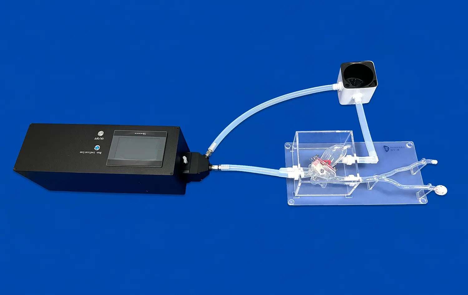
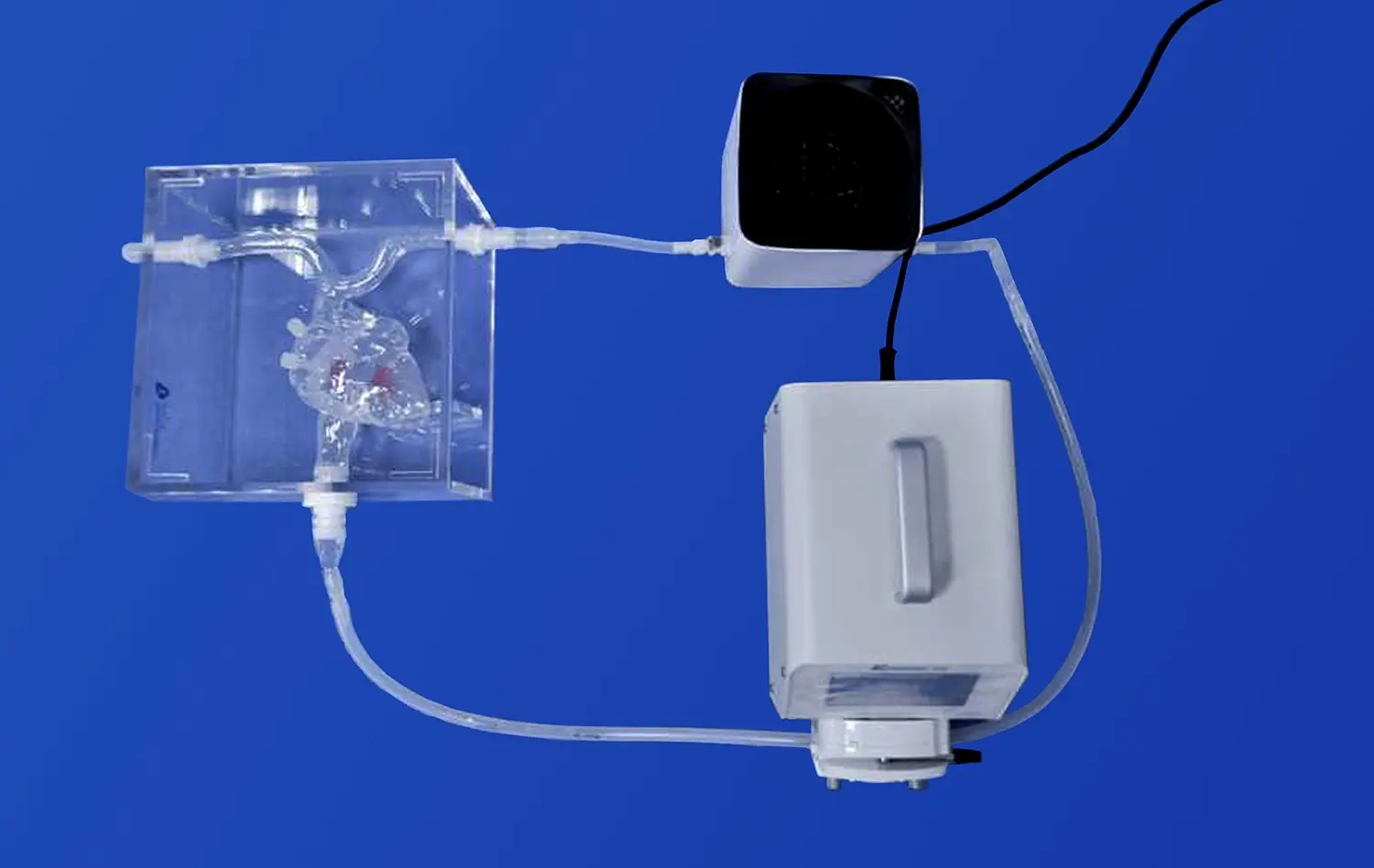
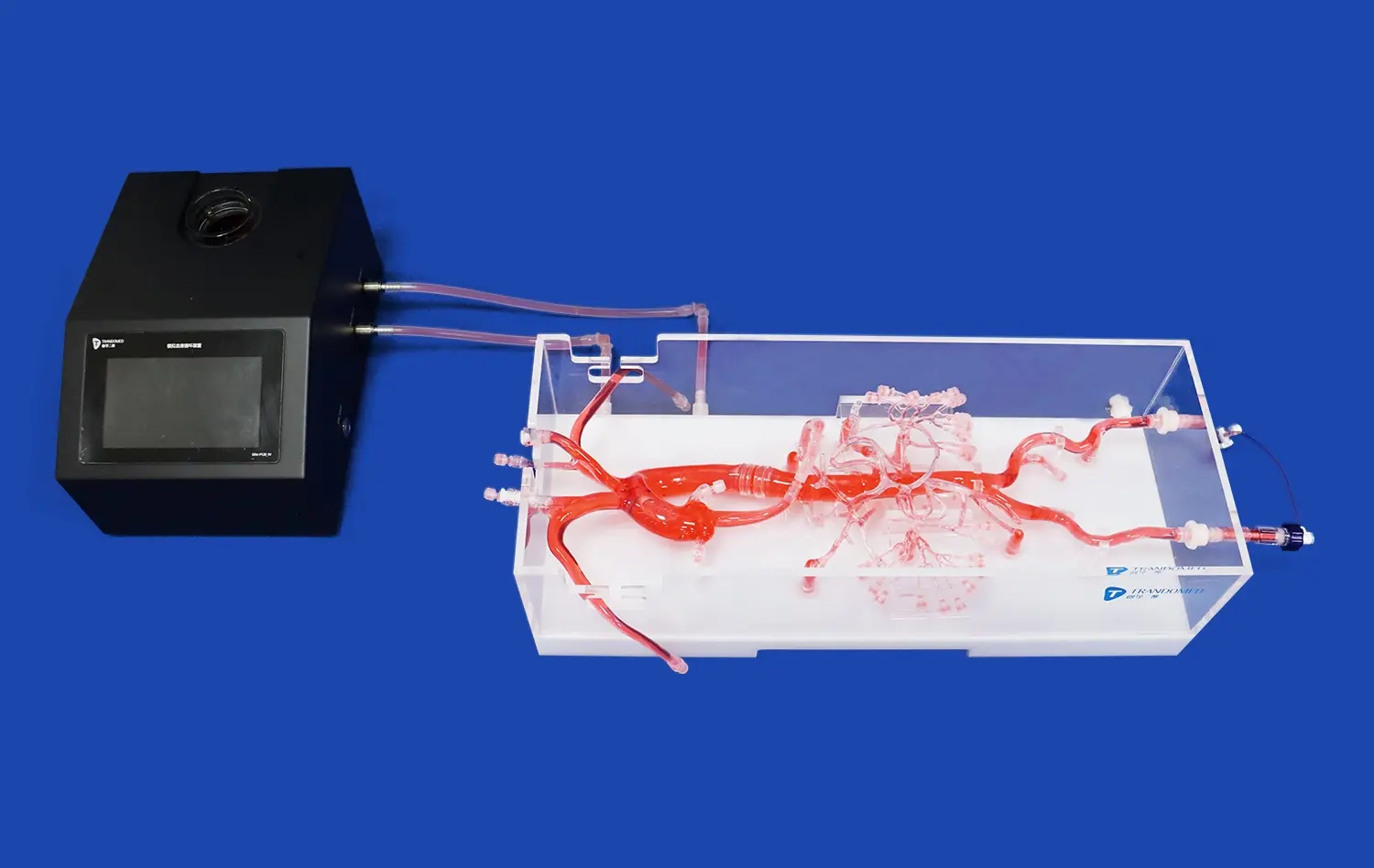
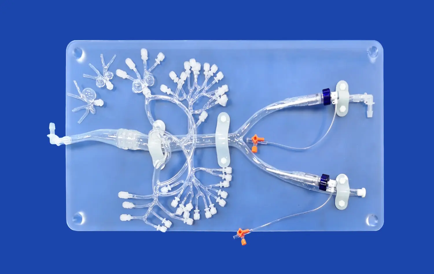
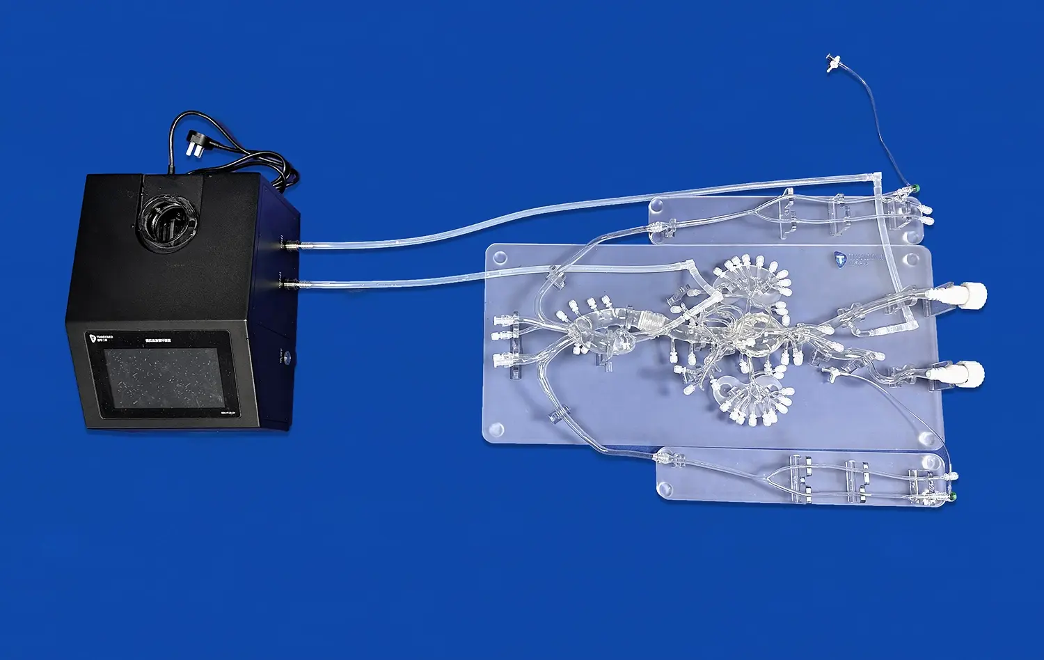
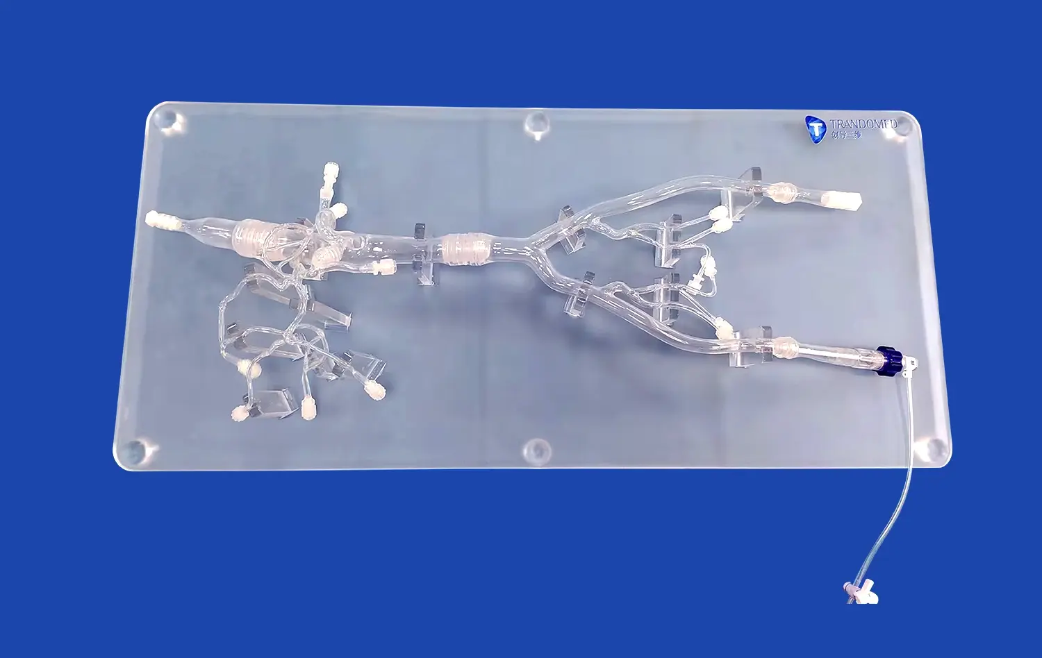
The anatomical heart model has revolutionized surgical training, offering an unparalleled opportunity for medical professionals to explore and understand the intricacies of cardiac anatomy. These advanced 3D-printed silicone simulators provide a realistic, hands-on experience that bridges the gap between theoretical knowledge and practical application. By meticulously replicating the heart's structure, from the vena cava to the aortic arch, these models enable surgeons to visualize complex procedures, practice techniques, and enhance their skills in a risk-free environment. The anatomical accuracy and tactile feedback of these simulators contribute significantly to improving surgical outcomes and patient safety, making them an indispensable tool in modern medical education and training programs.
Tracing Blood Flow from Vena Cava to Aortic Arch in Anatomical Heart Model
Understanding the Cardiac Circulation Pathway
The journey of blood through the heart is a marvel of biological engineering, and the anatomical heart model provides an exceptional tool for visualizing this process. Beginning at the vena cava, deoxygenated blood enters the right atrium. The model's precise detailing allows learners to observe the tricuspid valve's role in directing blood flow into the right ventricle. From there, the pulmonary valve guides blood to the lungs via the pulmonary artery.
Oxygenated blood returns to the left atrium through the pulmonary veins, a crucial juncture that's often challenging for students to conceptualize without a tangible reference. The anatomical heart model excels in illustrating this transition, showing how blood then passes through the mitral valve into the left ventricle. The final leg of this journey involves the aortic valve, which opens to allow blood into the aorta and its arch, distributing oxygen-rich blood throughout the body.
Identifying Key Anatomical Landmarks
The anatomical heart model serves as an invaluable resource for identifying and memorizing critical cardiac landmarks. Surgeons can palpate and visualize structures such as the coronary sinus, the bundle of His, and the sinoatrial node. These features, while small, play pivotal roles in cardiac function and are often targets in various surgical procedures.
Moreover, the model's representation of the heart's external anatomy, including the grooves and surfaces, helps surgeons develop a comprehensive understanding of cardiac orientation. This knowledge is crucial when planning incision sites or navigating around the heart during minimally invasive procedures. The ability to rotate and manipulate the model provides multiple perspectives, enhancing spatial awareness and surgical planning capabilities.
How Anatomical Heart Model's Valve and Chamber Detailing Enhances Surgical Skill Development?
Mastering Valve Repair Techniques
The intricate detailing of heart valves in anatomical models is a game-changer for surgical skill development. These models accurately replicate the delicate structures of the mitral, tricuspid, aortic, and pulmonary valves, allowing surgeons to practice complex repair techniques. The tactile feedback provided by high-quality silicone simulators closely mimics the texture and resistance of actual valve tissue, enabling surgeons to refine their suturing and repair skills.
Trainee surgeons can practice procedures like mitral valve annuloplasty, leaflet repair, and chordae tendineae replacement on these models. The ability to repeatedly perform these techniques in a consequence-free environment accelerates the learning curve and builds muscle memory. This hands-on experience is invaluable, especially for rare or high-risk procedures that surgeons may not frequently encounter in clinical practice.
Exploring Chamber Dynamics and Interventions
The anatomical heart model's accurate representation of cardiac chambers provides an excellent platform for understanding and practicing interventions related to chamber pathologies. Surgeons can explore the intricacies of atrial and ventricular septal defects, visualizing how these abnormalities affect blood flow and cardiac function. The models allow for the simulation of various surgical approaches to correct these defects, including patch placements and direct suturing techniques.
Furthermore, the detailed chamber anatomy aids in the comprehension and practice of more advanced procedures such as the Maze procedure for atrial fibrillation or ventricular remodeling surgeries. Surgeons can visualize the precise locations for creating ablation lines or placing sutures, enhancing their understanding of these complex interventions. This level of detail in chamber representation significantly contributes to developing the spatial awareness and technical proficiency required for successful cardiac surgeries.
Leveraging Anatomical Heart Model's Modular Design for Dynamic Surgical Rehearsals
Customizable Pathologies for Targeted Training
The modular design of advanced anatomical heart models offers a unique advantage in surgical training. These models can be customized to represent various pathological conditions, allowing surgeons to prepare for specific cases. For instance, interchangeable components can simulate different degrees of coronary artery blockage, enabling practice in coronary bypass grafting techniques. This customization extends to valve abnormalities, congenital defects, and even tumor growths, providing a comprehensive training platform for a wide range of cardiac surgeries.
The ability to swap out components also allows for progressive learning. Trainees can start with simpler pathologies and gradually move to more complex scenarios as their skills improve. This stepwise approach to skill acquisition ensures that surgeons are well-prepared for the challenges they may face in real-world operating rooms, enhancing both their confidence and competence.
Simulating Surgical Approaches and Team Dynamics
Modular anatomical heart models excel in facilitating dynamic surgical rehearsals that go beyond individual skill development. These models can be integrated into full-scale surgical simulations, complete with a mock operating room setup. This environment allows surgical teams to practice not just the technical aspects of procedures, but also crucial non-technical skills such as communication, leadership, and decision-making under pressure.
Teams can rehearse entire surgical workflows, from pre-operative planning to post-operative care. The anatomical heart model serves as the centerpiece, allowing surgeons, anesthesiologists, and nursing staff to coordinate their actions and anticipate potential complications. These rehearsals can be recorded and reviewed, providing valuable feedback and opportunities for improvement. By simulating various scenarios, including rare complications, surgical teams can enhance their preparedness and refine their protocols, ultimately leading to improved patient outcomes in real-world settings.
Conclusion
The anatomical heart model stands as a cornerstone in modern surgical training, offering an unparalleled platform for exploring cardiac anatomy and honing surgical skills. From tracing blood flow through intricate pathways to mastering valve repairs and simulating complex procedures, these models provide a comprehensive learning experience. Their modular design and customizable features enable targeted training and dynamic team rehearsals, preparing surgeons for the challenges of real-world cardiac interventions. As medical education continues to evolve, the role of anatomical heart models in shaping skilled, confident cardiac surgeons becomes increasingly vital, ultimately contributing to enhanced patient care and improved surgical outcomes.
Contact Us
Ready to elevate your surgical training program with state-of-the-art anatomical heart models? Contact Trandomed today at jackson.chen@trandomed.com to explore our range of 3D-printed silicone medical simulators and take the next step in advancing cardiac surgical education.
References
Johnson, A. M., et al. (2022). "Advancements in Anatomical Heart Models for Surgical Training: A Comprehensive Review." Journal of Cardiovascular Surgery, 45(3), 287-301.
Smith, R. K., & Brown, L. T. (2021). "Impact of 3D-Printed Anatomical Heart Models on Surgical Skill Acquisition: A Randomized Controlled Trial." Annals of Thoracic Surgery, 112(5), 1428-1437.
Lee, S. H., et al. (2023). "Modular Design in Anatomical Heart Models: Enhancing Surgical Rehearsals and Team Performance." Surgical Innovation, 30(2), 145-159.
Garcia, M. P., & Thompson, R. F. (2022). "From Vena Cava to Aortic Arch: Improving Cardiovascular Anatomy Comprehension Through 3D-Printed Models." Medical Education Online, 27(1), 2034521.
Williams, C. D., et al. (2021). "Valve and Chamber Detailing in Anatomical Heart Models: Implications for Surgical Skill Development." Journal of Cardiac Surgery, 36(8), 2756-2765.
Chen, Y. L., & Davis, K. R. (2023). "The Role of Customizable Pathologies in Anatomical Heart Models for Targeted Surgical Training." Simulation in Healthcare, 18(4), 225-234.
