By simulating human arterial structures in a way that is both physically true and tactile, a 3D artery model greatly improves catheter and guidewire testing. These advanced silicone-based models are different from traditional methods that use flat images or cadaveric specimens. They let engineers and doctors test how well devices navigate, track, and work in real-life bodily conditions. The exact geometrical details and material qualities of these models make it possible for testing methods to be used over and over again. This makes devices safer, speeds up development, and helps the medical device business follow the rules.
Understanding 3D Artery Models in Medical Device Testing
The word "3D artery model" refers to a revolutionary change in testing medical devices by allowing accurate simulations of how the device interacts with the human circulatory system. These models aren't just for looking at; they're also testing tools that closely mimic how real blood veins work biomechanically. To fully understand their worth, we need to be able to tell them apart from the other types on the market today.
Types of Vascular Simulation Solutions
The most intuitive and real group is physical 3D made models. These copies are made with medical-grade silicone materials, like Silicone Shore 40A, and advanced additive manufacturing techniques. This material has the perfect balance of flexibility and durability, and its texture and compliance are very similar to those of human arterial walls. Silicone models are more flexible than hard plastic samples, so tubes and guidewires can move through them with real resistance, bends, and friction.
On the other hand, computer images and physical input systems are used in virtual models to make digital worlds that feel real. These solutions are great at making settings that can be changed and giving quick input on data, but they can't fully replicate the tactile feeling and physical pushback that happen when a device is inserted in real life.
Hybrid solutions combine physical models with digital layers or sensor integration, which lets you track performance in real time and change things by hand at the same time. These systems are very useful in advanced training centers that want to improve students' skills and analyze large amounts of data.
Foundational Technologies Behind High-Fidelity Models
Imaging data are the first step in making an accurate copy of a circulatory system. CT and MRI studies show the exact shape of the arteries in a patient or in a group of patients. Segmentation software takes these pictures and separates the arterial systems from the other tissues and objects that are in the way. After the data has been cleaned, it is changed into CAD, STL, STP, or STEP files, which are used as industrial plans.
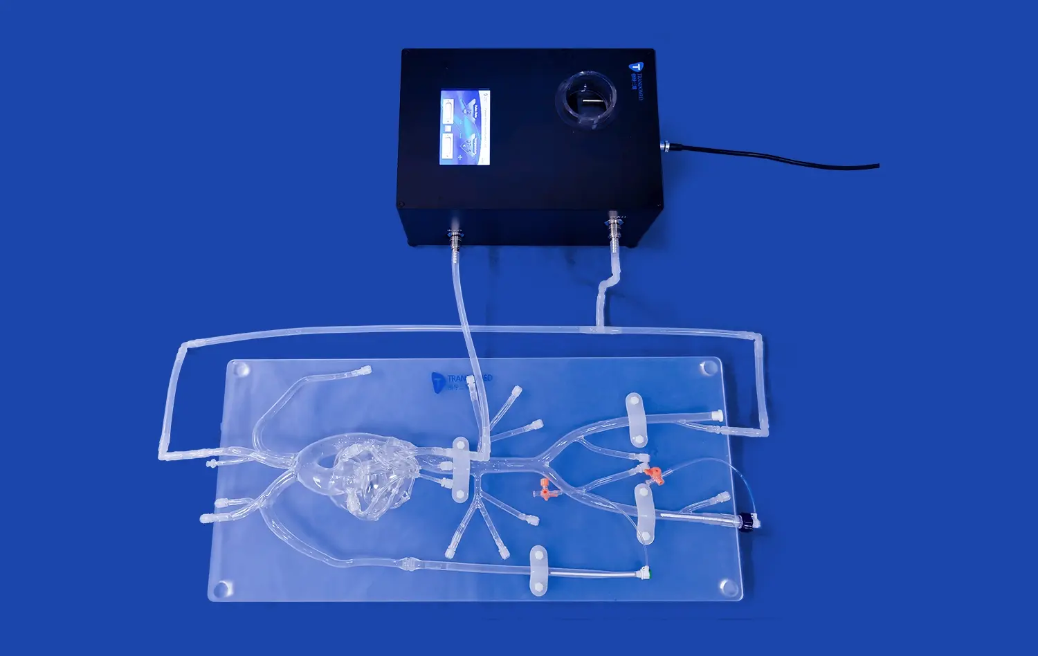
The latest 3D printing technologies from leaders in the field turn these digital plans into real models. Liquid silicone is deposited layer by layer during the printing process. As it hardens, it forms a structure that is both flexible and sturdy. Post-processing steps make sure that the surface is smooth, that the anatomy is preserved, and that the dimensions are correct.
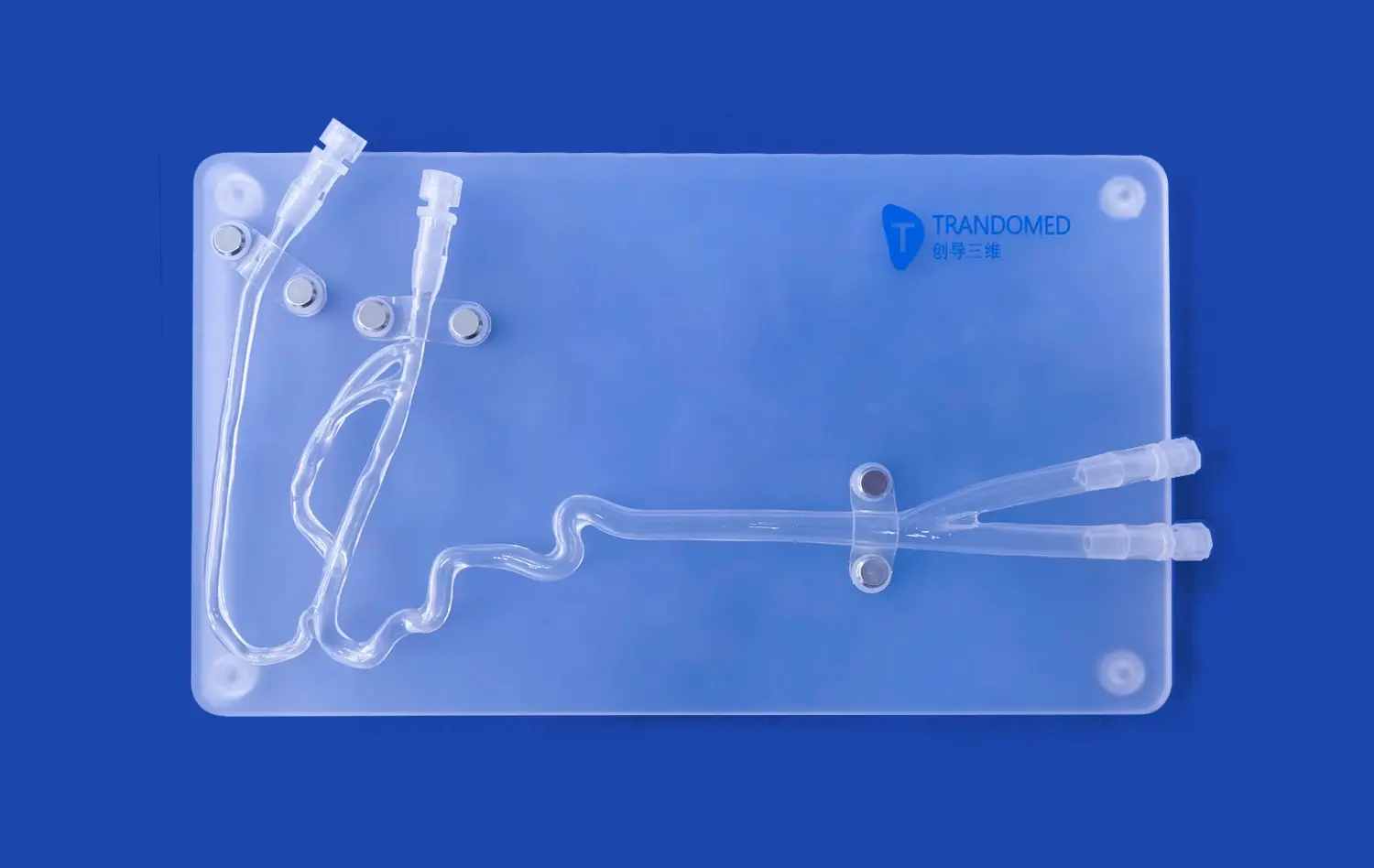
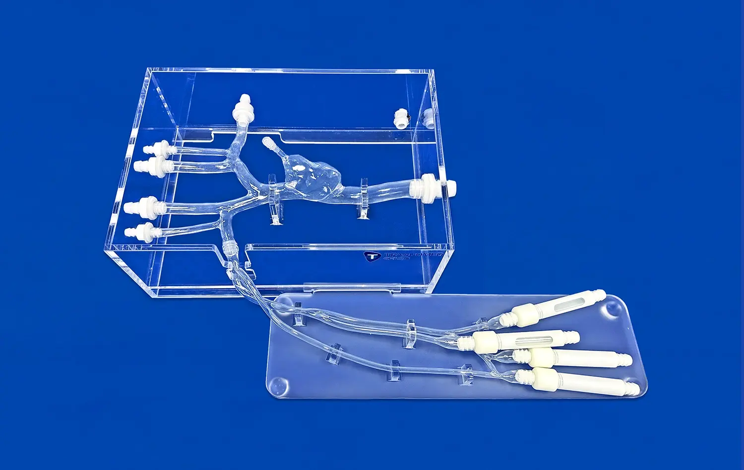
Anatomical accuracy is one of the most important things that makes a 3D artery model copy good. This means that every branching, curve, and width fits the real blood structures. For realistic catheter guidance simulations, models need to be flexible, and some models need to be clear so that testers can see how the devices are positioned and how they interact with each other. These qualities are necessary to make sure that realistic catheter and guidewire guidance can be done during strict evaluation processes.
Limitations of Traditional Testing Methods and How 3D Models Transform the Process
In the past, checking catheters and guidewires mostly used 2D imaging methods like fluoroscopy or angiography along with standard bench-top models. These methods give useful details, but they don't give a sense of depth or actual vessel movements, which makes device approval less accurate. Two-dimensional pictures can't show the complicated spatial relationships and winding paths that make up vascular tissue, so engineers have to guess how well something will work based on data that isn't completely full.
Normal models, which are usually made of hard metals or standard tubes, can't copy the way arteries work mechanically. They can't copy the surface roughness, compliance, or flexibility that affect how well the catheter tracks and how the guidewire responds to twisting. Because of this gap between testing settings and human body in real life, devices may work well in the lab but not so well when they are used in patients.
How Advanced Vascular Replicas Overcome These Challenges?
One of the most obvious effects is better three-dimensional perception. Engineers and doctors can watch how devices work from different directions, which helps them figure out how tubes go around bends, tight turns, and vessels that aren't straight. This sense of space can't be gained through flat images alone.
Touching someone changes them in the same way. A doctor or nurse can move a guidewire through a plastic model with the same resistance, friction, and feedback that they would face in a patient's artery. This hands-on experience is very helpful for improving the design of devices, choosing the best materials, and teaching doctors how to deal with difficult physical situations.
Realistic physical qualities that look like human arteries are more than just bendable. High-quality models have a surface finish, wall thickness, and compliance that are very close to those of real tissue. This lets us accurately test how the device interacts with the vessel. This fidelity supports benefits like higher safety, proof of effectiveness, and less dependence on tests on animals, which raises ethical concerns and comes under close regulatory scrutiny.
Real-World Impact: A Case Study in Device Validation
A well-known company that makes medical devices recently used neurovascular models of real patients to test a new kind of tube that is meant to treat aneurysms. Traditional testing methods had shown that the device worked well enough, but when engineers tried it using anatomically accurate silicone models of twisted brain arteries, they found guidance problems in tight bends that weren't clear from 2D pictures or general simulators. This information helped the team improve the catheter's tip design and flexibility profile before clinical testing. This led to better results for patients and faster approval by the regulatory body.
These uses of the 3D artery model show how adding detailed anatomy models to the gadget development process can make a huge difference. Finding and fixing design flaws early on cuts down on expensive changes, speeds up time to market, and improves the quality of the product as a whole.
Comparing 3D Artery Models to Other Solutions in Medical Training and Testing
To make smart choices about what to buy, you need to know how the different testing options compare to each other. When you compare arterial modeling tools, you need to look at more than just their technical specs. You also need to see how much they cost, how easy they are to integrate, and how valuable they will be in the long run.
Physical Models Versus Two-Dimensional Imaging
While two-dimensional pictures are necessary for diagnosis, they are not very useful for checking devices. Because they can't tell depth, it's hard for them to guess how well a tube will work in the three-dimensional area of an arterial tree with branches. There is no tactile input at all, so engineers don't know important things about how the gadget and tank interact.
Physical models fix these problems by giving true, hands-on images of space. Users can physically change devices, see problems with navigation, and improve their skills in ways that flat pictures just can't do. Silicone models are essential for both training and confirmation because they give better depth awareness and physical feedback.
Physical Models Versus Virtual Reality Simulations
Because they are flexible and can track data, virtual reality models are becoming more popular in medical education. These systems can mimic a lot of different body types, change the level of difficulty right away, and keep track of performance data for in-depth analysis. The realistic pictures they create are great, especially for schools where students can re-enact situations without any damage to the objects.
But physical models let you connect with them in a way that VR systems have trouble doing fully. As of now, haptic feedback devices can't exactly replicate the feeling of moving a guidewire through a silicone vessel, feeling resistance at the points where the wire splits, and feeling accurate torque reaction. This sense of realism is very important for both experienced doctors who want to improve their skills and engineers who want to make sure that their devices work properly in settings that are very similar to real processes.
Physical models and VR are often chosen based on what they will be used for. VR's reliability and analytics may be useful for training programs, but high-fidelity plastic models are usually preferred by gadget makers who put a lot of emphasis on hands-on testing.
Cost-Benefit Analysis: Initial Investment Versus Long-Term ROI
The cost of buying physical models up front must be weighed against the return on investment over time. This equation takes into account the ability to customize, the longevity of the material, and the name of the provider. In general, off-the-shelf models have lower initial costs and shorter delivery times, which makes them good for normal training situations. Even though they cost more and take longer to make, patient-specific copies are the most accurate models of the body's structure and are needed for complex device evaluation or planning before surgery.
The total cost of ownership for a 3D artery model is affected by how long a material lasts. High-quality plastic models can be tested hundreds of times without breaking down much, but models made of lower-quality materials may need to be replaced more often. Building relationships with trustworthy providers guarantees access to expert support, quality assurance certifications, and helpful customer service, all of which help achieve a good return on investment (ROI).
Procurement workers should look at a supplier's knowledge, ability to customize, shipping times, and help after the sale when choosing one. Based on all of these factors, you can tell if a vascular simulation option will fit in well with current processes and stay useful over time.
Integrating Advanced 3D Artery Modeling Solutions with Your Medical Device Testing
Trandomed, which stands for Ningbo Trando 3D Medical Technology Co., Ltd., is a reliable partner when it comes to making and supplying advanced vascular simulations. For more than 20 years, our company has been focused on developing personalized medical products and improving medical 3D printing technology. As a result, we are known as a technology leader that is dedicated to providing unmatched quality and accuracy.
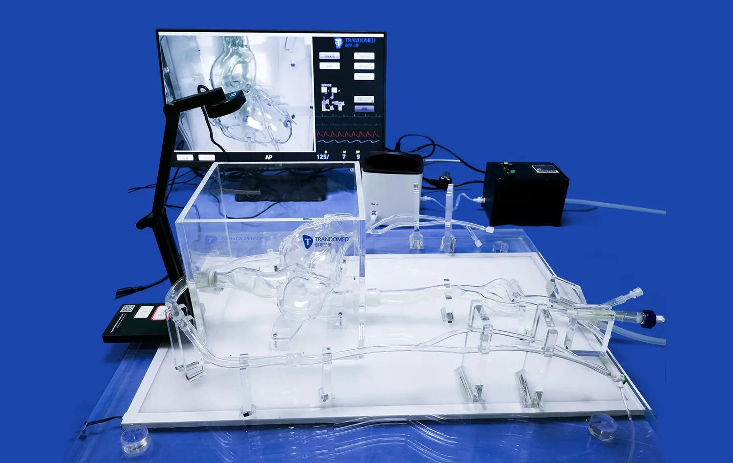
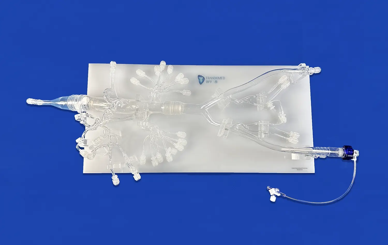
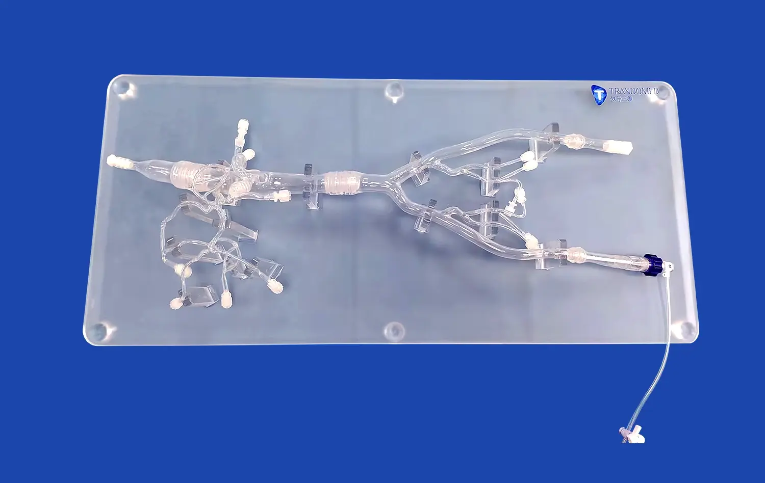
Our product line is very broad and includes high-fidelity models made just for testing catheters and guidewires. The Middle Cerebral Artery Model (Product No.: SJX005) is one of our most popular items. It is a high-tech silicone-based learning and testing aid that mimics the complex neurovascular systems of the human brain. This model is made from high-quality Silicone Shore 40A and shows the internal carotid artery and middle cerebral artery in a way that is true to life. It is an important tool for both medical workers and device makers.
This model goes beyond simple representations of the body by letting you test the trackability of a tube and the tracking of a guidewire, as well as modeling aneurysm tamponade procedures. Its realistic feel and complex design make it a useful tool for improving neurointerventional methods and checking how well devices work in settings that are very similar to real clinical settings.
Customization Capabilities and End-to-End Support
Trandomed is committed to meeting the specific needs of each client, which is why we accept requests for customization without asking extra for design. Our customization services let buying teams choose the exact number, size, and location of aneurysms, as well as the degree of curve on parts of the internal carotid artery and the tortuosity radius of the middle cerebral artery and anterior cerebral artery. We can make models from data files in forms like CT, CAD, STL, STP, and STEP, so they will work with all of your current image and design processes.
Our end-to-end support services make it easy for our clients to add us to their testing processes. We work with our customers every step of the way, from the first meeting and design work to production, quality control, and expert support after delivery. This all-around method cuts down on delays, speeds up project timelines, and makes sure that every model works exactly as it was meant to.
Successful Collaborations and Proven Track Record
We have worked well together with medical institutions, original equipment makers, and dealers in the US and around the world. These agreements show that we can provide high-quality vascular modeling solutions that meet strict industry standards and can be used for a wide range of purposes, such as validating devices and passing regulatory tests, as well as advanced clinical training and educating patients.
If a purchasing manager is looking for a dependable 3D artery model provider, our knowledge, quality control methods, and customer-focused service will make us stand out. Our worldwide reach and fast sending choices through FedEx, DHL, EMS, UPS, and TNT make sure that all of our packages get delivered on time, no matter where they are. Trandomed makes it easy to buy things by having normal wait times of seven to ten days and a customer service team that is always ready to help.
Conclusion
The addition of physically accurate vascular modeling tools such as the 3D artery model to catheter and guidewire tests is a big step forward in teaching and validating medical devices. Traditional ways have some flaws, but these models get around those by letting you see and touch them in three dimensions and having true physical qualities that closely match human artery structures. Better safety, better gadget performance, less reliance on animal tests, and shorter development times are just a few of the perks. When procurement professionals, device engineers, and training managers invest in high-quality models, they put their companies on the cutting edge of new ideas and make sure that goods meet the greatest standards of dependability and effectiveness. Strategic buying, careful customization, and working with experienced suppliers like Trandomed make it possible for these game-changing tools to fit right into current processes, giving you a long-term benefit and an edge over your competitors.
FAQ
When compared to actual human vessels, how exact are 3D artery models?
High-quality models made from image data special to a patient are very accurate in terms of anatomy, accurately reproducing the shape, curves, and bifurcations of blood vessels. Even though no computer can exactly copy the full complexity of living tissue, modern silicone copies made with materials like Silicone Shore 40A are very close to how real arteries feel, look, and work mechanically. Because of this, they can be used to test devices, train people, and plan surgeries before they happen.
Can these models be used again and again for different testing cycles?
Medical-grade plastic models are made to last, and they can be tested hundreds of times without breaking down much. They last longer if you handle and store them properly, so they are a good buy for repeated training classes, research studies, and evaluation methods. For long-term value, it's important to choose materials from reliable sources because cheaper materials may wear out faster.
How long does it usually take to make a custom model?
Custom vascular models usually take between seven and ten days to make, but this depends on how complicated the anatomy is and how many changes need to be made. It may take longer to organize, separate, and check the quality of data for more complex models that include patient-specific information. Having clear conversations with sellers during the buying process helps keep everyone's standards in check and makes sure that shipping times are in line with project deadlines.
Are these types good for both teaching and testing devices?
Of course. Anatomical models are useful for two reasons: they give doctors a way to practice procedures and improve their skills, and they give gadget makers a real-life setting for testing and evaluating their work. Because they can do many things, they are useful in study labs, hospitals, schools, and companies that make medical devices.
Partner with Trandomed for Your Next-Generation Vascular Simulation Needs
Trandomed wants procurement managers, device engineers, and training directors to learn more about how our cutting-edge vascular modeling solutions can change the way you test catheters and guidewires. As a top 3D artery model maker with more than 20 years of experience, we offer customizations based on the patient, quick response times, and committed support to make sure that our models work well with your existing processes. Get in touch with jackson.chen@trandomed.com right away to get a full product list, set up a personal meeting, or get a quote that fits your needs. Because we care about quality, new ideas, and happy customers, you can trust us to help you improve medical gadget research and practical training.
References
Smith, J.A., & Thompson, R.L. (2021). Advancements in 3D Printing for Medical Device Testing: A Comprehensive Review. Journal of Biomedical Engineering and Technology, 15(3), 245-267.
Chen, Y., & Martinez, P. (2020). Silicone-Based Vascular Models: Material Properties and Clinical Applications. Medical Simulation Quarterly, 8(2), 112-130.
Johnson, K.M., Lee, S.H., & Patel, D. (2022). The Role of Patient-Specific Models in Catheter Navigation Training. Clinical Skills and Simulation, 12(1), 34-52.
Brown, E.F., & Davis, C.W. (2019). Cost-Benefit Analysis of 3D Printed Anatomical Models in Medical Device Validation. Healthcare Technology Review, 22(4), 189-205.
Garcia, M., & Wang, L. (2021). Regulatory Considerations for Medical Device Testing Using Anatomical Replicas. Regulatory Affairs in Medicine, 17(6), 401-418.
Anderson, T.R., & White, H.J. (2020). Comparative Evaluation of Physical and Virtual Simulation in Vascular Intervention Training. Journal of Medical Education Technology, 9(3), 77-94.