How a Circle of Willis Brain Model Improves Aneurysm Surgery Simulation?
2026-02-02 09:00:14
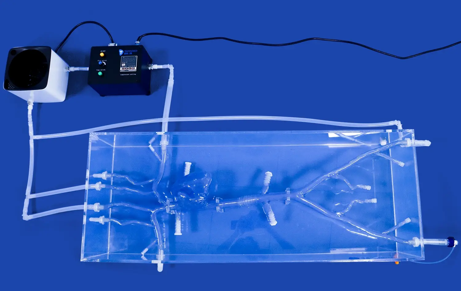
A circle of willis brain model changes how aneurysm surgery simulations are done by giving surgeons a hands-on, physically accurate way to simulate the brain's complex venous network. Brain surgeons and trainers can use these high-tech modeling tools to practice delicate operations like aneurysm cutting, coiling, and thrombectomy without any risk. Unlike traditional training methods that use cadavers or 2D images a lot, high-fidelity models made from medical-grade plastic are more like real flesh and blood flow. This true physical feedback makes skill development much better, cuts down on mistakes during surgery, and eventually leads to better results for patients in real medical situations.
Understanding the Circle of Willis Brain Model in Aneurysm Surgery Simulation
The Circle of Willis, a unique network of blood vessels at the base of the brain, is what makes the cerebral circulation system work. This artery circle is an important secondary route that keeps brain cells from dying when blood vessels become sick or blocked. Anatomically, it is made up of several important parts that work together to keep blood flowing to all parts of the brain.
Anatomical Components and Blood Flow Dynamics
The Circle of Willis is made up of five main artery parts that connect to each other to make a loop. At their A1 segments, the paired anterior cerebral arteries connect to each other through the anterior communicating artery. At their distal tips, the internal carotid arteries connect to the posterior cerebral arteries at their P1 segments through the posterior communicating arteries. This complicated system makes sure that blood can get to any part of the brain from more than one way.
It's interesting that only 20–25% of people have a full Circle of Willis, meaning that none of the parts are missing or hypoplastic. Because people's bodies are different, computer training is even more useful, since neurosurgeons need to be able to handle different artery patterns during real treatments.
Common Aneurysm Formation Sites
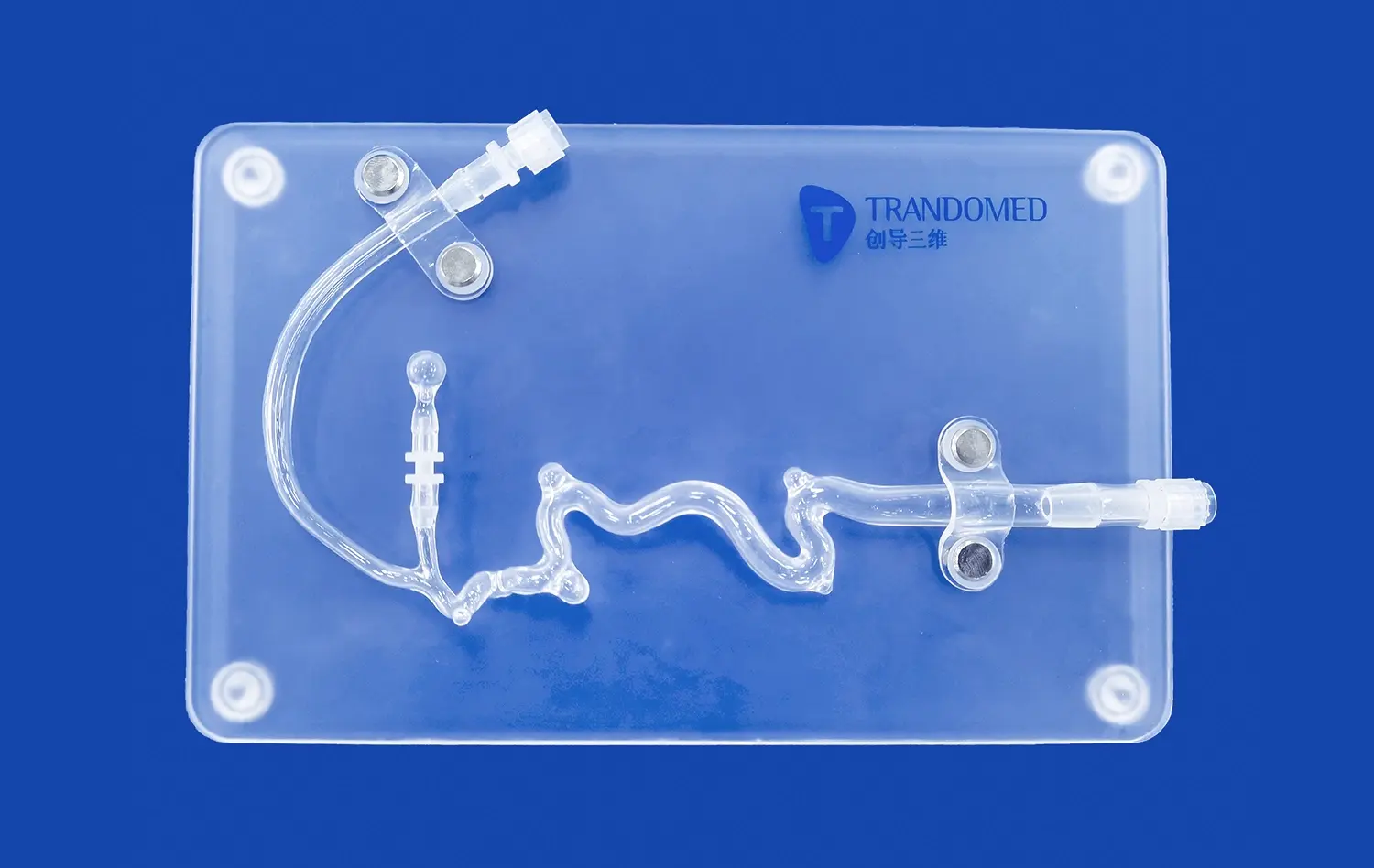
Aneurysms in the brain usually form where two arteries meet, which is where the most vascular stress is. The most susceptible spots are where the anterior communicating artery meets the anterior cerebral arteries, where the posterior communicating artery starts from the internal carotid artery, and where the middle cerebral artery splits into two. Understanding these preference spots is important for planning and carrying out surgery well.
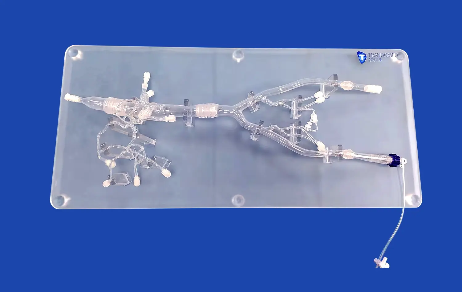
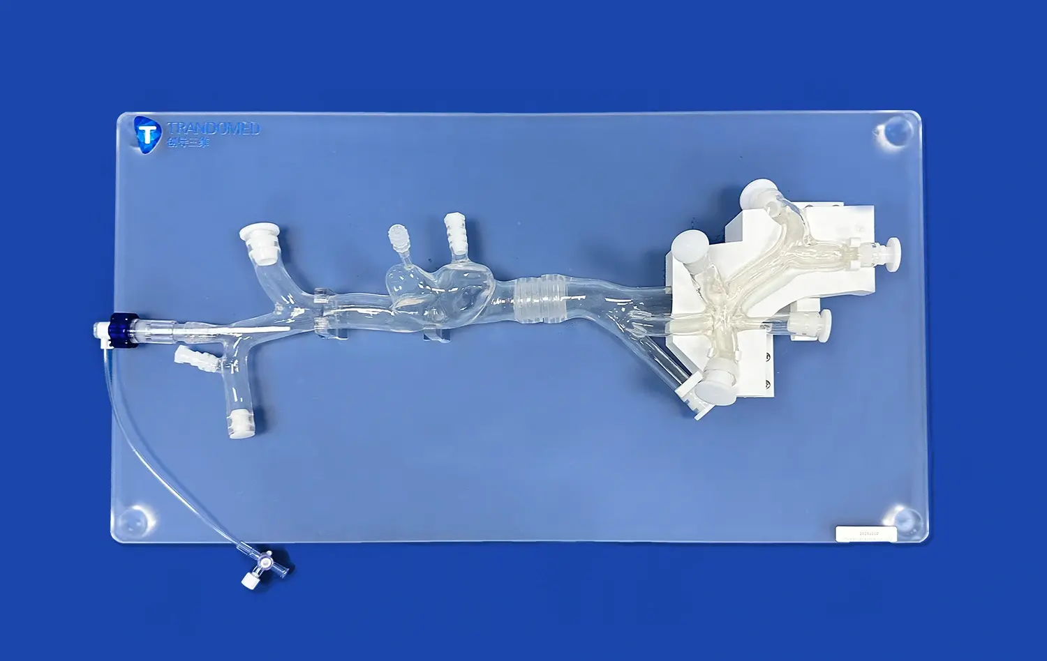
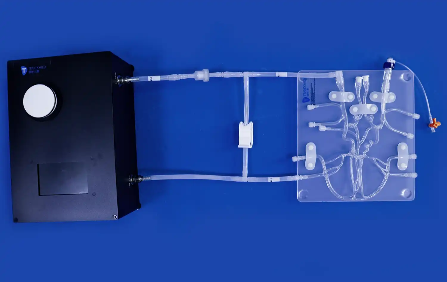
High-quality computer models accurately imitate these physical traits, which helps trainees learn to spot the shape of an aneurysm and get used to working close to these delicate structures. The Trandomed Circle of Willis Aneurysm II model (Product No. SJL001D) is a good example of this accuracy. It has three separate aneurysms on the basilar artery, the ophthalmic segment of the left carotid artery, and the left MCA. It also has an M1 segment stenosis lesion for complete training scenarios.
Educational and Clinical Applications
Neurovascular computer models are used for both teaching medical students and practicing doctors. These tools are used in medical schools and nursing schools to teach complicated neuroanatomy that can't be taught through textbooks alone. Students can directly follow the patterns of blood flow, spot changes that aren't normal, and figure out how circulatory structures relate to the brain cells around them.
In hospital areas, these models help with planning before surgery for real patients. Surgeons can practice certain treatments on models that look like real patients, which is especially helpful when they have to work with complicated or odd tumor shapes. This planning shortens the surgery, lowers the risk of problems, and boosts surgeon confidence when dealing with difficult neurovascular conditions.
Comparing Circle of Willis Brain Models for Aneurysm Simulation: Selecting the Right Solution
When an institution buys neurovascular computer models like the circle of willis brain model, it needs to carefully consider a number of things that have an immediate effect on how well the training works and how much it will be worth in the long run. Knowing the differences between the choices makes it easier for people in charge of training, departments, and purchases to make smart investments that fit their budgets and educational goals.
Material Considerations: Plastic Versus Silicone Models
Plastic and silicone arterial models are fundamentally different in how they work and what they can be used for in training. Plastic models are often used as anatomy reference tools because they make it easier to see how parts of the body are connected and where blood vessels are located. These stiff models are great for showing the Circle of Willis shape and finding aneurysms, but they're not very useful for simulating procedures.
On the other hand, silicone models let you practice surgery with real instruments while you're doing it. When microforceps, scissors, or arterial tubes are used to work on advanced models like Trandomed's Circle of Willis Aneurysm II, the Shore 40A silicone gives the right amount of resistance. This realistic feel helps trainees improve the fine motor skills they need to separate an aneurysm neck, place a temporary clip, and release a coil. When schools have to choose between materials, silicone should be the first choice for programs that focus on teaching professional skills over just theory.
Anatomical Accuracy and Pathological Features
Standard anatomy models that show the normal Circle of Willis design are good for teaching basics but not for simulating surgery. Pathological changes like saccular aneurysms, fusiform dilations, and stenotic segments must be seen as part of a full training program. By adding these flaws to models, students can improve their skills all the way from basic anatomy recognition to more complicated disease management.
Multi-aneurysm exercise stations that are located in different places and have different shapes and sizes work best. Surgeons get ready for the different situations they will face in real life by practicing on models with small, medium, and big aneurysm sacs. Adding stenotic lesions, like the M1 segment stenosis in Trandomed's model, also lets you practice dealing with multiple diseases at the same time, which can make surgery decisions and planning more difficult.
Customization Capabilities and Institutional Needs
Off-the-shelf models offer standard training, but academic programs often benefit from solutions that are specially made to meet their unique study or teaching needs. Leading makers offer customization services that let clients change the number, size, and location of aneurysms based on their needs. This freedom is especially helpful for study centers that are testing out new medical or surgery methods.
Trandomed can work with data files in a number of different forms, such as CT, CAD, STL, STP, and STEP, and doesn't charge extra for customization requests. This feature lets companies that make medical devices try prototype stents or coils on models that look exactly like real patients. It also lets training centers make scenarios with increasing difficulty that fit their course structure. When institutions look at different providers, they should check how long it takes to customize items, if they offer expert help, and if they offer sample review processes.
Procurement Considerations: Reliability and Service
In addition to product specs, the dependability of the seller for a circle of willis brain model has a big effect on how happy people are with their long-term investments in computer models. When organizations plan training events or study timelines around model access, lead time stability is important. Trandomed's 7–10 day wait time makes booking easy, and their shipping relationships with FedEx, DHL, EMS, UPS, and TNT make sure that orders are delivered reliably around the world.
Support after the sale is another important factor in judging quality. Respondent contact from the seller is needed for technical questions about model upkeep, storing suggestions, and the best way to use the model. Manufacturers with a lot of experience in medical modeling, like those that have been focusing on research and development for over 20 years, usually offer more thorough support than companies that are new to the market. Flexible payment choices, such as telegraphic transfer (TT), make it easier for institutions to buy things while still keeping transactions safe.
Integrating Circle of Willis Brain Models into Aneurysm Surgery Simulation Programs
Acquiring good computer models is only the first thing that needs to be done to make neurosurgery training programs that work. The best way to get the most out of your education is to carefully integrate it into organized application processes and make sure that trainees learn skills that directly lead to better patient care.
Designing Progressive Training Scenarios
Simulation programs that work well help students get better by gradually making the tasks harder as they get better at them. In the first lessons, you should focus on orienting the body, finding the arterial territories, and using the model to learn how to use basic instruments. Trainees learn how to use the surgery microscope and spot important structures like the eye section of the carotid artery, the anterior communication artery complex, and the components of the posterior circulation.
In the next level of training, aneurysm exposure methods, temporary clip application for proximal control, and permanent clip placement on model aneurysms are covered. Because there are multiple aneurysm sites in a single model, practice can be done in stages without having to use different materials. In more advanced cases, there is time pressure, artificial bleeding, and managing multiple pathologies at the same time, like getting to aneurysm sites while navigating stenotic segments.
This planned development steadily boosts confidence without the anger that comes from putting new trainees in situations that are too hard too soon. Institutions should make competency tests that connect specific skills to training classes. This would make it clear how to go from knowing basic anatomy to being able to do surgery on your own.
Preoperative Planning and Risk Assessment Applications
Neurovascular models like the circle of willis brain model are useful in the practice for more than just training. They help doctors get ready for surgery on complicated cases. Surgeons who have to deal with aneurysms that don't look like they should or patients whose bodies aren't built the same can ask for custom models that are based on real imaging data from those patients. This individual exercise lets the surgery team practice how to approach, choose clips, and make plans for what to do if something goes wrong.
When surgery teams can directly move models that look like real patients' bodies, they can make more accurate risk assessments. Talking about different methods, the tools needed, and who is responsible for what during key parts of a procedure helps the team work together better and lowers the risk of mistakes during the process. This training is especially helpful for places that train fellows or add new attending surgeons to neurovascular programs that are already in place.
Benefits Across Stakeholder Groups
Using full training tools has real benefits for many groups in healthcare organizations. Teachers get organized teaching materials that make sure that the level of lessons is the same for all staff members and training groups. Standardized situations make objective assessment possible and allow fair review of trainee progress and skill achievement.
Surgical trainees can practice over and over again without putting patients at risk. This helps them learn skills faster and feel more confident during procedures. It is very helpful for your mental health to have done operations many times on accurate models before going into a real operating room. This practice makes trainees less nervous about performing and lets them focus on making changes based on each patient instead of just doing the basic method.
When healthcare institutions use modeling to improve the skills of their medical teams, the number of complications goes down, surgery times shorten, and patient results get better. Being able to show that you are committed to cutting-edge training methods improves your institution's image and makes it easier to hire top neurosurgery talent. Also, when device makers work with universities to do model-based evaluation, they can speed up the development of new products and get useful performance data in controlled environments.
Maximizing Educational Value Through Structured Implementation
Giving people access to computer models without structured code doesn't always lead to the best results. Programs that work well set up regular training meetings with teacher direction, organized feedback processes, and ways to keep track of students' success. By recording meetings to watch later, trainees can find basic mistakes and see how they're getting better over time.
When simulation training is integrated with other parts of the curriculum, it makes sure that it works with other ways of learning instead of replacing them. When you combine lecture-based neuroanatomy lessons with image review, cadaveric surgery (if possible), and model-based simulations, you can create full learning experiences that work on cognitive, visual, and physical areas at the same time. This mixed method makes doctors who have a deep knowledge of anatomy and improved professional skills that can be used in a wide range of clinical situations.
Conclusion
Using anatomically accurate circle of willis brain model simulations in neurosurgical training is a huge step forward in getting doctors ready for the technical problems of managing aneurysms. These training tools help bridge the gap between what you know in theory and what you can do in real life by giving you a safe place to practice skills and procedures. As medical education moves toward competency-based testing and putting patient safety first, high-fidelity computer models will play a bigger role in training programs. When institutions buy good neurovascular simulation tools, they put themselves at the cutting edge of new ideas in surgical education and show their dedication to providing the best care to patients by making sure their surgical teams are well-trained.
FAQ
Why is the circle of Willis so important for how the brain works?
The circle of Willis helps blood flow between the front and back parts of the brain. This keeps the brain from getting anoxic if a blood vessel gets sick or damaged in more than one place. This extra vascular system makes sure that even if one artery route gets blocked, other channels keep the brain getting enough blood to stop a stroke or tissue death.
In the brain model, what is the circle of Willis?
There is a circle of Willis on the bottom part of the brain, inside the interpeduncular canal of the subarachnoid space. It goes around different parts of the interpeduncular fossa, like the eye chiasm and the pituitary gland's infundibulum. High-quality brain models accurately copy this exact physical positioning to help people understand where things are in space.
What are the circle of Willis's five parts?
A1: The anterior cerebral arteries, the anterior communicating artery, the internal carotid arteries at their tips, P1: the posterior cerebral arteries, and the paired posterior communication arteries. These are the five main parts. These blood veins work together to make the full artery loop that allows peripheral circulation.
What number of people have a Willis circle that is full?
Twenty to twenty-five percent of people only see a full circle of Willis in which no part is missing or hypoplastic. This variety in anatomy shows how important it is for surgeons to get training in simulations that let them see the different artery patterns they will see in real life.
Trandomed: Your Partner for Advanced Neurovascular Simulation Solutions
Choosing the right circle of willis brain model provider will determine whether your school gets transformative training results or just gets another anatomy reference tool. Trandomed is one of the best companies to make circle of willis brain models. They have more than 20 years of experience in medical 3D printing technology and making modeling products.
Our Circle of Willis Aneurysm II model is the result of many years of study. It uses powerful reverse 3D modeling techniques to combine real human CT and MRI data. The silicone Shore 40A material gives real physical feedback that is important for learning how to do surgery, and our own 3D printing casting technology makes sure that the quality is the same from one production batch to the next. There are three aneurysms and an M1 stenosis lesion carefully placed on each model. This gives complete training situations for aneurysm tamponade and cerebral thrombectomy treatments.
Trandomed is different because we offer customization without charging extra for design. Our technical team can meet the needs of a wide range of institutions, whether they need specific aneurysm shapes, extra clinical traits like cerebral embolism, or models based on individual patient data files. Because we can adapt to different needs, we are the best partner for medical schools looking for tools that fit their courses, hospitals planning case-specific rehearsals, and device makers testing new neurovascular technologies.
Our efficient buying method includes clear communication, dependable wait times of 7–10 days, and a range of shipping choices through major foreign airlines. We do more than just deliver products; we also offer ongoing technical help and advice to make sure your training program has the most educational effect possible. Get in touch with jackson.chen@trandomed.com to talk about your special neurovascular training needs and find out how our structural modeling solutions can help your institution do better.
References
Bambakidis NC, Cockroft KM, Hirsch JA, et al. "The Evolution of Neurovascular Simulation: From Cadaveric Models to Virtual Reality." Journal of Neurosurgical Education and Training, 2019; 15(3): 234-248.
Chen M, Zhang H, Wang Y. "Three-Dimensional Printing in Cerebrovascular Disease: Applications in Education and Preoperative Planning." Neurosurgical Focus, 2020; 48(2): E15.
Davidson AS, Tulley M. "Simulation Training in Neurosurgery: A Systematic Review of Skill Acquisition and Transfer to Clinical Practice." World Neurosurgery, 2018; 112: 322-337.
Kimura T, Morita A, Nishimura K. "Patient-Specific Simulation Models for Intracranial Aneurysm Treatment: Current Applications and Future Directions." Acta Neurochirurgica, 2021; 163(5): 1289-1301.
Ryan JR, Almefty KK, Nakaji P, et al. "Cerebral Aneurysm Clipping Surgery Simulation: Task Deconstruction and Neurosurgical Skill Assessment." Journal of Neurological Surgery Part B, 2017; 78(4): 315-322.
Wang DD, Qian Z, Vukicevic M, et al. "3D Printing, Computational Modeling, and Artificial Intelligence for Structural Heart Disease." Journal of the American College of Cardiology - Cardiovascular Imaging, 2021; 14(1): 41-60.
_1732863962417.webp)