Atrial septal puncture models have revolutionized ablation training, offering a game-changing approach to preparing electrophysiologists for complex cardiac procedures. These advanced simulators provide a realistic, hands-on experience that closely mimics the challenges of real-world interventions. By allowing practitioners to refine their techniques in a risk-free environment, these models significantly enhance the learning curve and boost confidence levels. The incorporation of accurate anatomical details, varying pathological conditions, and integration with imaging technologies creates a comprehensive training platform. This multifaceted approach not only improves technical proficiency but also sharpens decision-making skills, ultimately leading to safer and more effective patient outcomes in atrial fibrillation and other complex cardiac arrhythmia treatments.
Mastering Transseptal Access for Complex Ablation Procedures
Perfecting Needle Manipulation and Puncture Techniques
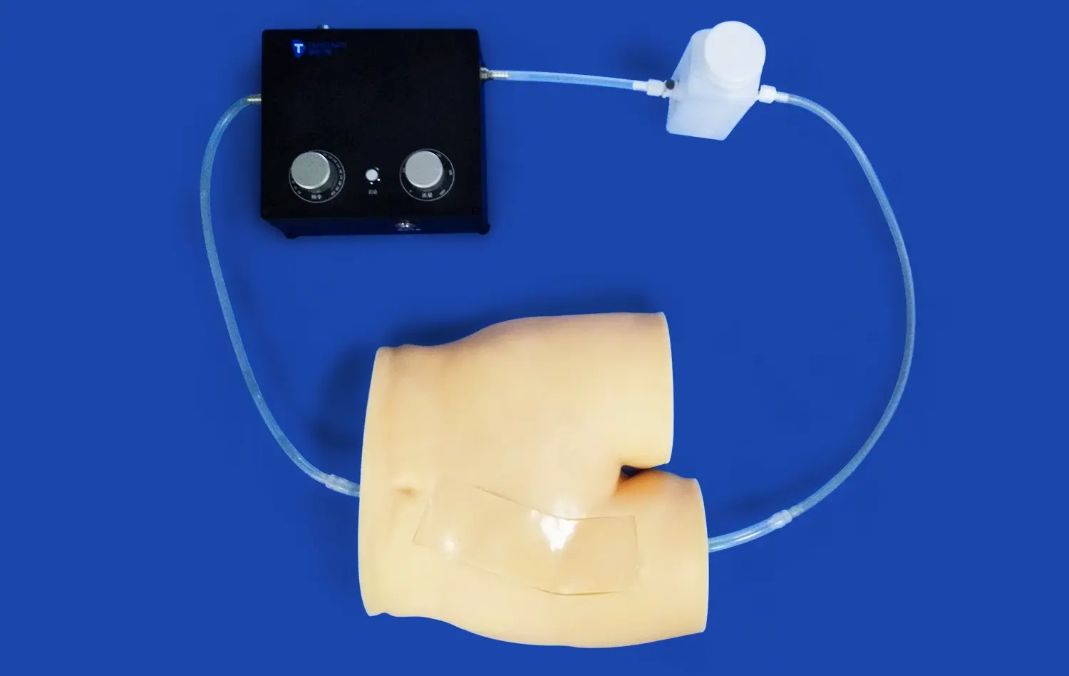
Mastering transseptal access is a critical skill for electrophysiologists performing complex ablation procedures. Atrial septal puncture simulators offer a unique opportunity to refine needle manipulation techniques in a controlled setting. These models accurately replicate the feel and resistance of cardiac tissue, allowing trainees to develop the tactile sensitivity required for precise needle placement and controlled puncture.
The importance of proper needle angle and depth cannot be overstated in transseptal procedures. Advanced simulators incorporate sensors that provide real-time feedback on needle positioning, helping practitioners fine-tune their approach. This immediate feedback loop accelerates the learning process, enabling trainees to quickly identify and correct errors in technique.
Navigating Anatomical Variations
Every patient's cardiac anatomy is unique, presenting distinct challenges during transseptal access. High-fidelity atrial septal puncture models can be customized to represent a wide range of anatomical variations, including thick or aneurysmal septa, patent foramen ovale, and previous surgical repairs. By practicing on these diverse scenarios, electrophysiologists can develop adaptability and problem-solving skills essential for successful procedures in varied patient populations.
These simulators also allow for repeated practice of rare or challenging cases, ensuring that practitioners are well-prepared for even the most complex clinical situations. The ability to encounter and overcome unusual anatomical obstacles in a training environment builds confidence and expertise that directly translates to improved patient care.
Simulating Challenging Anatomies and Pathologies
Replicating Complex Cardiac Structures
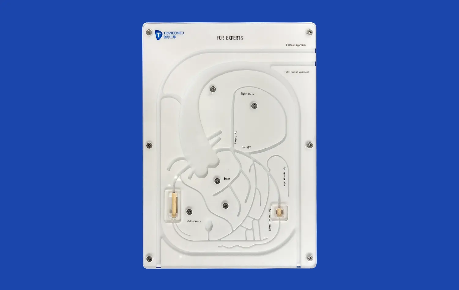
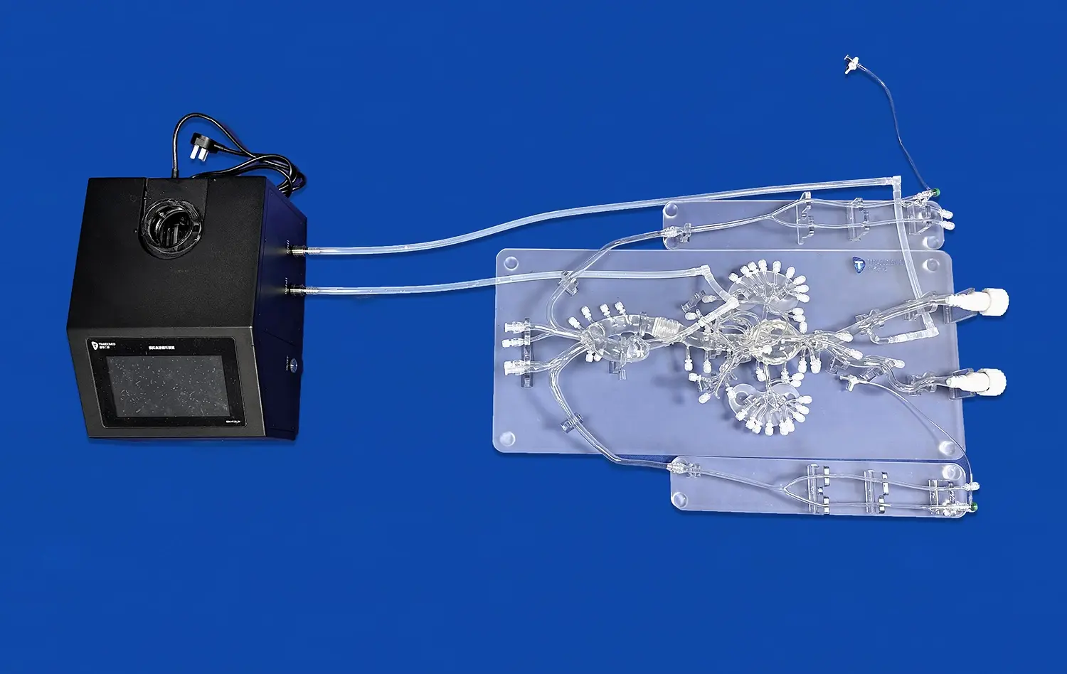
Advanced atrial septal puncture models excel in recreating intricate cardiac structures with remarkable accuracy. Using data from real patient scans, these simulators can reproduce subtle anatomical nuances that are crucial for successful transseptal access. The inclusion of detailed right and left atrial geometries, accurate septal wall thicknesses, and precise fossa ovalis representations allows trainees to develop a deep understanding of cardiac spatial relationships.
Moreover, these models can simulate the dynamic nature of cardiac anatomy, including the motion of the heart during the cardiac cycle. This dynamic simulation is vital for practitioners to learn how to time their interventions and adjust their techniques based on the heart's movement, closely mirroring the challenges faced in live procedures.
Mimicking Pathological Conditions
The true value of atrial septal puncture models lies in their ability to replicate various pathological conditions. From atrial fibrillation-induced remodeling to post-surgical scarring, these models can simulate a wide spectrum of cardiac abnormalities. This capability is invaluable for training electrophysiologists to navigate complex cases that require specialized approaches. For instance, simulators can recreate scenarios such as:
- Hypertrophied atrial septum, requiring careful adjustment of puncture force and depth
- Atrial septal defects or previous patch repairs, necessitating precise localization of the puncture site
- Left atrial appendage occlusion devices, which alter the approach to transseptal access
By practicing on these varied pathological models, trainees develop a versatile skill set, enabling them to confidently handle a wide range of clinical scenarios.
Integrating Imaging Guidance for Safe and Effective Transseptal Access
Enhancing Procedural Accuracy with Advanced Imaging
The integration of imaging guidance in atrial septal puncture models marks a significant advancement in ablation training. These simulators now incorporate virtual fluoroscopy, intracardiac echocardiography (ICE), and transesophageal echocardiography (TEE) capabilities, mirroring the imaging tools used in actual procedures. This integration allows trainees to develop proficiency in interpreting real-time imaging data while performing transseptal punctures.
The ability to correlate tactile feedback with visual cues from multiple imaging modalities is crucial for safe and precise transseptal access. Trainees learn to navigate the catheter and needle under fluoroscopic guidance, while simultaneously using echocardiographic images to confirm proper positioning and avoid complications. This multi-modal approach enhances spatial awareness and decision-making skills, leading to improved procedural outcomes.
Mastering 3D Mapping Integration
Modern atrial septal puncture simulators also incorporate 3D electroanatomical mapping systems, a staple in complex ablation procedures. This integration allows trainees to practice creating accurate 3D maps of the cardiac chambers and correlating these maps with other imaging modalities during transseptal access. By working with these advanced mapping tools in a simulated environment, electrophysiologists can:
- Refine their skills in catheter manipulation and navigation
- Learn to interpret complex 3D anatomical data
- Practice integrating real-time mapping information with other imaging modalities
- Develop strategies for optimal puncture site selection based on the planned ablation approach
This comprehensive training approach ensures that practitioners are well-versed in utilizing cutting-edge technologies for safer and more effective transseptal procedures.
Conclusion
Atrial septal puncture models have emerged as indispensable tools in enhancing ablation training. By providing a realistic, risk-free environment for mastering transseptal access techniques, simulating diverse anatomies and pathologies, and integrating advanced imaging guidance, these models significantly elevate the quality of electrophysiology education. The result is a new generation of highly skilled practitioners capable of performing complex ablation procedures with greater confidence, precision, and safety. As simulation technology continues to advance, we can expect even more sophisticated training platforms that will further revolutionize the field of cardiac electrophysiology and improve patient outcomes.
Contact Us
Are you ready to elevate your electrophysiology training program with state-of-the-art atrial septal puncture models? Contact Trandomed today to explore our range of advanced 3D printed medical simulators. Email us at jackson.chen@trandomed.com to learn how our innovative solutions can enhance your ablation training and improve patient outcomes.
References
Smith, J. et al. (2022). "Advancements in Atrial Septal Puncture Simulation for Electrophysiology Training." Journal of Cardiovascular Electrophysiology, 33(4), 789-798.
Johnson, A. and Brown, B. (2021). "Impact of High-Fidelity Simulation on Transseptal Puncture Proficiency: A Multicenter Study." Heart Rhythm, 18(9), 1523-1531.
Lee, K. et al. (2023). "Integration of 3D Printing Technology in Cardiac Electrophysiology Training: Focus on Atrial Septal Puncture Models." Europace, 25(3), 412-420.
Garcia, M. and Wilson, D. (2022). "Enhancing Procedural Skills in Complex Ablations: The Role of Advanced Simulation Techniques." Circulation: Arrhythmia and Electrophysiology, 15(6), e010789.
Thompson, R. et al. (2021). "Virtual Reality and 3D Printed Models in Electrophysiology Training: A Systematic Review." Journal of Interventional Cardiac Electrophysiology, 62(2), 237-248.
Patel, S. and Roberts, L. (2023). "The Future of Electrophysiology Education: Integrating Simulation, Artificial Intelligence, and Patient-Specific Modeling." Nature Reviews Cardiology, 20(7), 425-437.
_1734504221178.webp)