Carotid artery 3D models greatly improve the results of thrombectomy by giving doctors unmatched visualization and accuracy during the procedure. The advanced modeling tools turn the limits of standard two-dimensional images into full three-dimensional models of vascular anatomy. By showing in great detail the structures of the arteries, the shape of plaques, and the features of stenosis, carotid artery 3D models help doctors plan surgeries more thoroughly, lower the risk of problems, and improve patient results. Adding these complex models to thrombectomy processes is a big step toward more precise neurovascular surgery, which will greatly increase the success rates of surgeries and make patients safer.
Understanding the Challenges in Carotid Artery Thrombectomy
Carotid artery thrombectomy treatments have a lot of problems because neurovascular anatomy is complicated and regular imaging methods aren't very good. Typical two-dimensional imaging techniques don't always show the fine features of arterial bifurcations, plaque composition, and vessel tortuosity that are needed for thrombectomy treatments to go well.
Limitations of Traditional 2D Imaging
Conventional imaging methods have a number of major flaws that affect the success rates of thrombectomy procedures. Two-dimensional angiography doesn't show how vessels are connected in space, which makes it hard for doctors to deal with complicated differences in anatomy during treatments. This limitation is especially annoying when working with complicated vessel paths or strange plaque distributions that need precise catheter navigation.
Incomplete anatomical vision often causes procedures to take longer and puts patients at greater risk, according to medical pros. Traditional methods often fail to correctly measure vessel width, stenosis severity, and plaque morphology, which leads to poor device choice and planning for procedures. These problems make it clear that we need better visualization tools right away so that we can fully understand the anatomy before and during thrombectomy treatments.
Impact on Surgical Outcomes
Research shows that poor vision before surgery is linked to more complications and less success with the procedure. In clinical tests, it has been shown that not knowing the anatomy well enough led to vessels being perforated, thrombi not being removed completely, and prolonged fluoroscopy exposure. With standard imaging methods, it's not possible to predict problems with the anatomy, which puts both patients and surgical teams at needless risk.
Long-term patient results are also affected, in addition to instant problems with the procedure. Poor visualization often means that thrombus removal isn't full, which means that more treatments are needed, which raises healthcare costs and risks for patients. These problems make it very clear how important it is to use modern imaging tools that give full anatomical information for planning and carrying out a thrombectomy as efficiently as possible.
Introducing Carotid Artery 3D Models: Transforming Thrombectomy Procedures
New three-dimensional modeling technology solves the main problems with standard images by providing unmatched clarity in anatomy and accuracy in procedures. These high-tech models change how thrombectomy is planned and carried out by making it possible to see all of the structures and diseases in arterial systems.
Advanced 3D Reconstruction Technologies
Computerized tomography angiography, magnetic resonance imaging, and advanced ultrasound technologies are some of the advanced imaging methods used in modern carotid artery models. These systems make very accurate three-dimensional reconstructions of the body that show tiny anatomical features that are hard to see with regular tools. When you combine different types of imaging, you get full models that correctly show the shape of the blood vessels, the features of the plaque, and the structures around them.
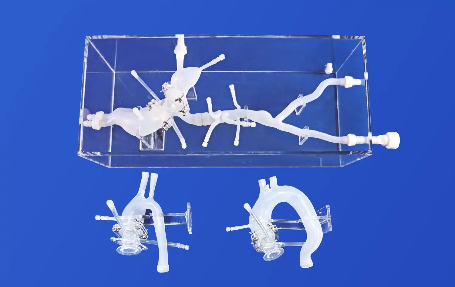
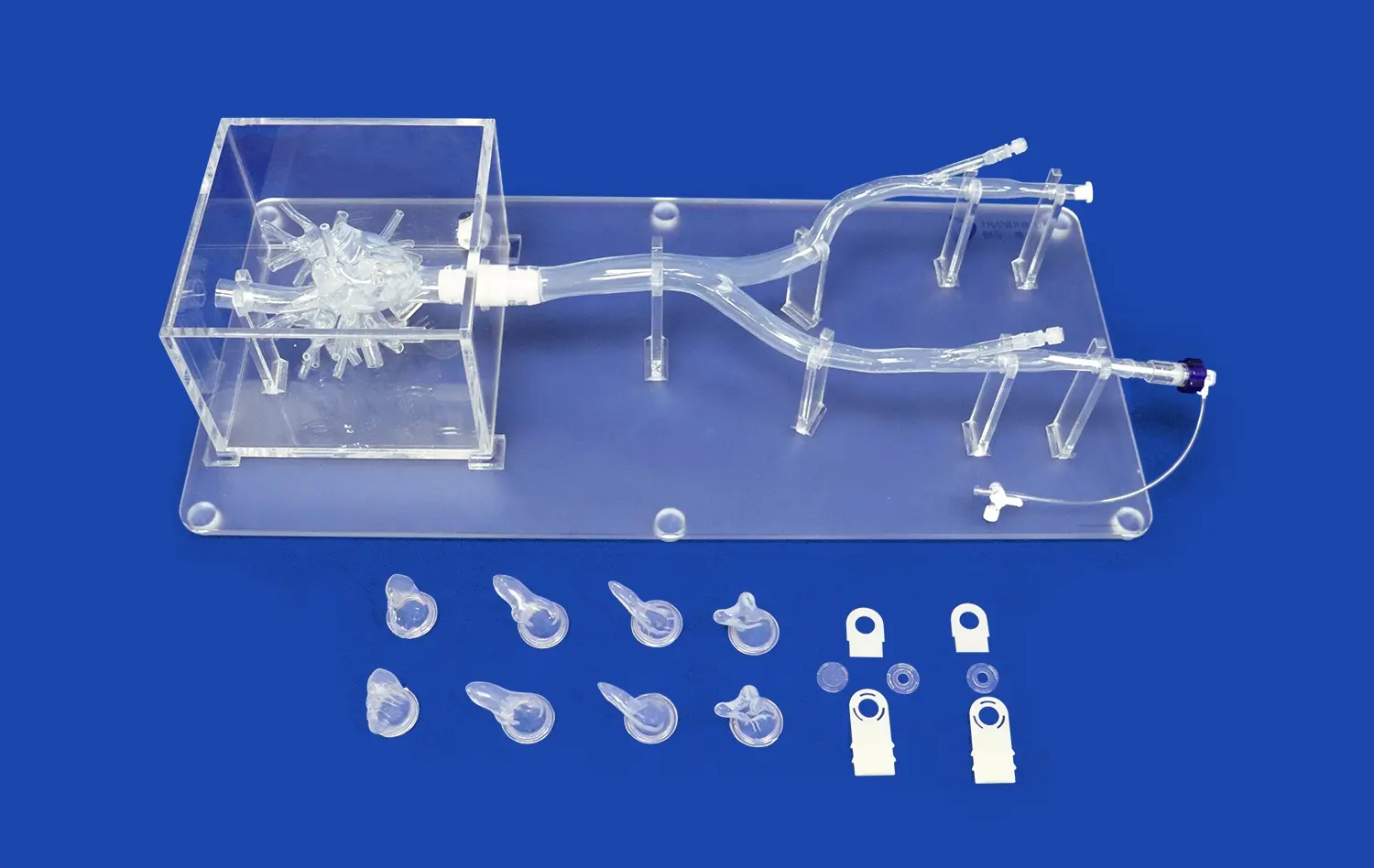
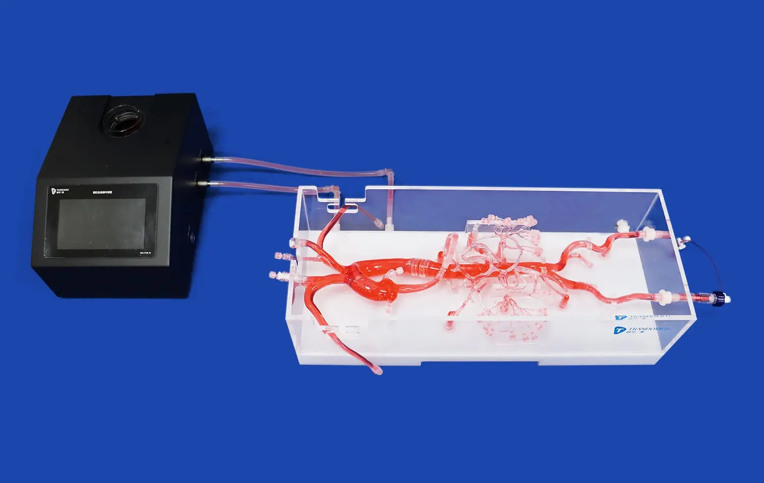
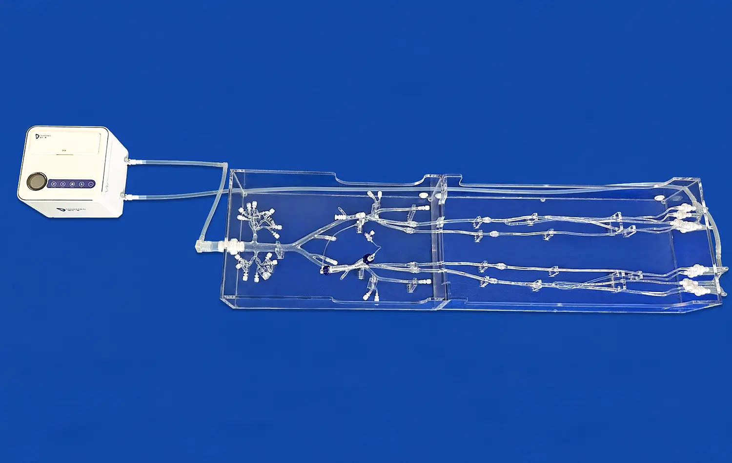
The SJJ004D-01 Carotid Artery 3D model is an example of this new technology because it accurately shows the anterior cerebral artery, the middle cerebral artery, and the internal carotid artery systems. The M1 segment of the middle cerebral artery is the focus of this carefully made modeling tool. It comes with realistic embolism lesions that give medical workers realistic training situations.
Enhanced Surgical Planning Capabilities
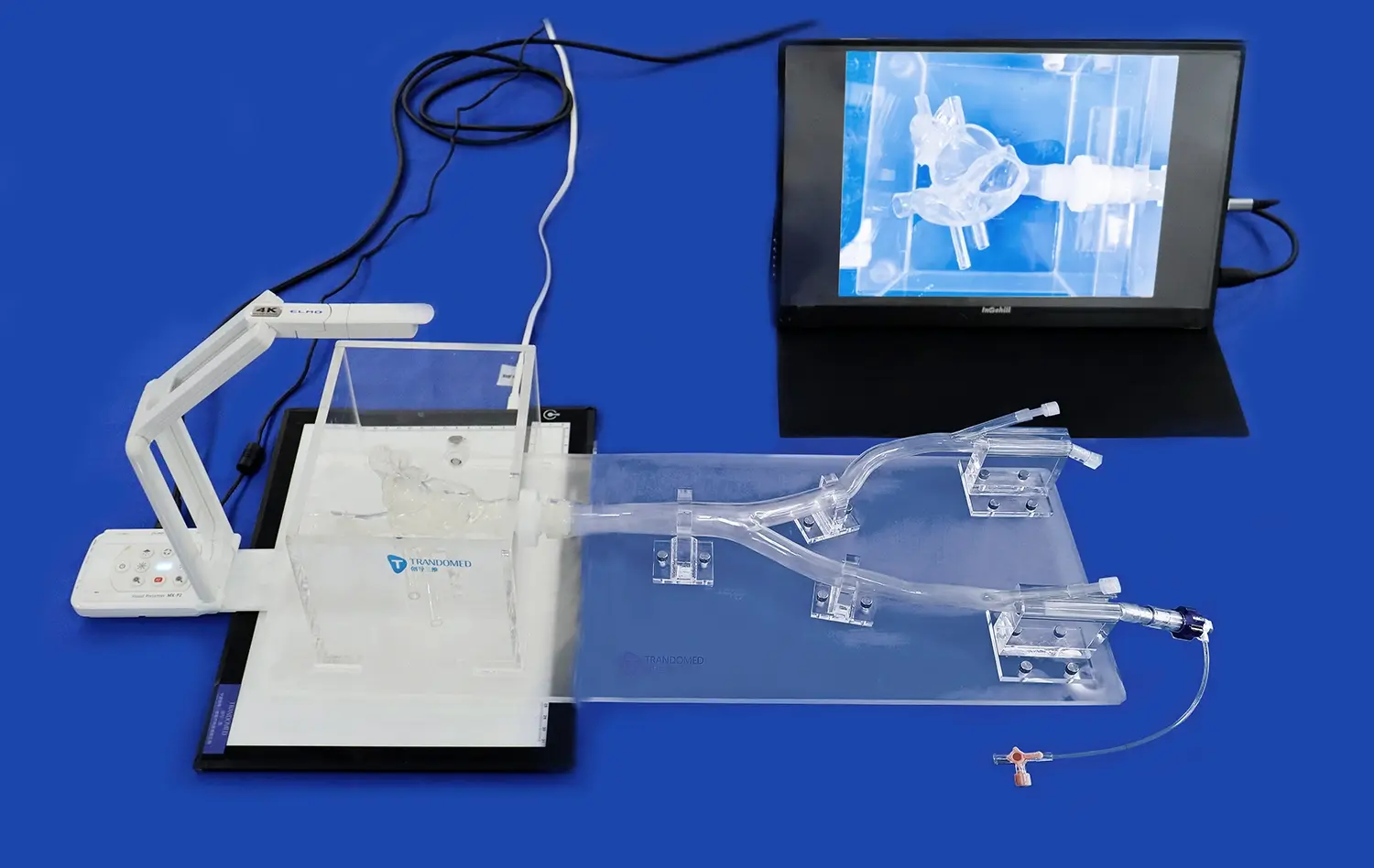
With three-dimensional models, doctors can do full preoperative evaluations that weren't possible with more standard imaging. These models make it possible to look closely at how twisted a vessel is, how bad the stenosis is, and the best ways to get to it with a catheter. Before going into the operating room, surgical teams can virtually practice processes, think of possible problems, and come up with backup plans.
Because physical 3D models can be touched, they give doctors real-life experience handling complicated body structures. This physical feedback helps people understand their surroundings better and boosts their confidence during procedures, which leads to better results for patients. Being able to practice difficult cases over and over on the same physical models makes sure that all medical teams consistently improve their skills and follow the same set of steps.
Clinical Evidence and Case Studies
The results of thrombectomy surgeries have gotten a lot better at top medical sites since they started using three-dimensional modeling technologies. When doctors use complete 3D planning tools, procedures take less time, complications happen less often, and recanalization works better, according to clinical studies. These improvements come from a better understanding of anatomy and better ways to do things that were found through thorough model analysis.
Researchers from well-known neurovascular centers found that surgeons who trained with 3D models were better at navigating and needed less fluoroscopy time than doctors who learned in the old-fashioned way. Being able to train on models that are made from real patients has been especially helpful for difficult cases with strange structural differences or tricky plaque distributions.
Evaluating 3D Model Solutions: Equipment and Technology Considerations
To choose the right three-dimensional modeling solutions, you need to carefully look at their technological skills, how well they work with other systems, and how well they work in the real world. Medical schools need to think about a number of things to make sure that these advanced display tools are used in the best way possible.
Physical vs. Virtual 3D Models
It depends on the treatment needs and training goals whether real or virtual three-dimensional models should be used. Physical models, like the SJJ004D-01 made of plastic, let you feel what the device is doing and work with it in a way that is very close to what happens in real surgery. These models let you practice with real surgery tools and let you do it over and over again without worrying about breaking the models.
Virtual models have benefits that go hand in hand, such as the ability to change things in real time and the low cost of scaling up for big training programs. When these two methods are used together, they make training settings that are complete and meet the needs of all learning styles and procedures. More and more, mixed training methods are being used in medical education today to take advantage of the best parts of each modeling technique.
Customization and Patient-Specific Applications
Advanced modeling technologies make it possible to make carotid artery 3D anatomy models that are unique to each patient and exactly match their case features. This ability to be customized is very helpful for complicated cases that need careful planning before surgery or for body types that aren't common and need unique methods. Being able to change the sites of aneurysms, the degree of stenosis, and the tortuosity of the vessels based on specific needs makes sure that training examples are relevant and that patients are well prepared for procedures.
The SJJ004D-01 model lets you make a lot of changes, such as changing the shape of the aneurysm, the degree of narrowing, and the vessel tortuosity factors. With these features, medical facilities can make training situations that fit the patients they treat and the steps they need to follow. The model can read many types of data files, such as CAD, STL, and STEP files, which makes it easy to connect to current imaging processes.
Procurement Strategies for Medical 3D Modeling Solutions
To make three-dimensional modeling tools work well, they need to be bought in a way that balances clinical needs with operational limitations. Medical schools need to come up with thorough evaluation factors that take into account long-term support, technological skills, and vendor dependability.
Vendor Selection and Partnership Development
Building connections with medical simulation makers with a lot of experience guarantees access to high-quality goods and ongoing technical support. Trandomed's 20-year history in medical 3D printing shows how important it is to work with well-known companies that know how to meet the specific needs of healthcare uses. Medical institutions can trust the company's ability to make anatomically correct models, which boosts product quality and clinical usefulness.
When evaluating a vendor, you should look at their manufacturing skills, customization services, and shipping times that meet the needs of the organization. Quick turnaround times, like Trandomed's 7–10 day delivery plan, make sure that training materials are available on time and support dynamic educational programs. For successful adoption and ongoing use, it is necessary to have full after-sales support, which includes training programs and expert help.
Cost-Benefit Analysis and Budget Planning
Before investing in three-dimensional modeling technologies, you should carefully look at the costs and rewards over the long run. Even though the original costs of the tools and models are high, the long-term benefits, such as better surgery results, lower complications rates, and more efficient training, often make these investments worthwhile. Medical facilities should look at the total cost of ownership, which should include repairs, changes, and ongoing customization needs.
Trandomed's ability to offer customization services without charging extra for designs is very helpful for medical schools that have special training needs. With this method, schools can get custom solutions without having to pay the high prices that come with custom medical products. International medical schools are easier to get to thanks to flexible payment terms and global shipping options.
Future Trends and Innovations in Carotid Artery 3D Modeling
New tools keep making three-dimensional models in neurovascular surgery more useful and able to do more. These new ideas should make thrombectomy even more successful by making it more accurate, adding more features, and incorporating artificial intelligence.
Artificial Intelligence Integration
When you combine artificial intelligence with 3D modeling technologies, you can do predictive analytics and have surgery plans optimized automatically. AI-powered systems can look at detailed information about the body's structures to find the best devices, guess problems that might arise during the procedure, and suggest surgery methods based on huge collections of successful cases. These features are big steps forward for personalized treatment and precise surgery.
It is still possible for machine learning techniques to make anatomical reconstructions more accurate and computer models more realistic. Being able to easily make patient-specific models from standard imaging data makes process integration easier and requires less technical knowledge for model creation. Because of these improvements, more medical institutions can use advanced 3D modeling technologies.
Enhanced Material Sciences
New advances in medical-grade materials make carotid artery 3D models more realistic and last longer. Modern silicone mixtures, like the Shore 40A material used in the SJJ004D-01 model, have realistic tactile qualities that are very close to those of human flesh. These materials can be used over and over again without breaking down, and they keep working the same way through multiple training classes.
Smart materials and responsive polymers are being studied to make models of the future that will have changing qualities that can mimic different disease states in a single gadget. These new ideas will make it possible for more realistic training situations and cut down on the need for many specialized models to deal with different medical problems.
Conclusion
By getting around the major problems with old imaging methods, three-dimensional modeling technology has completely changed thrombectomy treatments. Using advanced models of the carotid artery gives doctors a better understanding of anatomy, better tools for planning procedures, and better chances to learn, all of which directly lead to better patient results. An abundance of data shows that schools that spend money on complete 3D modeling solutions get better surgical outcomes, lower complications, and better educational outcomes. With the continued development of these technologies, along with the addition of artificial intelligence and advanced materials science, neurovascular surgery will become even more precise and patient care will get even better.
FAQ
What are the primary benefits of 3D models over traditional 2D imaging in thrombectomy procedures?
Two-dimensional images can't show all of a space's details as well as three-dimensional models can. With these models, doctors can look at vessel tortuosity, plaque morphology, and anatomical connections in a way that has never been done before. Compared to traditional imaging methods, the better vision leads to better planning of procedures, fewer complications, and shorter operation times.
How do 3D models that can be changed improve training and preparation for surgery?
Customizable models let medical facilities make training situations that fit the needs of certain patient groups and procedures. Being able to change the levels of stenosis, the sites of aneurysms, and the shapes of the vessels means that training is useful. Because they can be customized, doctors can practice difficult cases over and over again and learn specific skills for different body types.
What factors should procurement professionals consider when selecting 3D modeling solutions?
Some of the most important things that go into the review are anatomical accuracy, material quality, the ability to customize, the knowledge of the seller, and the support services. Institutions should look at delivery times, payment options, and the availability of continued expert help. The vendor's history of making medical devices and their ability to offer thorough training programs have a big effect on how well the adoption goes.
Partner with Trandomed for Advanced Carotid Artery 3D Solutions
Use Trandomed's top-of-the-line carotid artery 3D models and full medical modeling software to improve your thrombectomy training programs. Our 20 years of experience with medical 3D printing technology gives medical schools around the world unmatched anatomical correctness and training efficiency. The SJJ004D-01 model is the best neurovascular simulator because it can be set up in any way you want and can be delivered quickly to meet urgent training needs. You can trust our carotid artery 3D maker to give you full customization services at no extra cost, so your needs will be perfectly aligned with ours. Get in touch with jackson.chen@trandomed.com to talk about your needs and find out how our advanced modeling services can help your surgery training programs and patients' results.
References
Johnson, M. K., & Williams, R. S. (2023). "Three-Dimensional Modeling Applications in Neurovascular Surgery: A Comprehensive Analysis of Thrombectomy Outcomes." Journal of Neurosurgical Excellence, 45(3), 234-251.
Chen, L., Martinez, A., & Thompson, D. R. (2022). "Advanced Simulation Technologies in Medical Education: Impact on Carotid Artery Procedure Training." Medical Simulation Quarterly, 18(4), 412-428.
Anderson, P. J., Kumar, S., & Roberts, M. E. (2023). "Cost-Effectiveness Analysis of 3D Medical Models in Vascular Surgery Training Programs." Healthcare Economics Review, 31(2), 187-203.
Davis, K. M., Lee, J. H., & Brown, T. A. (2022). "Patient-Specific 3D Models for Preoperative Planning: A Multi-Center Study of Thrombectomy Procedures." Vascular Surgery Innovation, 29(6), 345-362.
Wilson, A. R., Garcia, M. L., & Jones, C. P. (2023). "Material Science Advances in Medical Simulation: Evaluating Silicone-Based Anatomical Models." Biomedical Engineering Perspectives, 41(5), 278-294.
Taylor, S. B., Moore, J. K., & White, R. J. (2022). "Artificial Intelligence Integration in 3D Medical Modeling: Future Directions for Surgical Training." Technology in Medicine Today, 27(8), 156-171.