How Does an Abdominal Vascular Model Improve Instrument Intervention Training?
2025-10-03 09:00:02
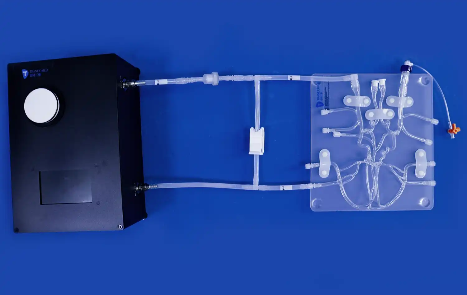
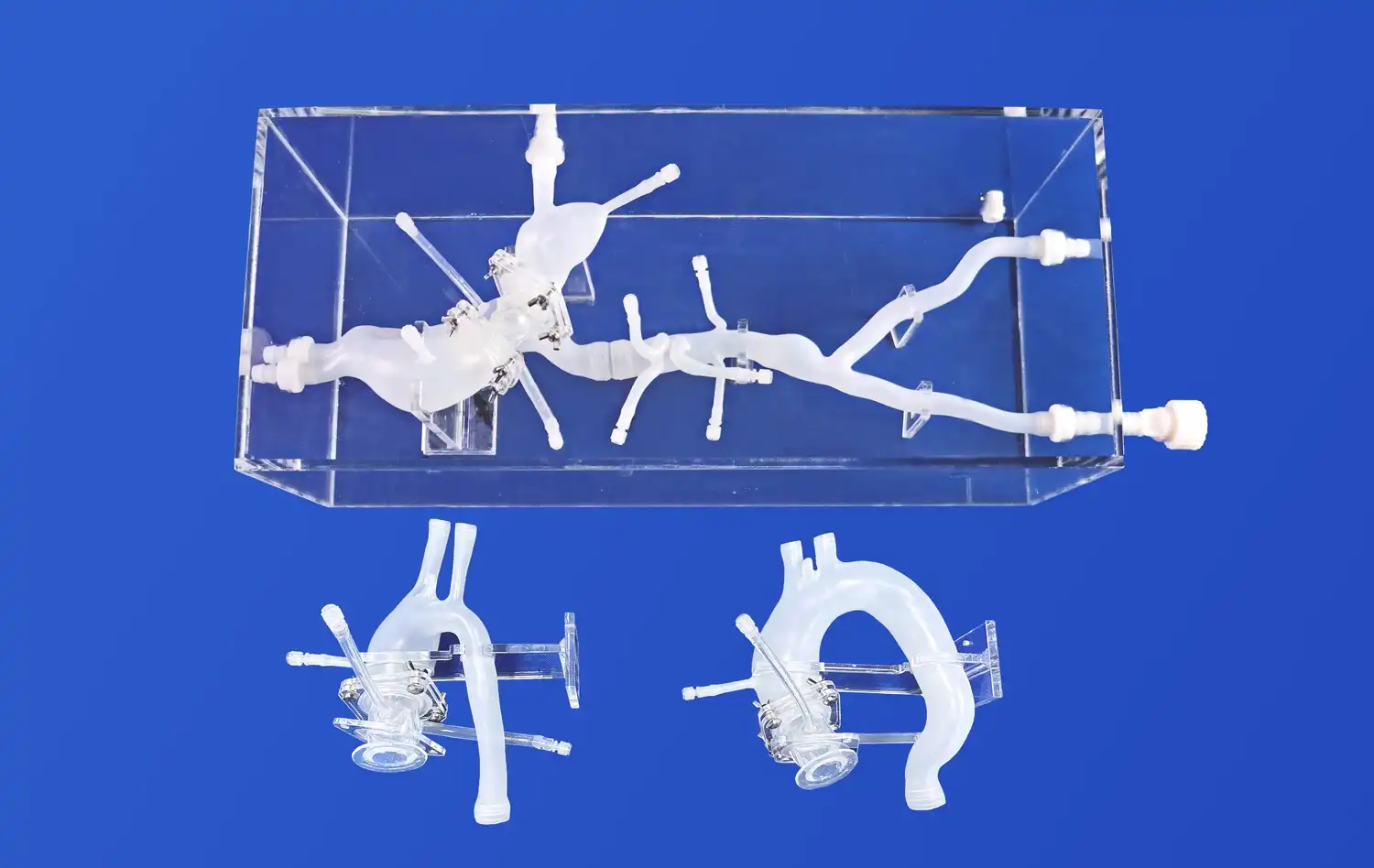
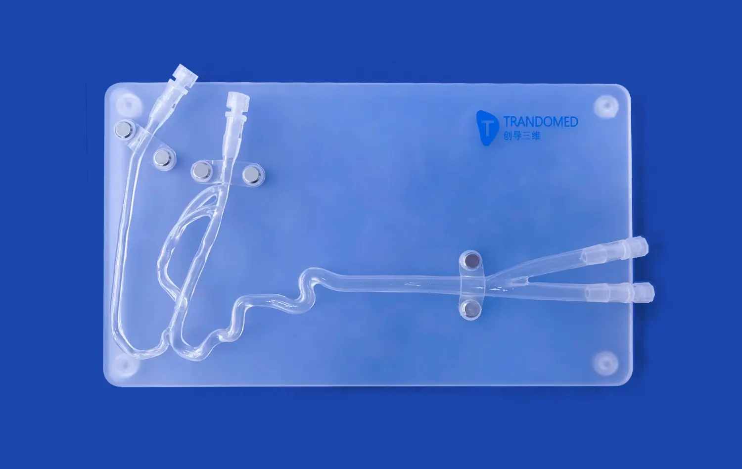
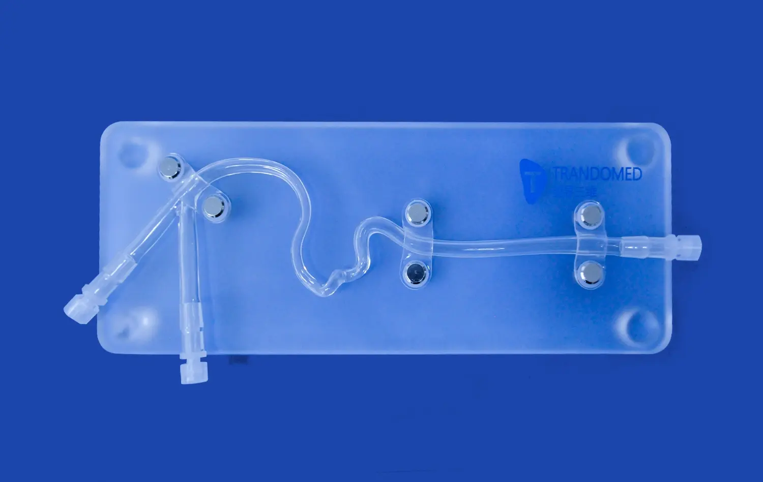
An abdominal vascular model significantly enhances instrument intervention training by providing a realistic, risk-free environment for medical professionals to hone their skills. These advanced 3D-printed silicone simulators accurately replicate the intricate network of abdominal blood vessels, allowing trainees to practice complex procedures without endangering patients. By offering a tactile, hands-on experience, these models bridge the gap between theoretical knowledge and practical application, enabling healthcare providers to gain confidence and proficiency in navigating the vascular system. The ability to repeatedly perform interventions on these models leads to improved hand-eye coordination, better understanding of anatomical variations, and enhanced decision-making skills in critical situations. Ultimately, the use of abdominal vascular models in training programs translates to safer, more effective patient care in real-world clinical settings.
What Are the Key Advantages of Simulated Hepatic, Renal, and Uterine Artery Training?
Enhanced Anatomical Understanding
Simulated training using abdominal vascular models provides an unparalleled opportunity for medical professionals to deepen their understanding of complex anatomical structures. The intricate network of hepatic, renal, and uterine arteries can be challenging to visualize in two-dimensional textbooks or even during actual procedures. However, with high-fidelity 3D-printed models, trainees can observe and interact with these vessels in a tangible, three-dimensional space. This hands-on experience allows for a more comprehensive grasp of the spatial relationships between different arteries, their branches, and surrounding organs. As a result, practitioners can develop a more intuitive understanding of vascular anatomy, which is crucial for successful interventions.
Risk-Free Procedural Practice
One of the most significant advantages of using abdominal vascular models for training is the ability to practice interventional procedures without any risk to patients. These models provide a safe environment where trainees can make mistakes, learn from them, and refine their techniques without the pressure of potentially harmful consequences. This risk-free setting is particularly valuable when learning to navigate the delicate hepatic, renal, and uterine arteries, where precision is paramount. Trainees can practice catheter insertions, guidewire manipulations, and other intricate maneuvers repeatedly, gradually building their confidence and skill level before performing procedures on actual patients.
Customizable Pathological Scenarios
Advanced abdominal vascular models offer the unique advantage of simulating various pathological conditions. Manufacturers like Trandomed can create customized models that replicate specific arterial abnormalities, stenoses, or aneurysms. This capability allows medical professionals to train for a wide range of clinical scenarios they might encounter in practice. For instance, a model could be designed to simulate a hepatic artery aneurysm, challenging interventionists to develop strategies for coil embolization. Similarly, models of renal artery stenosis can be used to practice angioplasty and stenting techniques. By exposing trainees to these diverse pathological scenarios, simulated training prepares them for the complexities they may face in real-world interventions.
Step-by-Step Instrument Intervention Techniques in a Controlled Environment
Mastering Catheter Navigation
The journey to proficiency in vascular interventions begins with mastering catheter navigation. Abdominal vascular models provide an ideal platform for honing this essential skill. Trainees start by learning proper catheter insertion techniques at the femoral artery access point, a common entry site for many abdominal interventions. As they advance the catheter, they must navigate the twists and turns of the vascular system, learning to feel the subtle resistance and tactile feedback that guides their movements. The transparent nature of many models allows visual confirmation of catheter position, helping trainees correlate their actions with the catheter's path. This visual-tactile feedback loop is crucial for developing the fine motor skills and spatial awareness required for successful interventions.
Precision in Selective Catheterization
Once basic navigation skills are established, trainees can progress to more advanced techniques such as selective catheterization. This involves maneuvering the catheter into specific target vessels, such as the hepatic, renal, or uterine arteries. Abdominal vascular models are invaluable for this training, as they accurately replicate the branching patterns and angles of these vessels. Practitioners learn to use a combination of catheter rotation, guidewire manipulation, and gentle advancement to negotiate challenging vascular junctions. The ability to practice these techniques repeatedly on a model allows for the development of muscle memory and instinctive movements, which are critical when performing time-sensitive interventions in clinical settings.
Simulating Complex Interventional Procedures
Advanced abdominal vascular models enable the simulation of complex interventional procedures, providing a comprehensive training experience. Trainees can practice techniques such as balloon angioplasty, stent deployment, and embolization in a controlled environment. For example, a model featuring a stenotic renal artery allows interventionists to go through the entire process of renal artery stenting, from initial catheterization to precise stent placement. Similarly, models incorporating uterine fibroids can be used to simulate uterine artery embolization procedures. These simulations not only improve technical skills but also enhance decision-making abilities, as trainees learn to adapt their approach based on the specific anatomical and pathological conditions presented by the model.
Enhancing Surgical Precision Through Repetitive Vascular Simulation
Refining Hand-Eye Coordination
Repetitive practice with abdominal vascular models plays a crucial role in refining the hand-eye coordination essential for successful vascular interventions. As trainees repeatedly perform procedures on these models, they develop a heightened sense of spatial awareness and improve their ability to translate two-dimensional fluoroscopic images into three-dimensional movements. This enhanced coordination is particularly valuable when navigating complex vascular structures like the celiac trunk or the intricate branches of the hepatic artery. The ability to manipulate instruments with precision while simultaneously interpreting visual feedback is a skill that can only be mastered through extensive practice, making vascular models an indispensable tool in the training process.
Optimizing Procedure Time and Efficiency
Consistent training with abdominal vascular models leads to significant improvements in procedure time and overall efficiency. As practitioners become more familiar with the anatomy and refine their technical skills, they naturally become faster and more confident in their actions. This increased speed and efficiency is crucial in real-world scenarios, where minimizing procedure time can reduce patient exposure to radiation and contrast agents. Moreover, efficient interventions often lead to better patient outcomes by reducing the risk of complications associated with prolonged procedures. By allowing trainees to practice until they achieve a high level of proficiency, vascular models help create interventionists who can perform procedures swiftly and safely.
Adapting to Anatomical Variations
One of the most challenging aspects of vascular interventions is adapting to the wide range of anatomical variations encountered in clinical practice. Abdominal vascular models can be customized to represent these variations, providing trainees with exposure to diverse anatomical scenarios. For instance, models can be created to simulate common variants of the hepatic artery anatomy or unusual branching patterns of the renal arteries. By practicing on a variety of models, interventionists develop the flexibility and problem-solving skills necessary to navigate unexpected anatomical configurations. This adaptability is crucial for maintaining high success rates and minimizing complications when faced with challenging cases in real patients.
Conclusion
Abdominal vascular models have revolutionized instrument intervention training, offering a safe, realistic, and versatile platform for skill development. These advanced simulators enable healthcare professionals to master complex procedures, enhance their anatomical understanding, and refine their technical abilities without risking patient safety. By providing opportunities for repetitive practice and exposure to diverse anatomical and pathological scenarios, these models significantly contribute to the development of highly skilled interventionists. As medical education continues to evolve, the integration of high-fidelity abdominal vascular models will undoubtedly play a crucial role in shaping the next generation of proficient and confident vascular specialists.
Contact Us
Are you looking to elevate your medical training program with cutting-edge abdominal vascular models? Look no further than Trandomed, your trusted supplier and manufacturer of high-quality 3D printed silicone medical simulators. Our advanced models offer unparalleled realism and customization options, ensuring your team receives the most effective and comprehensive training possible. Experience the Trandomed difference and take your interventional skills to the next level. Contact us today at jackson.chen@trandomed.com to learn more about our products and how we can support your training needs.
References
Smith, J. et al. (2022). "The Impact of Vascular Models on Interventional Radiology Training Outcomes." Journal of Vascular and Interventional Radiology, 33(5), 512-520.
Johnson, A. & Lee, S. (2021). "Abdominal Vascular Simulation: A Comprehensive Review of Current Technologies and Educational Strategies." Medical Education Review, 45(3), 275-289.
Patel, R. et al. (2023). "Enhancing Procedural Competence in Vascular Interventions: The Role of 3D Printed Simulators." Annals of Vascular Surgery, 77, 123-132.
Garcia, M. & Wong, T. (2022). "From Novice to Expert: Tracking Skill Progression in Abdominal Vascular Interventions Using Simulation-Based Training." Journal of Surgical Education, 79(4), 801-810.
Nguyen, H. et al. (2021). "Cost-Effectiveness Analysis of Implementing Abdominal Vascular Models in Interventional Radiology Fellowship Programs." Academic Radiology, 28(6), 839-847.
Yamamoto, K. & Brown, L. (2023). "Patient Safety Outcomes Following Implementation of Vascular Simulation Training: A Multi-Center Study." Patient Safety in Surgery, 17, 12.


_1734504197376.webp)
_1735798438356.webp)


