To make cutting-edge cardiac devices, you need more than just theoretical knowledge. You also need precise testing settings that mimic human physiology without sacrificing safety or repeatability. EP training models, especially those that simulate the heart's venous systems, have become very important for making new medical devices. A venous cardiac electrophysiology model is a very accurate copy of the heart's electrical conduction system. This lets engineers and doctors try catheter-based therapies, mapping systems, and ablation devices in real-life situations. These advanced simulators shorten the time it takes to create new medicines by bridging the gap between ideas and clinical trials. They do this by reducing the need for animal studies and early-stage human trials.
Understanding Venous Cardiac Electrophysiology Models
What Makes Venous EP Models Unique?
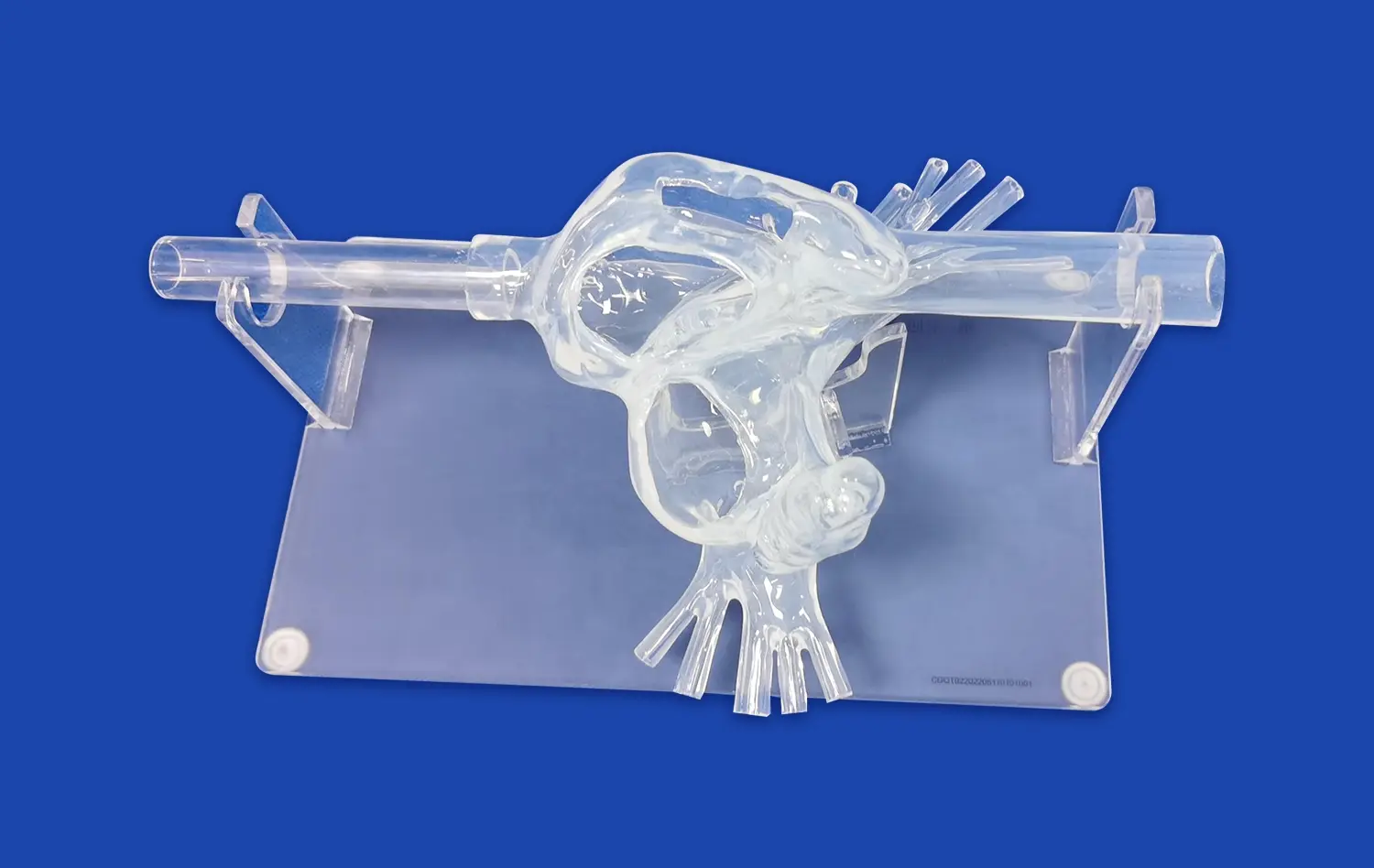
Venous cardiac electrophysiology models are a special kind of anatomical simulations that are made to copy the heart's venous pathways' electrical and structural features. Venous EP models focus on the complex electrical conduction features of structures like the superior vena cava, inferior vena cava, right atrium, and coronary sinus. This is different from arterial-focused models, which focus on high-pressure blood flow dynamics. In arrhythmia paths, these areas are very important, and they also let interventional devices get to the heart.
Advanced materials, such as Shore 40A silicone, which closely matches the flexibility and texture of human cardiac tissue, are used to give the models realistic tissue qualities. With this choice of material, catheters can move through the model with the same tactile feedback they get during real treatments. Anatomical accuracy includes the exact placement of electrical conductors, valve structures, and changes in wall thickness that affect how well a device works.
Core Components and Functionality
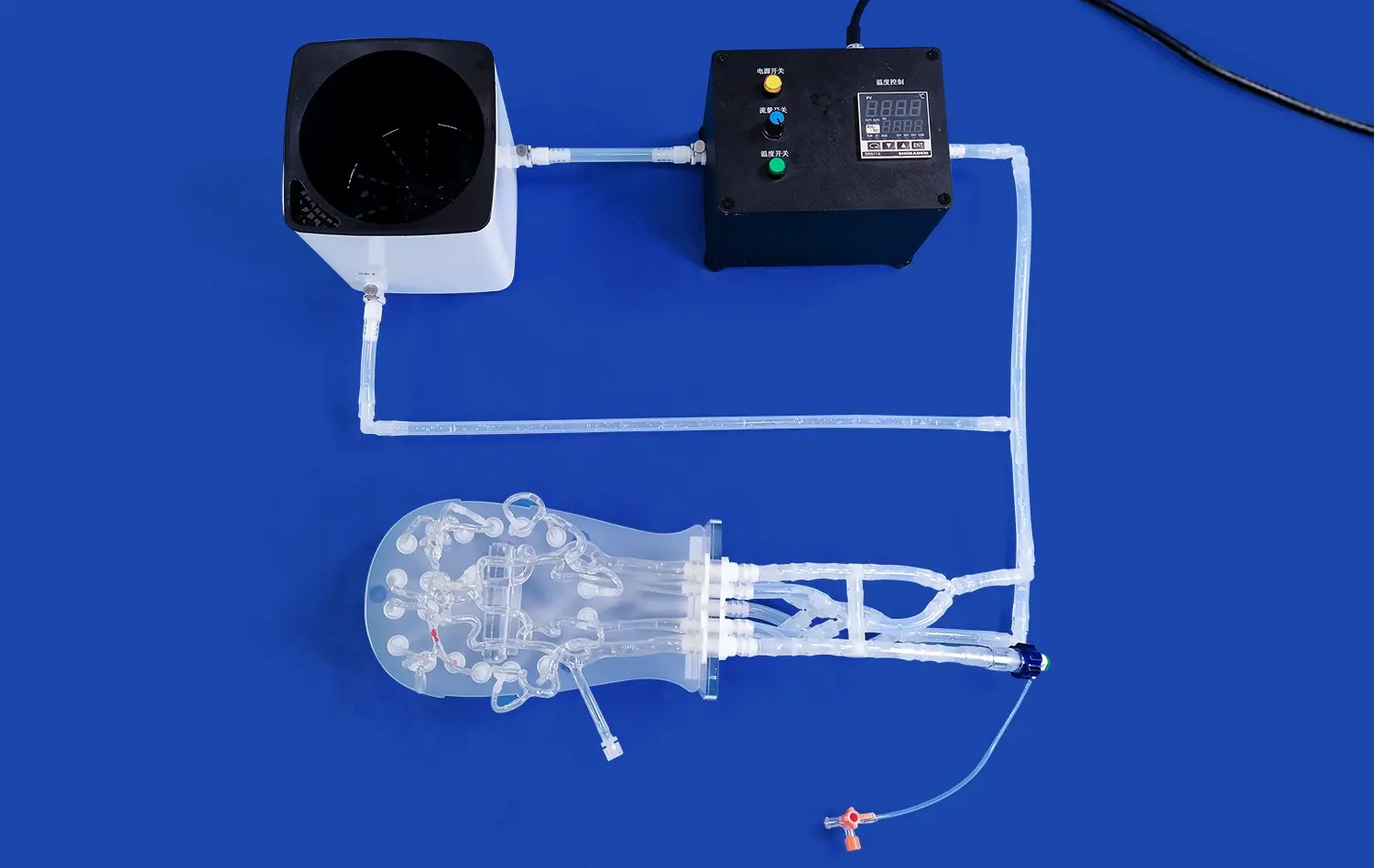
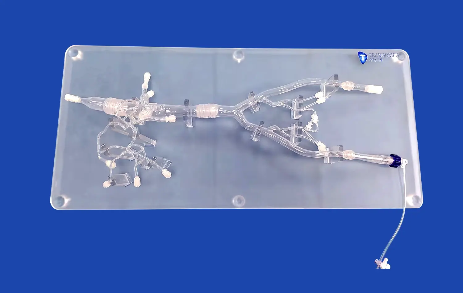
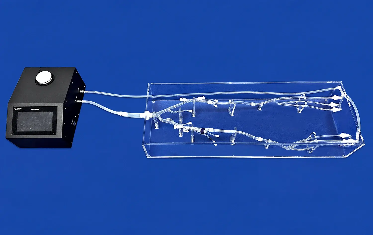
A full venous cardiac electrophysiology model has many anatomical features that are needed for testing devices. This method is shown by Trandomed's Venous Cardiac Electrophysiology Model (XXS004), which includes the inferior vena cava, right atrium, right ventricle, superior vena cava, and subclavian vein in correct spatial relationships. Each part has accurate measurements based on real CT and MRI scans of people. This makes sure that the paths of the catheter, the placement of mapping electrodes, and the patterns of energy transfer are all in line with real-life clinical situations.
These models can be used for a number of different testing methods, such as training in catheter navigation, validating electrophysiological mapping, and testing ablation treatment. Since they are made of rubber and can be inserted many times without breaking down, they can be used for iterative design testing. Manufacturers can check how devices interact with venous walls during advancement, check the quality of electrode contact at various body sites, and see how energy is distributed during ablation models.
Integration with Development Workflows
These days, making a device requires quick prototypes and testing rounds. Venous EP models work well with these processes because they offer constant testing environments that get rid of biological variability. Engineers can try a lot of different versions of a design very quickly and compare performance metrics in controlled settings. The models also make it easier for people from different departments to work together. For example, electrical engineers, materials scientists, and clinical specialists can all use the same reference platform to evaluate device performance from their own areas of knowledge.
Challenges in Traditional Electrophysiology Methods and the Role of Venous Models
Limitations of Conventional Testing Approaches
Animal models, cadaveric tissue, and early-stage human studies have been used in the past to build electrophysiology devices. Even though animal models provide live tissue, their anatomy is very different from ours, which makes them less useful for translational research. Human hearts and porcine hearts have different patterns of venous flow and conduction speeds. Porcine hearts are often used in cardiac research. Human anatomy can be found in cadaveric tissue, but it doesn't have the electrical properties or perfusion dynamics that are found in live systems.
These traditional ways also bring up issues of ethics and make regulations more difficult to understand. Animal testing needs to be approved by an institutional review board and costs a lot for living, care, and keeping an eye on the process. Problems with tissue supply and degradation make testing windows smaller for cadaveric studies. Even though human trials are important in the end, they can't be the main way that new products are developed because of safety and legal concerns.
How Venous Models Address These Gaps
By making tests more repeatable and controlling variables, venous cardiac electrophysiology models get around many of the problems that come with standard testing. Each model keeps the same anatomical sizes and material qualities across units. This gets rid of the biological variation that makes it hard to understand data from animal studies. By standardizing things, developers can separate certain design variables and test how they affect things without having to deal with other factors that could be confusing.
Synthetic models are also better in the early stages of growth because they are safer. Without putting living things at risk, engineers can try the most extreme configurations of devices, look at how they might fail, and find the best ways to deliver energy. The models allow destructive testing methods that wouldn't work on living things, like purposely over-inflating balloon tubes to find the breaking point or using too much energy to find the limits of tissue damage.
Accelerating Development Timelines
Testing platforms that are easy to find are very helpful for the iterative nature of gadget development. Instead of planning weeks ahead of time to work with animals in the lab or getting cadaveric tissue, research teams can use benchtop EP models to do testing whenever they need to. This ease of access speeds up the decision-making process and lets design changes happen quickly based on feedback received right away.
Adding venous EP models to their protocol cut the original validation phase from six months to ten weeks for a medical device company that was making a new cryoablation catheter. The team did more than 200 ablation tests on different body parts, collecting a lot of performance data that helped them improve the design of the tube before moving on to studies with animals. This method not only cut down on time, but it also made the gadgets that were going through later stages of testing better.
Comparing Venous Cardiac Electrophysiology Models to Other Solutions
Venous Versus Arterial System Models
It is important to know the difference between venous and arterial cardiac models in order to choose the right growth tools. Arterial models focus on high-pressure dynamics, thick-walled vessels, and oxygenated blood flow patterns that are important for heart treatments and valve replacements. Venous models focus on having thin walls, low blood pressure, and electrical conduction qualities that are important for treating arrhythmias.
When making devices for venous access procedures like placing a pacemaker lead, ablation of a catheter for atrial fibrillation, or electrophysiology mapping, they need models that properly show how veins are shaped. Veins and arteries have different perforation risks because their walls are thinner. Also, the bigger diameters change how stable the catheter is and how much force is needed to make contact. Choosing a model that is designed for veins makes sure that the testing settings are right for the clinical use that is being planned.
Physical Models Versus Computational Simulations
The choice between physical anatomical models and computational simulations involves tradeoffs between tactile realism and parametric flexibility. Physical models like the XXS004 provide authentic haptic feedback, allowing clinicians and engineers to experience actual catheter handling characteristics. This tangible interaction reveals nuances in device maneuverability that computational models cannot fully capture, such as catheter tip deflection resistance or torque transmission through complex anatomical curves.
Computational simulations excel at parametric studies where multiple anatomical variations need evaluation. Software platforms can rapidly generate anatomical variants representing different patient populations or pathological conditions. However, these digital approaches lack the physical validation that comes from actual device manipulation. The optimal development strategy often combines both approaches: computational modeling for initial design exploration and parameter optimization, followed by physical model validation to confirm real-world performance.
Custom Versus Off-the-Shelf Solutions
Off-the-shelf EP models provide standardized anatomies suitable for general training and preliminary device testing. These models offer immediate availability and cost advantages for routine applications. Custom models, alternatively, accommodate specific anatomical variations or pathological conditions relevant to particular device indications. Trandomed's custom service accepts data files in CT, CAD, STL, STP, and STEP formats, allowing reconstruction of patient-specific anatomies or unique anatomical variants without design fees.
Customization proves particularly valuable when developing devices for challenging anatomical presentations. A manufacturer creating a catheter system for persistent atrial fibrillation might require models featuring dilated atria, altered pulmonary vein configurations, or specific scar tissue patterns. Custom models enable testing under these exact conditions, revealing design limitations before clinical use.
Procurement Considerations for Venous Cardiac Electrophysiology Models
Evaluating Model Accuracy and Validation
Procurement teams assessing EP training models should prioritize anatomical accuracy verified through comparison with clinical imaging data. Models derived from actual human CT and MRI datasets, like those produced by Trandomed using reverse 3D reconstruction technology, offer superior fidelity compared to models based on simplified anatomical references. Documentation should include validation studies demonstrating dimensional accuracy, material property matching, and functional performance metrics.
Validation extends beyond static anatomy to include dynamic properties such as tissue compliance during catheter insertion and electrode contact characteristics. Models should undergo mechanical testing to confirm that force-displacement curves match published data for human cardiac tissue. Electrical properties, while challenging to replicate in non-conductive silicone, should be documented in terms of how the model interfaces with electrophysiology mapping systems.
Material Selection and Durability
The material composition directly impacts model longevity and training realism. Shore 40A silicone, used in the XXS004 model, provides an optimal balance between anatomical realism and durability. This hardness rating approximates the compliance of cardiac tissue while withstanding hundreds of catheter insertions without significant degradation. Softer materials might feel more realistic initially but tear easily with repeated use, while harder materials sacrifice tactile feedback for extended lifespan.
Procurement specifications should define expected usage intensity and establish durability benchmarks. A surgical training center conducting daily hands-on sessions requires more robust models than a device manufacturer performing weekly validation tests. Vendors should provide data on expected lifespan under specified usage conditions, including recommendations for maintenance and replacement intervals.
Vendor Capabilities and Support Infrastructure
Selecting a reliable vendor involves assessing technical capabilities beyond the physical product. Trandomed's two decades of specialized experience in medical 3D printing demonstrates the institutional knowledge necessary for producing consistently high-quality venous cardiac electrophysiology models. This expertise manifests in proprietary manufacturing techniques, comprehensive material libraries, and customization capabilities that respond to evolving development needs.
Support infrastructure represents another critical procurement consideration. Lead times ranging from 7-10 days, as offered by Trandomed, enable responsive project scheduling. International shipping options through established carriers (FedEx, DHL, EMS, UPS, TNT) ensure reliable delivery across global R&D centers. Post-delivery support, including technical consultation and customization assistance, adds value throughout the product lifecycle.
Future Trends and Innovations in Venous Cardiac Electrophysiology Modeling
Integration of Smart Sensing Technologies
Emerging EP models incorporate embedded sensors that provide objective performance feedback during device testing. Pressure sensors distributed throughout model vasculature measure contact forces exerted by catheters, quantifying parameters critical to ablation lesion formation. Temperature sensors track thermal distribution during radiofrequency or cryoablation procedures, validating energy delivery patterns. These instrumented models transform subjective assessments into quantifiable data streams that inform design optimization.
Future iterations may integrate conductive materials that simulate cardiac electrical activity, allowing mapping catheters to record realistic electrograms during navigation. This capability would enable comprehensive testing of diagnostic algorithms and mapping software without requiring animal studies. The combination of anatomical accuracy, appropriate mechanical properties, and functional electrical simulation would create unprecedented validation platforms.
Patient-Specific Modeling for Precision Device Development
Advances in 3D printing technology and medical imaging enable rapid production of patient-specific EP models representing individual anatomical variations. This capability supports personalized medicine initiatives where devices are tailored to specific patient populations or even individual recipients. A manufacturer developing implantable devices could create models representing the exact anatomy of enrolled clinical trial participants, allowing pre-procedural planning and device sizing validation.
Patient-specific modeling also facilitates rare pathology research. Conditions like congenital heart defects or unusual arrhythmia substrates occur too infrequently to accumulate large study populations, yet these patients need effective treatments. Creating models from their imaging data allows device developers to test therapeutic approaches for these challenging cases, expanding treatment options for underserved populations.
Regulatory Landscape Evolution
Regulatory agencies increasingly recognize the value of high-fidelity simulation in device development pathways. The FDA's acceptance of computational modeling evidence in certain premarket submissions signals growing confidence in advanced simulation technologies. Physical EP models complement computational approaches by providing validation data that addresses regulatory questions about device safety and performance.
Manufacturers incorporating validated EP models into their development and training programs demonstrate commitment to quality and safety. Documentation showing extensive benchtop testing on anatomically accurate models strengthens regulatory submissions by illustrating comprehensive risk mitigation strategies. As regulatory frameworks evolve, early adoption of advanced simulation tools positions manufacturers advantageously within increasingly competitive markets.
Conclusion
EP training models have fundamentally transformed medical device development by providing accessible, reproducible, and anatomically accurate testing platforms. The venous cardiac electrophysiology model specifically addresses the unique requirements of devices targeting cardiac arrhythmias through venous access routes. By overcoming limitations inherent in traditional testing methods, these sophisticated simulators accelerate innovation cycles, reduce development costs, and improve device safety profiles before clinical introduction. The integration of advanced materials, customization capabilities, and emerging smart technologies positions EP models as essential infrastructure for cardiovascular device innovation. Organizations committed to excellence in cardiac device development should prioritize partnerships with experienced vendors offering validated, customizable solutions that evolve alongside technological advancement.
FAQ
How do EP training models specifically benefit arrhythmia device development?
EP training models provide controlled environments where ablation catheters, mapping systems, and diagnostic tools can be tested against consistent anatomical benchmarks. The models replicate venous pathways and cardiac chambers where arrhythmogenic circuits originate, allowing engineers to optimize catheter steerability, electrode contact mechanics, and energy delivery parameters. This targeted testing reveals design strengths and limitations before expensive clinical trials, reducing development risk.
Can I request a trial or demonstration before purchasing?
Many vendors, including Trandomed, accommodate evaluation requests to ensure product suitability before commitment. Prospective clients can discuss specific testing requirements and explore whether standard models meet their needs or if customization would better serve their applications. Contacting jackson.chen@trandomed.com initiates this consultation process, where technical specifications, customization options, and delivery timelines can be addressed comprehensively.
What limitations should I expect with physical EP models?
Physical models cannot replicate all aspects of living cardiac tissue. They lack active electrical conduction, perfusion dynamics, and living tissue responses to energy application. These limitations mean physical models serve specific validation purposes within broader development programs that also include computational modeling, animal studies, and clinical trials. Understanding these boundaries helps procurement teams set appropriate expectations and design comprehensive testing protocols.
Partner with Trandomed for Advanced EP Model Solutions
Medical device innovation demands testing platforms that match the sophistication of the technologies being developed. Trandomed, as a leading venous cardiac electrophysiology model supplier, brings over two decades of specialized expertise in 3D printed medical simulators to support your development objectives. Our Venous Cardiac Electrophysiology Model (XXS004) combines anatomical precision derived from real human CT data, durable Shore 40A silicone construction, and comprehensive customization capabilities—all backed by rapid 7-10 day lead times and global shipping infrastructure. Whether you're refining catheter navigation systems, validating ablation technologies, or training clinical teams, our models provide the reliability and realism your projects require. Reach out to jackson.chen@trandomed.com to discuss how our customizable solutions can be tailored to your specific anatomical requirements and testing protocols, accelerating your path to clinical success.
References
Stevenson, W.G., & Soejima, K. (2020). Catheter Ablation for Ventricular Tachycardia: Practical Approaches and Clinical Outcomes. Cardiac Electrophysiology Clinics, 12(3), 345-358.
Trayanova, N.A., & Boyle, P.M. (2019). Advances in Computational Modeling of Cardiac Electrophysiology for Therapy Optimization. Journal of Cardiovascular Electrophysiology, 30(11), 2563-2575.
Hsu, J.C., & Miller, J.M. (2021). Simulation-Based Training in Electrophysiology: Current Applications and Future Directions. Heart Rhythm Society Educational Guidelines, 18(4), 612-625.
Chen, M., & Zhou, X. (2022). Material Science Innovations in Medical Simulation: Replicating Cardiac Tissue Properties. Biomedical Engineering Advances, 7(2), 89-103.
Natale, A., & Calkins, H. (2023). Device Development for Complex Cardiac Arrhythmias: Integrating Simulation Technologies. Circulation: Arrhythmia and Electrophysiology, 16(1), 78-92.
Food and Drug Administration. (2022). Guidance for Industry: Medical Device Development Tools—Qualification Process. Center for Devices and Radiological Health, Regulatory Documentation Series, Document #450-2022.
_1734507205192.webp)
_1732863713705.webp)