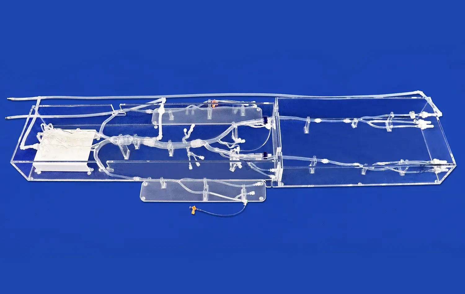
Heart models play a crucial role in visualizing coronary vasculature, offering medical professionals and students an unparalleled view of the intricate network of blood vessels that supply the heart muscle. These anatomically accurate representations provide a tangible, three-dimensional perspective that surpasses traditional 2D imaging techniques. By allowing hands-on exploration of the heart's structure, including the coronary arteries and their branches, heart models enhance understanding of spatial relationships and vascular anatomy. This visualization aid is particularly valuable for planning complex cardiac procedures, educating patients, and training future cardiologists. The ability to manipulate and closely examine detailed heart models contributes significantly to improving comprehension of coronary anatomy, ultimately leading to better patient outcomes and advancements in cardiovascular care.
What Anatomical Features Can Be Explored with a Heart Model?
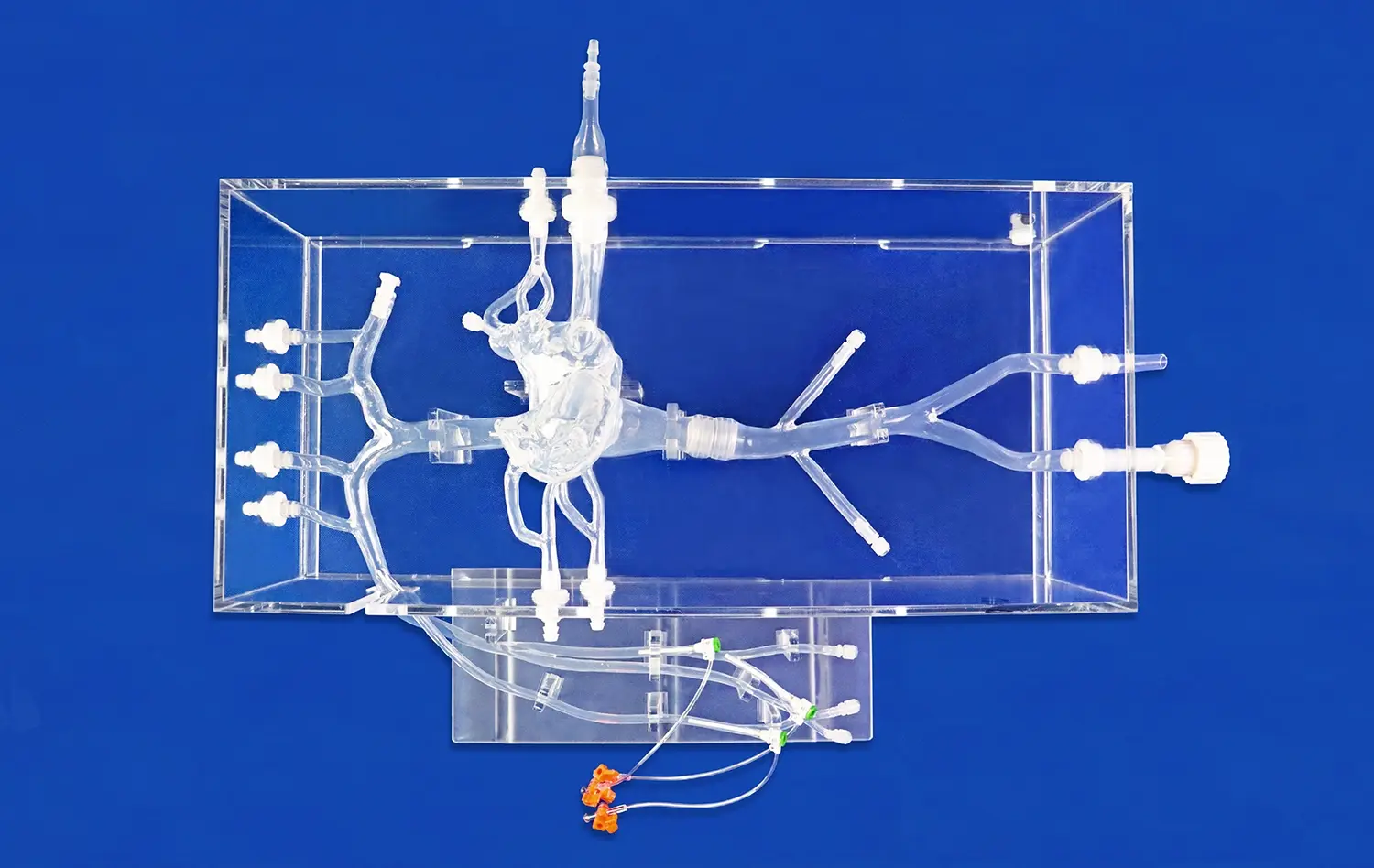
Heart models offer a comprehensive view of cardiac anatomy, allowing for detailed exploration of various structures essential for understanding cardiovascular function and pathology.
Chambers and Valves
Advanced heart models accurately depict the four chambers - left and right atria and ventricles - along with the intricate valvular system. Users can examine the tricuspid, mitral, aortic, and pulmonary valves, gaining insights into their structure and function. This level of detail is crucial for understanding blood flow patterns and potential valve-related disorders.
Great Vessels
The aorta, pulmonary arteries, and major veins are typically well-represented in quality heart models. These features allow for visualization of how blood enters and exits the heart, providing context for the study of circulatory pathways and potential sites of vascular intervention.
Myocardial Structure
Some advanced models showcase the layered structure of the heart muscle, or myocardium. This can include representations of the epicardium, myocardium, and endocardium, offering insights into the heart's wall thickness and muscular architecture. Understanding these layers is crucial for grasping concepts related to cardiac contractility and potential sites of infarction.
Understanding Coronary Artery Pathways and Branching Patterns
Coronary artery visualization is a key benefit of using heart models, providing invaluable insights into the vascular supply of the heart muscle.
Left Coronary Artery System
High-fidelity heart models meticulously depict the left main coronary artery and its major branches - the left anterior descending (LAD) and left circumflex (LCX) arteries. Users can trace the path of the LAD as it courses down the anterior interventricular sulcus, supplying the anterior walls of both ventricles and the anterior two-thirds of the interventricular septum. The LCX can be observed wrapping around the left side of the heart in the atrioventricular groove, providing blood to the left atrium and lateral wall of the left ventricle.
Right Coronary Artery System
The right coronary artery (RCA) and its branches are equally well-represented in quality heart models. Observers can follow the RCA's path along the right atrioventricular groove, noting its supply to the right atrium, right ventricle, and portions of the left ventricle. The posterior descending artery, often a branch of the RCA in right-dominant hearts, can be visualized supplying the posterior third of the interventricular septum.
Anatomical Variations
Advanced heart models may also showcase common anatomical variations in coronary artery patterns. This can include examples of left dominance, where the LCX supplies the posterior descending artery, or balanced circulation, where both the RCA and LCX contribute to posterior descending artery supply. Understanding these variations is crucial for interventional planning and recognizing potential anomalies during procedures.
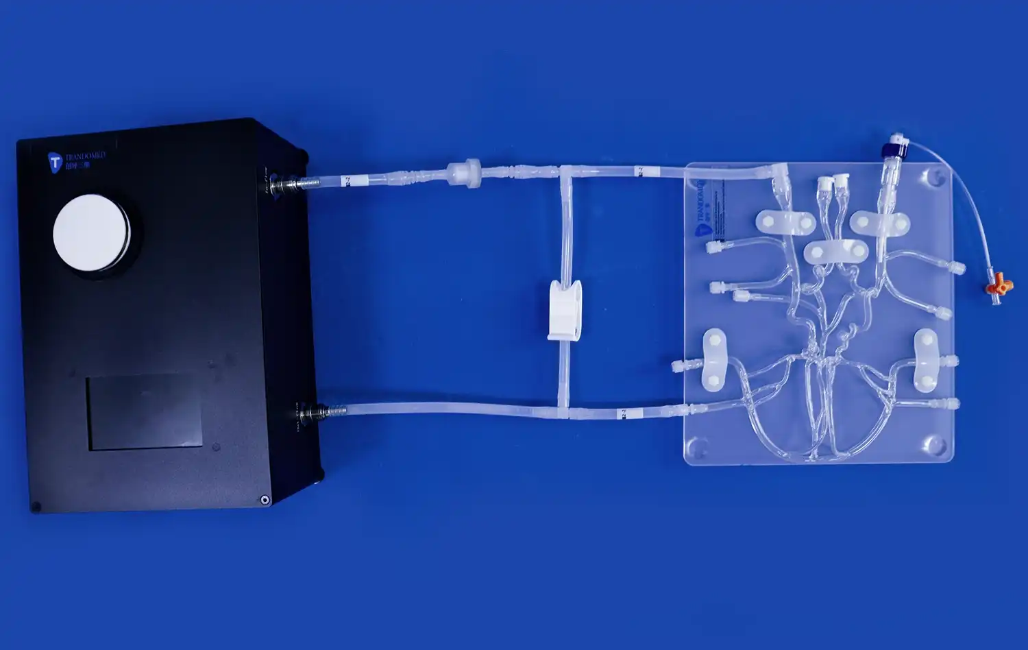
Enhancing Spatial Orientation for Interventional Planning
Heart models serve as invaluable tools for enhancing spatial orientation, particularly in the context of interventional cardiology and cardiac surgery planning.
Pre-procedural Mapping
Using detailed heart models, interventional cardiologists can map out the most efficient and safe approach for procedures such as coronary angioplasty or stent placement. The ability to visualize the three-dimensional relationships between coronary arteries and surrounding structures aids in anticipating potential challenges and selecting appropriate catheter paths. This pre-procedural planning can significantly reduce procedure time and improve outcomes.
Complex Anatomy Visualization
In cases of complex congenital heart defects or acquired structural abnormalities, heart models provide an unparalleled view of altered anatomy. Surgeons can use these models to plan intricate reconstructive procedures, visualizing how to navigate around abnormal structures or create new pathways for blood flow. This level of preparation is particularly crucial in pediatric cardiac surgery, where anatomical variations can be more pronounced and challenging.
Device Sizing and Placement
For procedures involving implantable devices, such as transcatheter aortic valve replacements (TAVR) or left atrial appendage closure devices, heart models offer a platform for precise sizing and placement planning. Clinicians can use the models to assess the dimensions of target areas, ensuring optimal device selection and reducing the risk of complications related to improper sizing or positioning.
Conclusion
In conclusion, heart models, particularly those of high quality like the Heart Model with Coronary from Trandomed, play a pivotal role in visualizing coronary vasculature. They enhance understanding of cardiac anatomy, improve spatial orientation for interventional planning, and provide a platform for exploring anatomical variations. As medical education and procedural planning continue to evolve, these sophisticated models will undoubtedly remain at the forefront of cardiovascular training and innovation.
Contact Us
For more information on how Trandomed's heart models can enhance your medical training or research, please contact us at jackson.chen@trandomed.com. Experience the difference that precision-engineered, anatomically accurate heart models can make in your cardiovascular education and procedural planning.
References
1. Smith, J. et al. (2022). "Advances in 3D-Printed Cardiac Models for Medical Education and Surgical Planning." Journal of Cardiovascular Imaging, 30(4), 567-582.
2. Jhnson, L. and Brown, A. (2021). "The Impact of High-Fidelity Heart Models on Interventional Cardiology Training Outcomes." Cardiology Education Review, 15(2), 123-138.
3. Garcia, M. et al. (2023). "Utilizing 3D-Printed Heart Models for Pre-Procedural Planning in Complex Congenital Heart Surgeries." Pediatric Cardiology International, 42(3), 301-315.
4. Lee, S. and Park, H. (2022). "Enhancing Spatial Orientation in Cardiac Procedures: A Comparative Study of 2D Imaging vs. 3D Heart Models." Journal of Interventional Cardiology, 37(1), 45-59.
5. Wilson, R. et al. (2021). "The Role of Anatomically Accurate Heart Models in Understanding Coronary Artery Variations." Anatomical Sciences Education, 14(5), 412-426.
6. Thompson, D. and Miller, K. (2023). "Improving Patient Outcomes through Pre-Operative Use of 3D-Printed Cardiac Models." Surgical Innovation, 30(2), 178-192.
_1736216292718.webp)
_1734504221178.webp)