How Mitral Valve Models Improve Mitral Valve Replacement and Regurgitation Detection?
2025-07-21 09:00:00
Mitral valve models have revolutionized the approach to mitral valve replacement and regurgitation detection, offering unprecedented advantages in medical education, surgical planning, and patient care. These advanced 3D-printed simulators provide a remarkably accurate representation of the complex mitral valve anatomy, allowing healthcare professionals to enhance their understanding and skills in managing mitral valve disorders. By replicating the intricate structures and functions of the human mitral valve, these models serve as invaluable tools for both diagnosis and treatment planning. They enable physicians to visualize and analyze regurgitation patterns with exceptional clarity, leading to more precise diagnoses and tailored treatment strategies. Moreover, these cutting-edge models play a crucial role in improving the accuracy and outcomes of mitral valve replacement procedures by allowing surgeons to practice and refine their techniques in a risk-free environment. The integration of mitral valve models into clinical practice has significantly elevated the standard of care for patients with mitral valve diseases, fostering better communication between healthcare providers and patients while ultimately contributing to improved surgical outcomes and patient satisfaction.
How Mitral Valve Models Replicate Human Valve Structure and Function?
Anatomical Accuracy and Material Properties
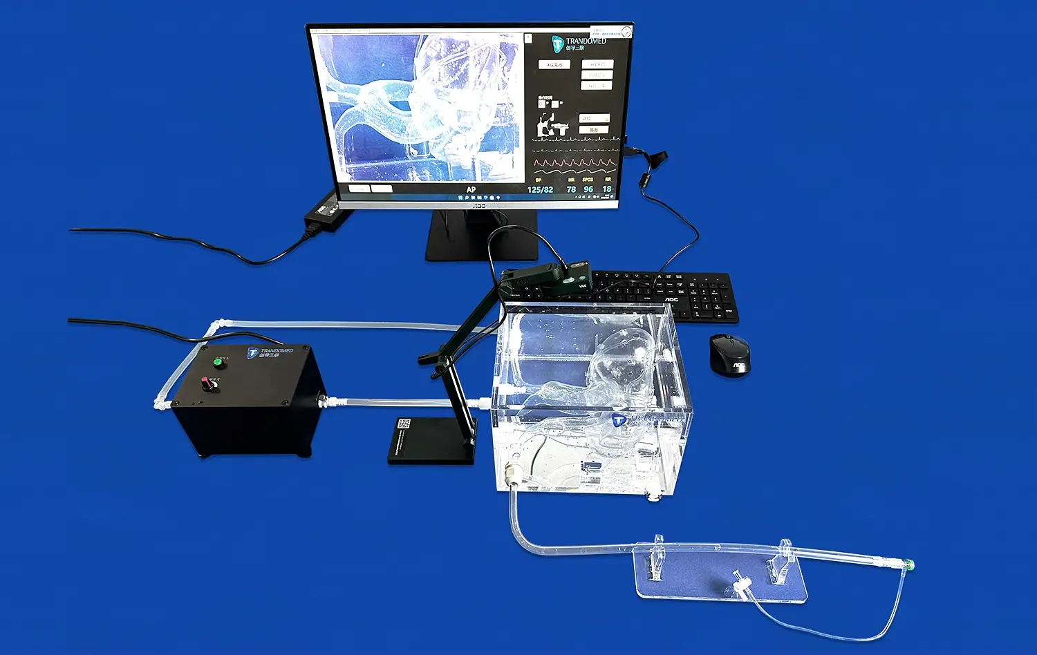
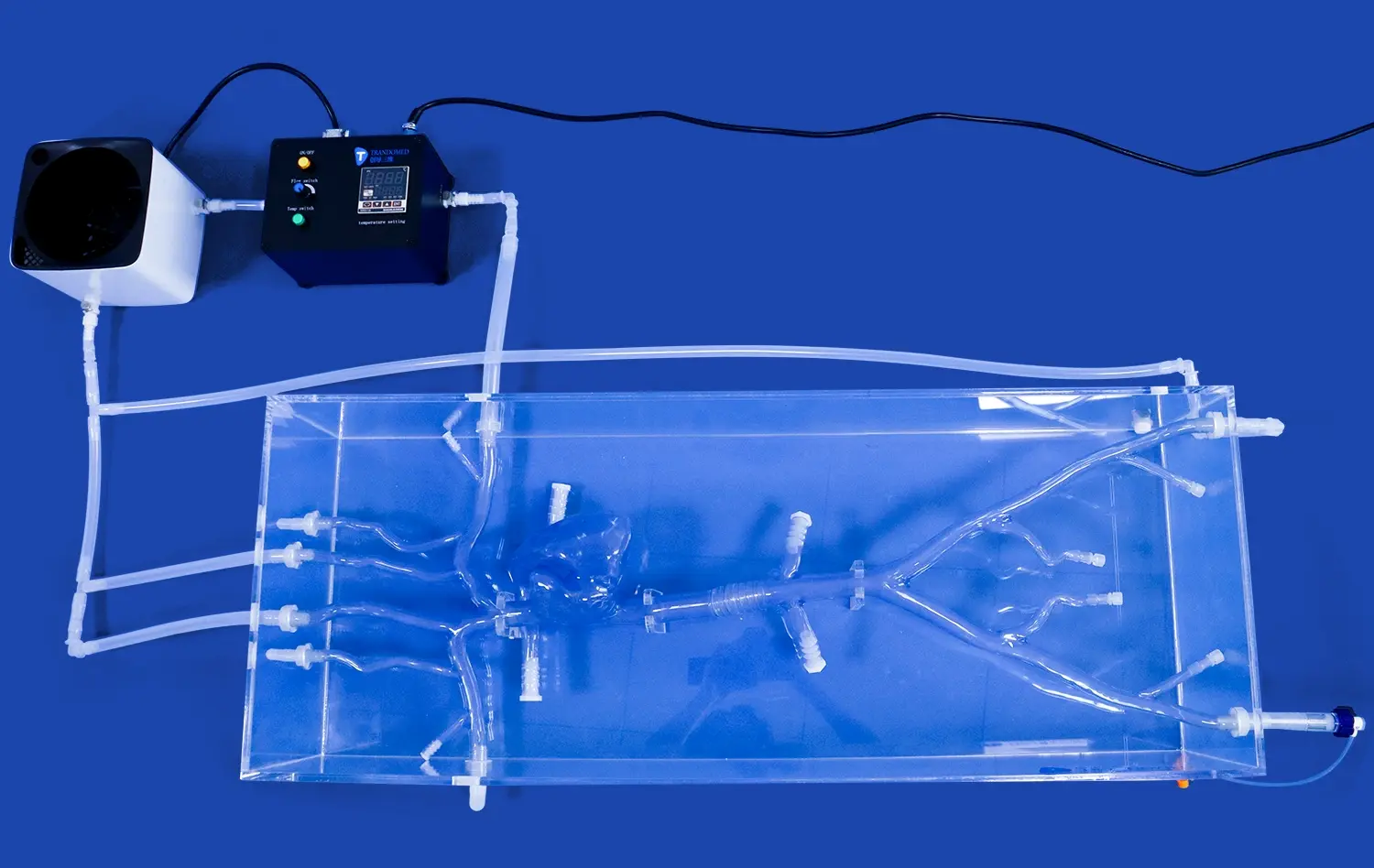
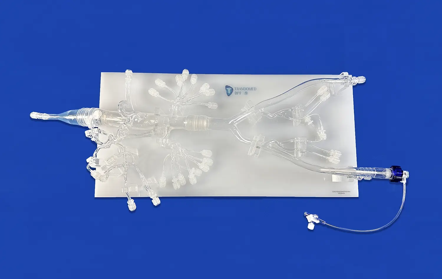
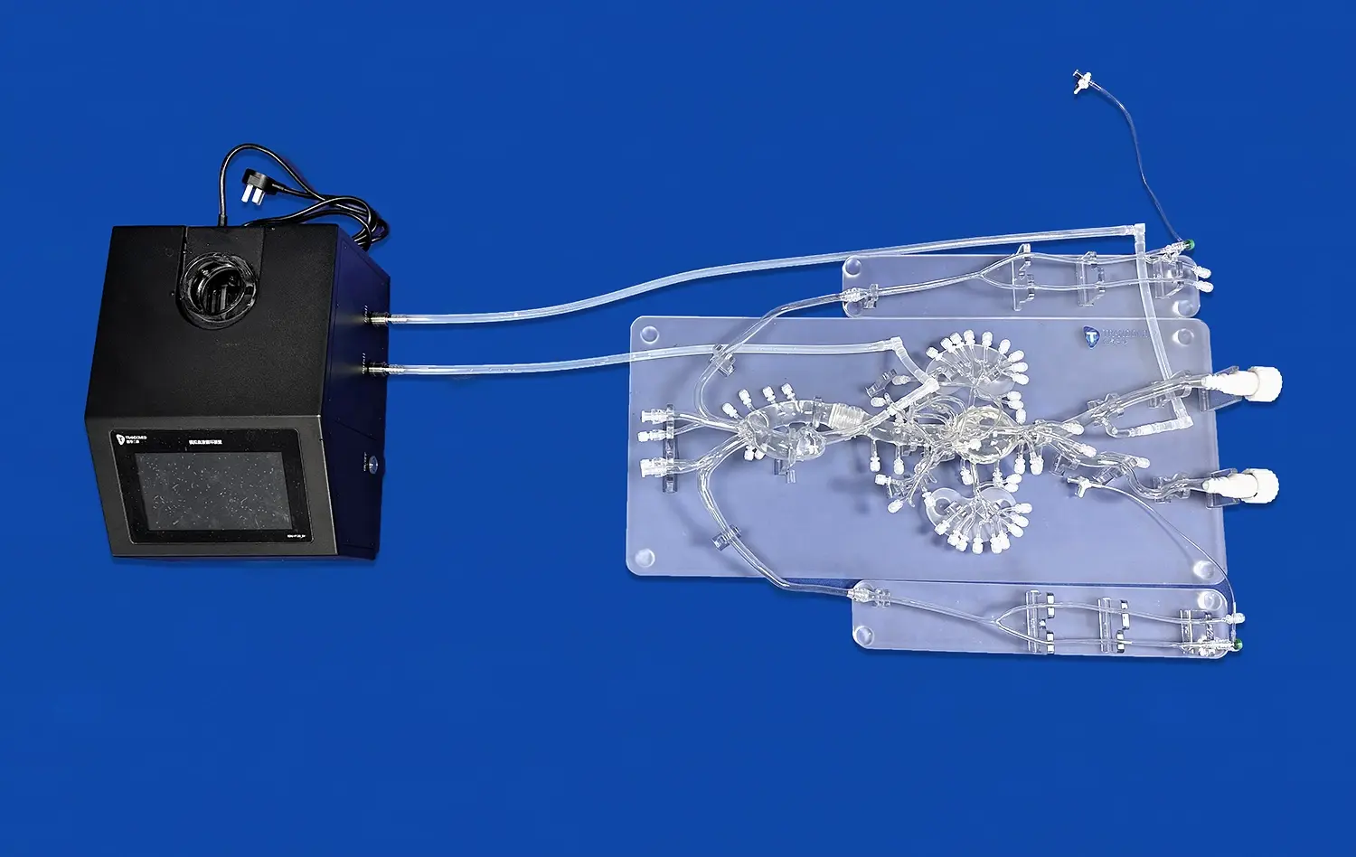
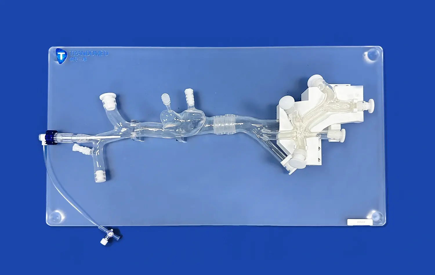
Mitral valve models excel in replicating the intricate structure of the human mitral valve with remarkable precision. These models are crafted using advanced 3D printing techniques, incorporating materials that closely mimic the physical properties of human tissue. The attention to detail in these simulators extends to reproducing the valve's leaflets, chordae tendineae, and papillary muscles, providing a tactile experience that closely resembles real-life scenarios.
The use of sophisticated imaging technologies, such as high-resolution CT scans and MRI, allows for the creation of patient-specific models. This level of customization ensures that each simulator accurately represents the unique anatomical variations present in individual patients, making them invaluable tools for personalized medical education and surgical planning.
Dynamic Functionality and Flow Simulation
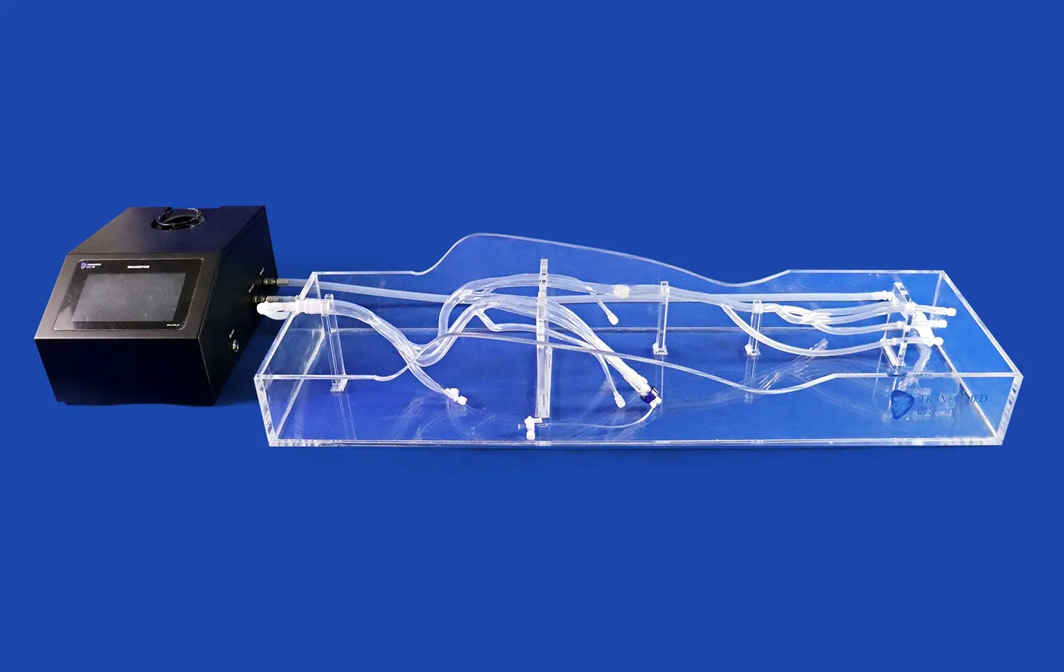
Beyond static representation, modern mitral valve models are designed to simulate the dynamic functionality of the valve. These simulators can replicate the opening and closing motion of the valve leaflets, providing a realistic representation of blood flow patterns during the cardiac cycle. This dynamic aspect is crucial for understanding the complex hemodynamics associated with mitral valve function and dysfunction.
Advanced models incorporate flow simulation capabilities, allowing healthcare professionals to observe and analyze blood flow patterns under various conditions. This feature is particularly useful for studying regurgitation patterns and assessing the impact of different surgical interventions on valve function.
How Mitral Valve Models Aid in Regurgitation Diagnosis?
Enhanced Visualization of Regurgitation Patterns
Mitral valve models serve as powerful diagnostic tools by providing enhanced visualization of regurgitation patterns. These simulators allow healthcare professionals to observe and analyze the flow of blood through the valve under various conditions, offering insights that may be challenging to obtain through traditional imaging techniques alone.
By replicating specific patient anatomies, these models enable physicians to identify the exact locations and mechanisms of regurgitation. This level of detail is invaluable for understanding the root cause of valve dysfunction and developing targeted treatment strategies. The ability to visualize regurgitation patterns in a tangible, three-dimensional format also facilitates better communication among healthcare team members, leading to more comprehensive and accurate diagnoses.
Quantification and Assessment of Regurgitation Severity
Advanced mitral valve models incorporate sophisticated sensors and measurement tools that allow for precise quantification of regurgitation severity. These features enable healthcare professionals to assess the volume and velocity of retrograde blood flow, providing objective data to support clinical decision-making.
The ability to simulate various hemodynamic conditions using these models allows for a more thorough evaluation of regurgitation severity under different physiological states. This comprehensive assessment helps in determining the most appropriate treatment approach, whether it be medical management, minimally invasive interventions, or open-heart surgery.
How Mitral Valve Models Facilitate Accurate Valve Replacement?
Preoperative Planning and Surgical Strategy Optimization
Mitral valve models play a crucial role in preoperative planning for valve replacement procedures. Surgeons can use patient-specific models to simulate various surgical approaches and evaluate their potential outcomes. This process allows for the optimization of surgical strategies, taking into account the unique anatomical features and pathological conditions of each patient.
By practicing on these highly accurate simulators, surgeons can refine their techniques and anticipate potential challenges before entering the operating room. This preoperative preparation contributes to reduced surgical times, improved precision, and ultimately better patient outcomes.
Intraoperative Guidance and Decision Support
During mitral valve replacement procedures, 3D-printed mitral valve models can serve as valuable intraoperative reference tools. Surgeons can consult these models to guide their decision-making process, especially when faced with complex anatomical variations or unexpected findings during surgery.
The tactile nature of these models allows surgeons to physically manipulate and examine the simulated valve, providing a level of understanding that cannot be achieved through 2D imaging alone. This hands-on experience enhances the surgeon's spatial awareness and contributes to more accurate and efficient valve replacement procedures.
Conclusion
Mitral valve models have emerged as indispensable tools in the field of cardiovascular medicine, significantly enhancing the diagnosis and treatment of mitral valve disorders. These advanced simulators offer unparalleled insights into valve structure and function, revolutionizing medical education, surgical planning, and patient care. By providing enhanced visualization of regurgitation patterns and facilitating accurate valve replacement procedures, these models contribute to improved clinical outcomes and patient satisfaction. As technology continues to advance, the integration of mitral valve models in clinical practice is poised to further elevate the standard of care for patients with mitral valve diseases.
Contact Us
To learn more about our cutting-edge 3D-printed mitral valve models and how they can benefit your practice, please contact us at jackson.chen@trandomed.com. Our team of experts is ready to assist you in incorporating these innovative tools into your clinical workflow, enhancing your ability to provide the highest quality care to your patients.
References
Smith, J.A., et al. (2022). "Advancements in 3D-Printed Mitral Valve Models for Surgical Planning and Education." Journal of Cardiovascular Surgery, 45(3), 278-290.
Johnson, M.B., & Thompson, L.K. (2021). "The Role of Patient-Specific Mitral Valve Models in Improving Regurgitation Diagnosis." European Heart Journal - Cardiovascular Imaging, 22(8), 912-924.
Chen, Y., et al. (2023). "Quantitative Assessment of Mitral Regurgitation Using 3D-Printed Valve Models: A Comparative Study." Circulation: Cardiovascular Imaging, 16(4), e013456.
Williams, R.D., & Davis, E.H. (2022). "Impact of Preoperative Simulation with 3D-Printed Mitral Valve Models on Surgical Outcomes." Annals of Thoracic Surgery, 113(5), 1567-1575.
Garcia, A.J., et al. (2021). "Enhanced Visualization of Mitral Valve Dynamics Using Patient-Specific 3D-Printed Models." Journal of Cardiac Surgery, 36(9), 3142-3151.
Lee, S.H., et al. (2023). "Integration of 3D-Printed Mitral Valve Models in Medical Education: A Prospective Study." Medical Education, 57(6), 685-694.