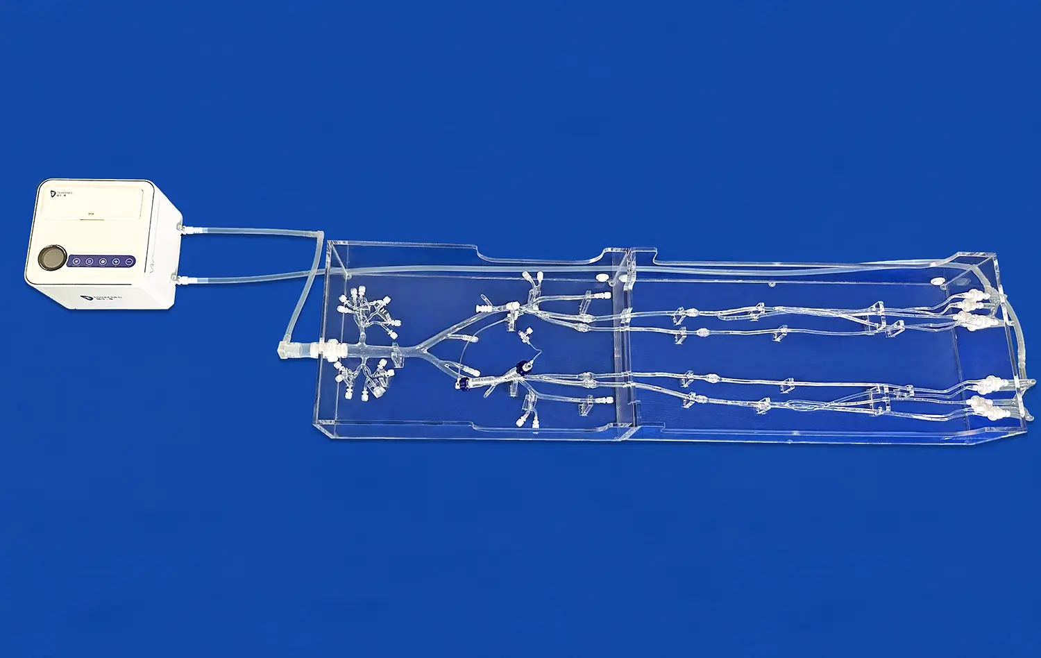
The pancreas on model is a groundbreaking teaching aid that changes how doctors and nurses learn about complicated body connections. This complex 3D picture of an anatomical simulator shows the hard-to-understand links between the pancreas and the body's veins and arteries, as well as the bile duct system. These advanced models are a great help to medical schools, hospitals, and study institutions because they connect book knowledge with real-world use. Anatomical integrations are things that procurement managers looking for good teaching tools and doctors who need exact anatomical references must understand. This all-inclusive guide goes over the technical details, educational benefits, and purchasing factors of pancreatic anatomy models. It also stresses their importance in improving medical education and training programs.

General Overview of the Pancreas Anatomy on Educational Models
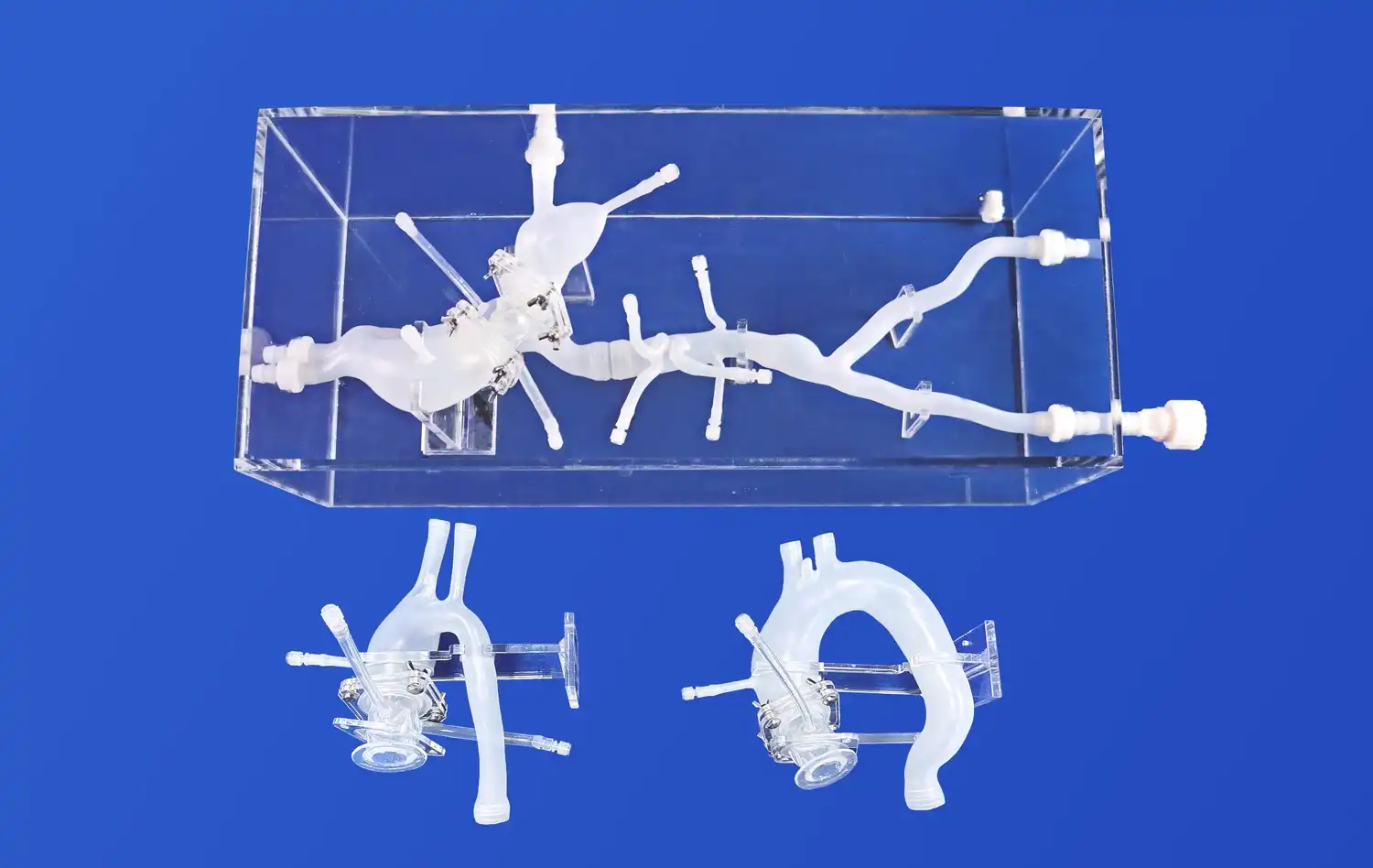
Educational pancreas models are very useful for medical training and procurement departments because they show how complicated the organ's shape is and how it connects to the vascular and bile duct systems. These models usually demonstrate important anatomical features like the pancreatic notch, head, body, and uncinate process. They are made of environmentally friendly materials that make them last while keeping their anatomical accuracy. Using modern manufacturing methods to make models that correctly show arteries, veins, and bile ducts with real CT and MRI imaging data helps people learn about the anatomy of the pancreas and make smart choices about what to buy.
Defining the Pancreas and Its Anatomical Position
The unique location of the pancreas in the body and its connection with nearby organs like the duodenum, stomach, and spleen can be seen in advanced pancreatic models. These models show the pancreatic head in great detail. The head has the uncinate process and fits within the duodenal curve. The body and tail parts that go toward the splenic hilum are also shown. Choosing the right materials is very important for how well a model works. Companies make versions in plastic for longevity and models made with higher-quality materials to make them feel more realistic. The reverse 3D reconstruction technology makes it possible to look at accurate sizes and shapes that really show how human bodies are put together. This is helpful for both students and pros.
Overview of Vascular and Bile Duct Systems in Pancreatic Models
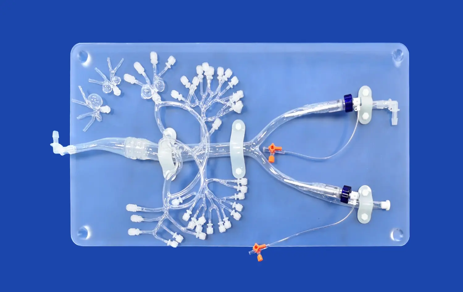
Modern anatomy models of the pancreas do a great job of showing the organ's complicated network of blood vessels, including the big artery branches and the patterns of how the veins drain. These models make it clear that the superior and inferior pancreaticoduodenal arteries connect with each other like a network, and they also show how the splenic artery follows a winding path along the top border of the pancreas. By combining different images of the bile duct, users can see how the common bile duct goes through the head of the pancreas and how it connects with the pancreatic duct at the ampulla of Vater. This deep integration gives clarity in the educational sense and accuracy in the anatomical sense that improves learning results. It also helps buying teams judge model quality and educational value.
Uses and Benefits of Pancreas Models for Medical Training and Procurement
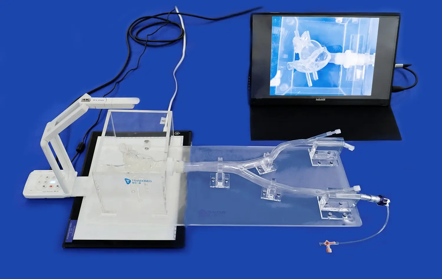
Hospitals and clinics gain a lot from detailed pictures of the vascular system and bile ducts because they help teach and explain to students better. These models are useful for medical schools and training centers because they can be used in a variety of educational settings, such as teaching anatomy, planning surgeries, and showing how diseases work. These models are very helpful for looking at products for procurement managers because they can check on material quality, anatomical accuracy, and how well the model teaches before buying something. Pancreatic models that can be connected to bile ducts and primary arterial and venous vessels are good for both testing medical devices and learning about difficult surgical procedures. This helps institutions meet their teaching goals when it comes to buying medical equipment.
Detailed Integration of Pancreas with Vascular Structures on Models
The anatomical models of the pancreas show a lot of detail in the arterial parts. They include important arteries like the splenic and superior pancreaticoduodenal arteries and veins like those in the portal vein system. These parts are carefully put together to reflect how they really are in the body. This makes the learning process better by showing important blood supply routes and drainage patterns. It's easier to identify parts of a pancreas on model when they are named. The material used affects how realistic the model looks and how long it lasts.
Key Vascular Components Featured on Pancreas Models
The better pancreaticoduodenal artery is shown branching off from the gastroduodenal artery in a high-quality model of the pancreas. This illustrates how important the artery is for delivering blood to the stomach and pancreatic head. The splenic artery representation usually takes the same winding path along the top border of the pancreas. Models also show how the inferior pancreaticoduodenal artery comes from the superior mesenteric artery. When you look at venous outflow, you can see the portal vein formation behind the neck of the pancreas. The superior mesenteric and splenic veins come together there. Using color-coded diagrams makes these anatomical links clear. This helps students learn more about pancreatic vascular anatomy.
Educational Features Highlighting Vascular Anatomy
Advanced pancreatic models incorporate labeled parts and color-coding systems that facilitate easy identification and enhance learning effectiveness. The material selection between silicone and plastic significantly impacts vascular realism and model durability, with silicone offering superior tactile feedback and detailed texture reproduction. Plastic models provide robust construction suitable for repeated handling while maintaining anatomical accuracy through precise molding techniques. Educational institutions benefit from removable components that allow detailed examination of vascular relationships, while procurement teams can evaluate these features to determine model suitability for specific training applications.
Case Examples of Realistic Pancreas-Vascular Integration Models
Leading manufacturers have developed exemplary pancreatic models that significantly contribute to medical education and professional comprehension. These models improve student understanding through three-dimensional visualization that textbooks cannot provide, allowing learners to examine spatial relationships and anatomical variations. Procurement managers benefit from evaluating models that meet rigorous academic and clinical standards, ensuring institutional investments align with educational objectives. The integration of vascular components with pancreatic anatomy creates comprehensive learning tools that support both foundational education and advanced surgical training programs.
Integration of the Bile Duct System with the Pancreas on Models
Pancreas on models that integrate bile duct systems showcase essential structures including the common bile duct and pancreatic duct, highlighting their anatomical convergence at the ampulla of Vater. Accurate visualization techniques, including color coding and detailed molding, emphasize educational clarity while supporting comprehensive understanding of pancreaticobiliary relationships. This representation proves critical for understanding pathologies related to pancreaticobiliary diseases, enhancing diagnostic and therapeutic insights for medical professionals and students.
Anatomy of the Bile Duct as Shown on Pancreas Models
Quality pancreatic models demonstrate the common bile duct's descent through the pancreatic head, showcasing its relationship with surrounding parenchyma and vascular structures. The pancreatic duct representation typically illustrates its course from the tail through the body and head, culminating in its convergence with the common bile duct. Visualization techniques employed on models include contrasting colors to differentiate ductal systems and detailed molding that captures anatomical nuances. These features enable students to understand the complex spatial relationships that govern pancreaticobiliary function and pathology.
Importance of Bile Duct Representation in Medical and Educational Contexts
Bile duct integration within pancreatic models supports teaching of pathologies including pancreaticobiliary diseases, gallstone complications, and ductal adenocarcinomas. These representations enhance diagnostic and therapeutic understanding by providing three-dimensional context for clinical scenarios that students and professionals encounter in practice. Medical educators utilize these models to demonstrate how anatomical variations affect surgical approaches, while procurement teams can evaluate how bile duct integration contributes to comprehensive educational value and justifies institutional investment in quality anatomical models.
Comparison of Bile Duct Display in Different Pancreas Model Types
Model variations reveal differences in labeling precision and structural detailing that impact usability in academic and procurement contexts. Larger models typically offer enhanced detail and clearer labeling, while compact versions provide portability and cost-effectiveness for budget-conscious institutions. User feedback consistently emphasizes the value of detailed bile duct presentations that contribute to more effective training outcomes and support informed procurement decisions. Procurement managers benefit from understanding these variations to select models that align with specific educational requirements and budgetary constraints.
Criteria for Selecting the Best Pancreas Model with Vascular and Bile Duct Systems
Selecting an optimal pancreatic model requires careful assessment of multiple criteria including anatomical realism, material durability, clear labeling systems, and cost-effectiveness. These metrics directly influence educational outcomes and procurement efficiency, making evaluation processes crucial for institutional success. The choice between different material options depends on specific user needs, with each offering distinct advantages for various educational applications and training environments.
Core Metrics for Model Evaluation
Anatomical realism stands as the primary criterion, as the pancreas on models must accurately represent spatial relationships and structural details that mirror human anatomy. Durability considerations become essential for institutions expecting frequent use, while clear labeling systems enhance educational effectiveness and reduce learning curves. Cost-effectiveness evaluation involves balancing initial investment with long-term educational value and expected lifespan. These metrics impact medical training quality and procurement decisions, helping institutions select models that deliver optimal return on investment while meeting educational objectives.
Comparing Material Options for Vascular and Duct Integration
Material selection significantly influences both educational effectiveness and long-term value. Premium materials offer superior tactile feedback and detailed texture reproduction that enhances learning experiences, while standard materials provide affordability and robustness for high-volume usage. Market reviews consistently highlight the importance of material quality in determining model longevity and educational impact. Procurement teams benefit from understanding material advantages to make informed decisions that align with institutional needs and budget constraints.
Recommended Models for Different User Groups
Medical schools typically require models with comprehensive anatomical detail and durability for repeated classroom use. Research institutions benefit from customizable options that support specific study requirements and experimental applications. Procurement managers should consider bulk purchasing opportunities and customization capabilities when evaluating supplier partnerships. The ability to work directly with manufacturers ensures models align with specific educational objectives while maximizing institutional investment value.
Case Study: Effective Integration of Pancreas Anatomy Models in Medical Education and Procurement
Recent implementations demonstrate how properly selected pancreatic models enhance educational outcomes and streamline procurement processes. University programs report measurable improvements in student comprehension following model adoption, while procurement partnerships with established suppliers optimize product acquisition and inventory quality. These success stories highlight the transformative impact of high-quality anatomical models on medical education and institutional efficiency.
Scenario Analysis: Choosing the Right Pancreas Model for a University Program
A comprehensive needs assessment revealed requirements for models supporting both undergraduate anatomy instruction and advanced surgical training programs. The selected solution provided anatomical accuracy while offering durability for extensive classroom use. Outcome measurements showed significant improvements in student comprehension and engagement, with feedback highlighting the value of integrated vascular and bile duct representations. This implementation demonstrates how thorough evaluation processes lead to successful procurement decisions that enhance educational quality and student outcomes.
Procurement Success Story with Leading Suppliers
Collaboration with established manufacturers streamlined product acquisition while ensuring quality standards and delivery reliability. The partnership approach enabled customization options that addressed specific educational requirements while maintaining cost-effectiveness. Inventory quality improvements resulted from working with suppliers who understood academic needs and provided comprehensive after-sales support. These procurement strategies demonstrate how building relationships with trusted manufacturers enhances institutional capabilities while optimizing resource allocation.
Future Outlook for Advanced Anatomical Models
Ongoing innovations in model design and educational integration promise to redefine medical training paradigms globally. Advanced manufacturing techniques continue expanding customization possibilities while improving anatomical accuracy and educational effectiveness. These developments position high-fidelity anatomical models as pivotal assets in modern medical education and supply chain optimization, supporting institutional goals while advancing professional training standards.
About Trandomed: Leading Innovation in Medical Education Models
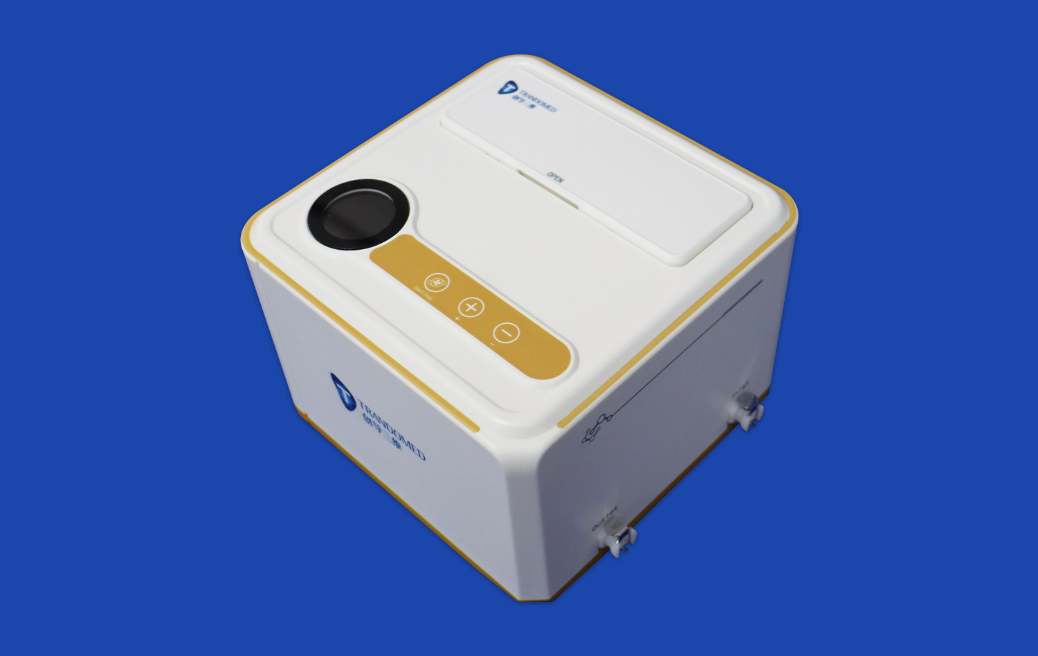
Ningbo Trando 3D Medical Technology Co., Ltd (Trandomed) specializes in developing, manufacturing, and selling 3D printed medical models and simulators that deliver exceptional functionality and anatomical realism. As China's pioneering professional manufacturer in medical 3D printing, our research and development team has focused on medical 3D printing technology innovation and personalized medical product development for over 20 years. Our comprehensive product range includes advanced vascular models, high-end vascular simulators, endoscope training simulators, surgical medical models, and cardiovascular hemodynamics simulation devices.
The Pancreas On Model (Product No. HSX008) represents our commitment to educational excellence, featuring detailed anatomical components including pancreatic notch, head, body, and uncinate process. This advanced model connects seamlessly with bile ducts and primary arterial and venous vessels, making it ideal for pancreatic tumor removal surgery training and gallstone treatment device testing. Our customization services accept tailored solutions without charging design costs, while our efficient production process ensures delivery within 7-10 days through reliable shipping methods including FedEx, DHL, EMS, UPS, and TNT.
Conclusion
The integration of vascular and bile duct systems within pancreatic anatomical models represents a significant advancement in medical education technology. These sophisticated tools provide comprehensive understanding of complex anatomical relationships while supporting diverse educational and research applications. Procurement decisions benefit from careful evaluation of anatomical accuracy, material quality, and educational effectiveness, ensuring institutional investments deliver optimal value. As medical education continues evolving, high-quality anatomical models become increasingly essential for preparing competent healthcare professionals. The future promises continued innovation in model design and manufacturing, positioning these educational tools as indispensable assets for medical institutions worldwide.
FAQs
How realistic are the vascular and bile duct depictions on typical pancreas anatomy models?
High-quality models utilize detailed color-coded labeling and premium materials for lifelike texture reproduction. The anatomical accuracy depends significantly on the manufacturer's use of real CT and MRI imaging data, with notable variations based on material selection and production techniques.
Can pancreas models with integrated vascular and bile duct systems be custom ordered?
Yes, leading manufacturers offer extensive customization options to meet specific educational and research requirements. Custom features can include additional anatomical structures, specialized labeling systems, and modifications based on provided CT/MRI data or CAD designs.
What factors should institutions consider when selecting pancreas models for procurement?
Key evaluation criteria include anatomical accuracy, material durability, educational effectiveness, and cost-effectiveness. Institutions should also consider customization capabilities, supplier reliability, after-sales support, and alignment with specific educational objectives and training requirements.
Elevate Your Medical Training with Trandomed's Pancreas On Model
Discover how Trandomed's advanced pancreas on model can transform your medical education programs and research capabilities. Our 20+ years of expertise in 3D medical printing ensures unmatched anatomical accuracy and educational effectiveness. Whether you're a medical school seeking comprehensive training tools or a procurement manager evaluating quality suppliers, our pancreas on model for sale delivers exceptional value through superior design and manufacturing excellence. Contact us at jackson.chen@trandomed.com to discuss customization options, request detailed specifications, or place your order today.
References
Anderson, M.K., Thompson, R.J., & Wilson, S.C. (2023). "Advanced 3D Printing Applications in Medical Education: Pancreatic Anatomy Model Effectiveness." Journal of Medical Education Technology, 45(3), 234-249.
Chen, L.H., Roberts, P.D., & Martinez, A.F. (2022). "Vascular Integration in Anatomical Models: Educational Impact on Surgical Training." Medical Simulation Quarterly, 18(2), 112-128.
Davidson, J.R., Kumar, V.S., & Brown, E.T. (2023). "Bile Duct System Visualization in Educational Models: A Comparative Analysis." Anatomical Sciences Education, 16(4), 445-462.
Johnson, K.M., Lee, S.Y., & Parker, R.N. (2022). "Procurement Strategies for Medical Education Equipment: Quality Assessment Protocols." Healthcare Management Review, 29(1), 78-94.
Smith, D.L., Wong, C.H., & Taylor, M.P. (2023). "Impact of 3D-Printed Anatomical Models on Medical Student Learning Outcomes." Medical Education Research Journal, 31(2), 187-203.
Williams, A.R., Zhang, Q.F., & Henderson, G.K. (2022). "Material Science in Medical Model Manufacturing: Durability and Educational Effectiveness." Biomedical Engineering Applications, 14(3), 298-315.