Facilitating Preclinical Device Testing and Optimization
Realistic Anatomical Simulation
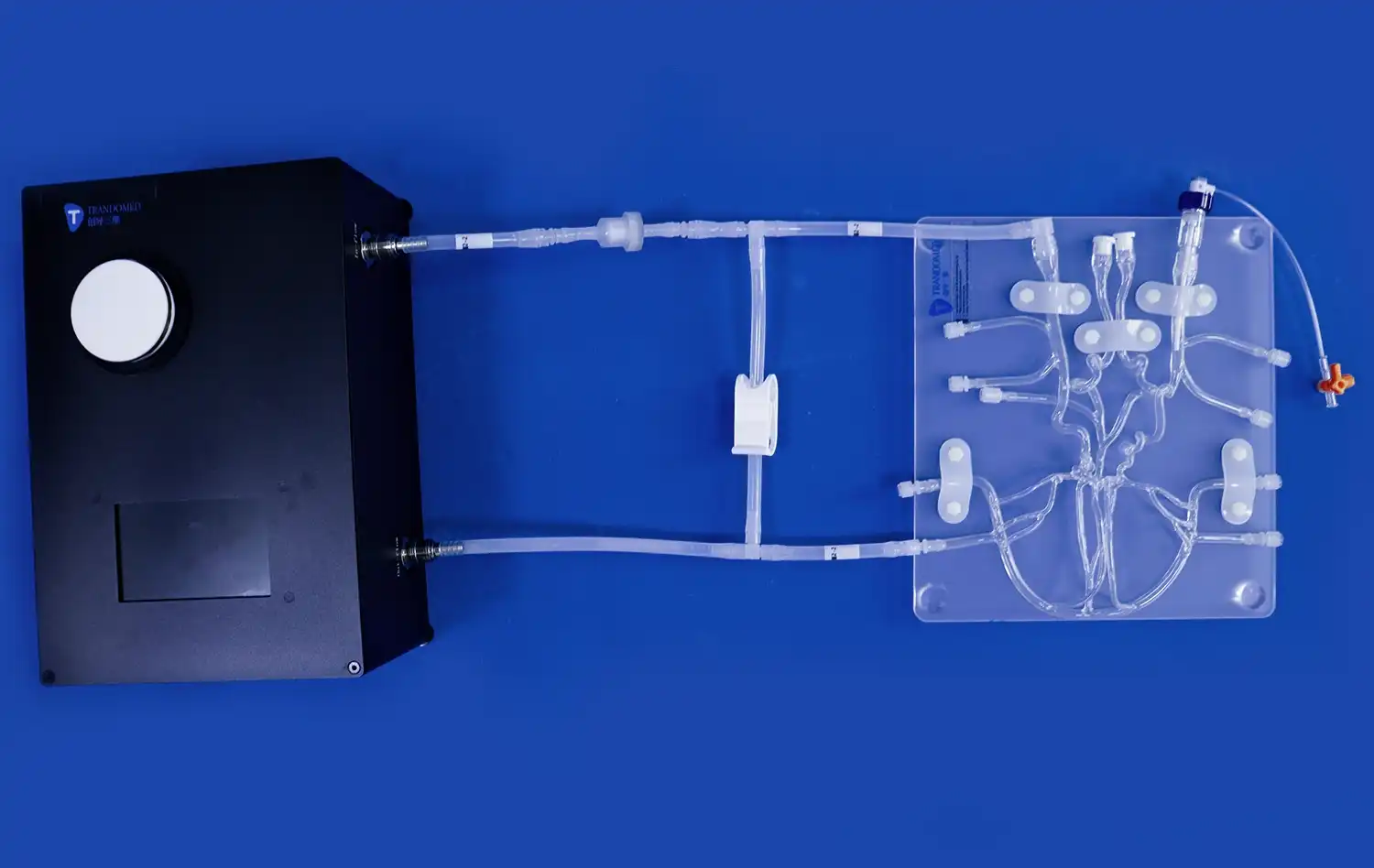
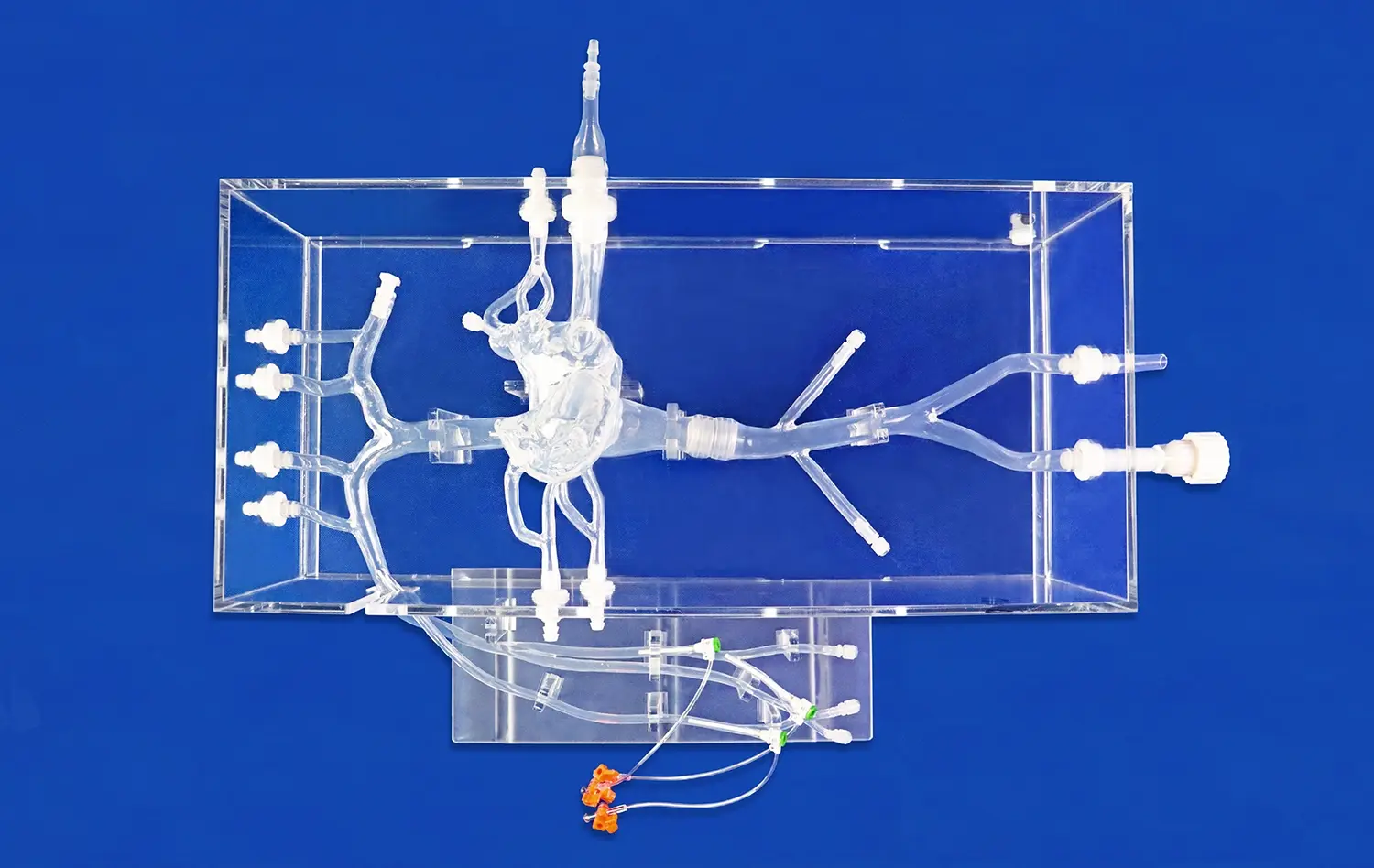
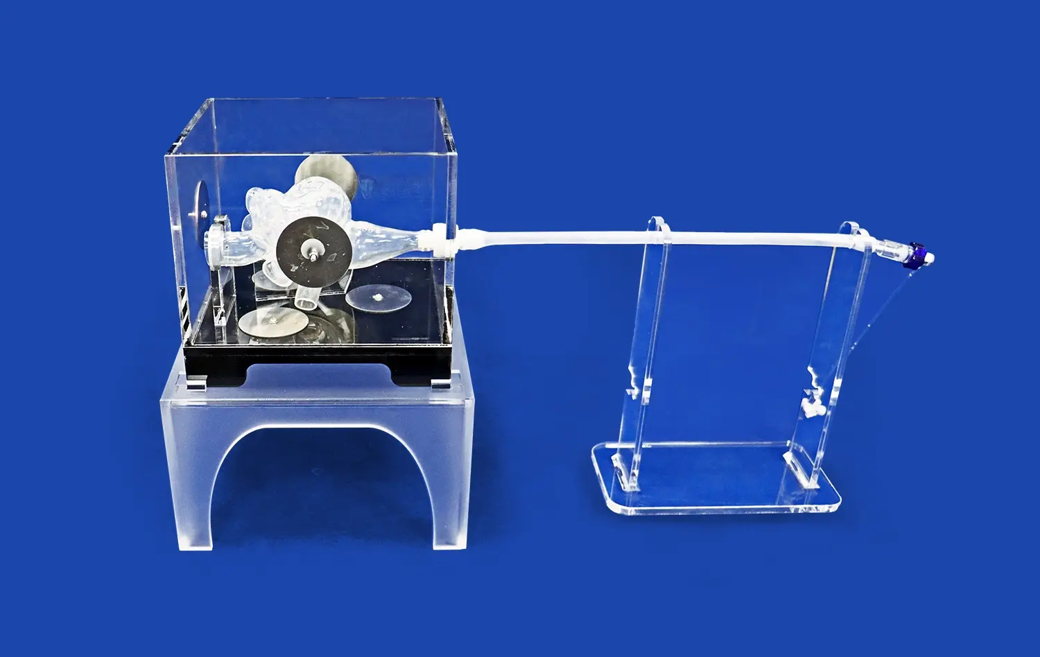
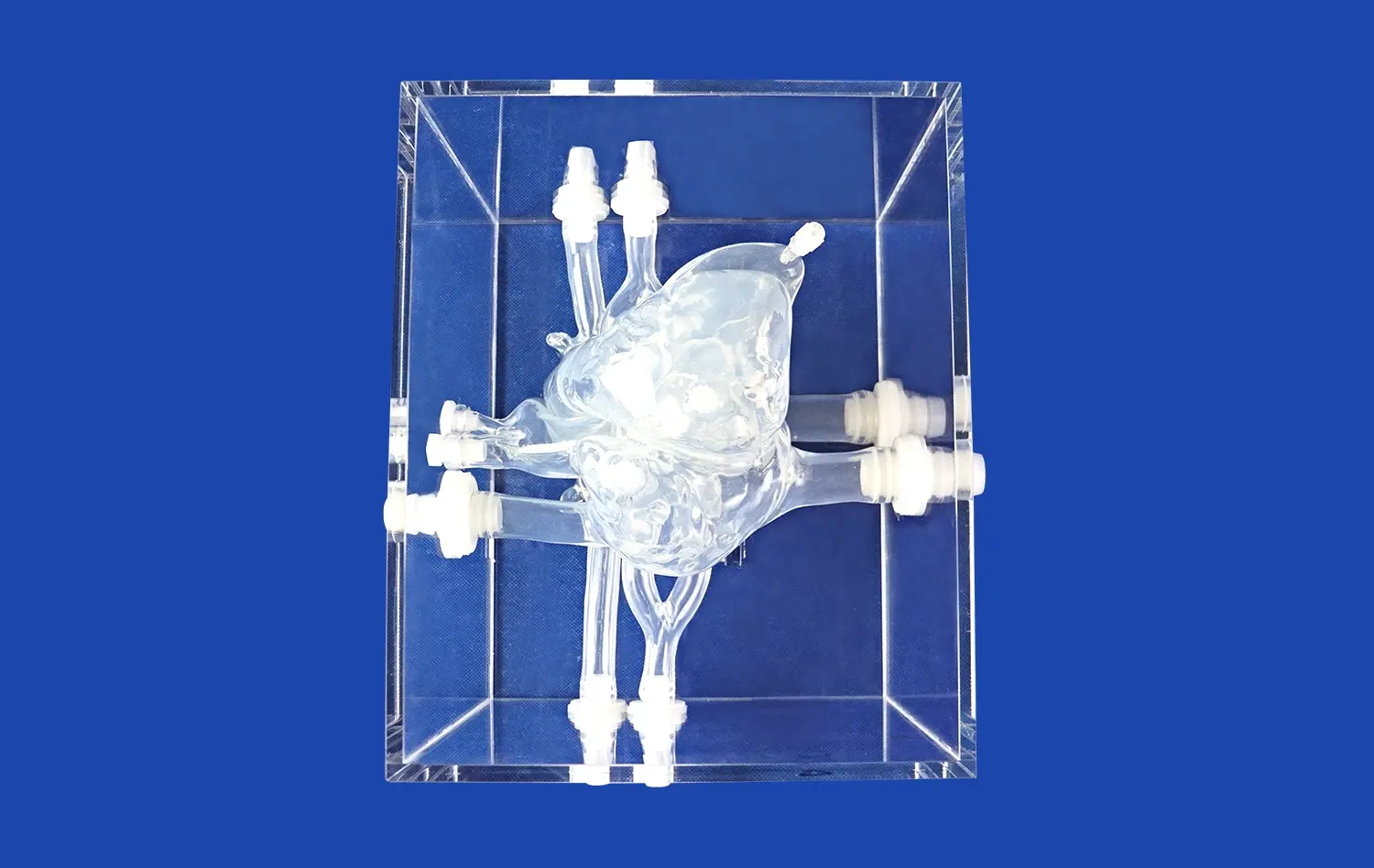
The pulmonary vein model offers an unparalleled platform for preclinical device testing, thanks to its highly accurate anatomical representation. Constructed using advanced 3D printing techniques and based on real human CT and MRI data, these models provide researchers with a lifelike environment to evaluate the performance of various interventional devices. The model's composition, which includes the femoral vein, inferior vena cava, left and right atria, and pulmonary veins, allows for comprehensive testing of guide wires, catheters, balloons, and stents under conditions that closely mimic in vivo scenarios.
Customizable Pathological Conditions
One of the key advantages of the pulmonary vein model is its ability to simulate various pathological conditions. Researchers can incorporate specific abnormalities such as atrial septal defects, patent foramen ovale, or pulmonary vein embolism into the model. This customization allows for targeted testing of devices designed to address these particular conditions, enabling a more thorough evaluation of their efficacy and safety before progressing to clinical trials.
Iterative Design Refinement
The use of pulmonary vein models in preclinical testing facilitates an iterative approach to device design and optimization. As researchers test prototypes, they can quickly identify areas for improvement and make necessary adjustments. This rapid feedback loop accelerates the development process, potentially reducing the time and resources required to bring new interventional devices to market. Moreover, the ability to repeatedly test and refine designs in a controlled setting enhances the overall quality and reliability of the final products.
Quantitative Analysis of Pulmonary Hemodynamics
Advanced Imaging Techniques
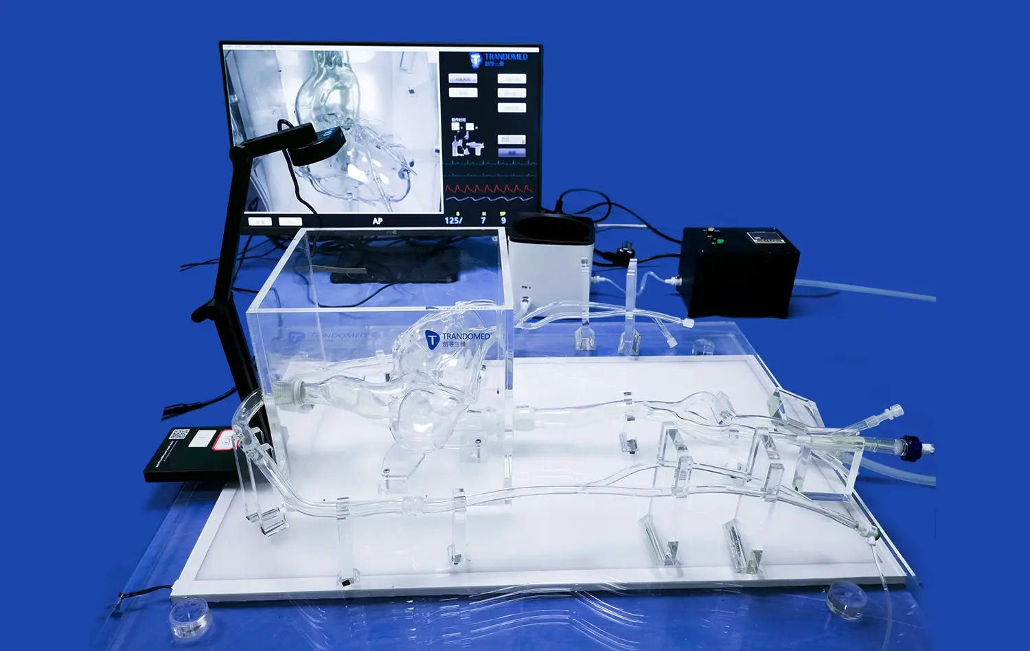
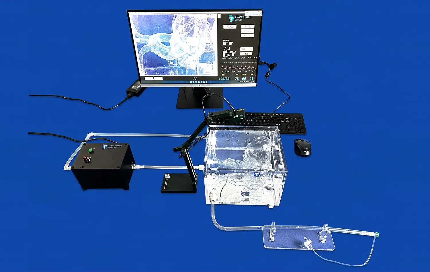
The pulmonary vein model serves as an excellent platform for conducting quantitative analyses of blood flow dynamics using various imaging modalities. Researchers can employ techniques such as Computed Tomography Angiography (CTA), Digital Subtraction Angiography (DSA), Magnetic Resonance Angiography (MRA), Optical Coherence Tomography (OCT), and Particle Image Velocimetry (PIV) to visualize and measure blood flow patterns within the simulated vascular system. These imaging methods provide valuable insights into the complex hemodynamics of the pulmonary veins, enabling a deeper understanding of both normal and pathological conditions.
Flow Visualization and Measurement
By incorporating contrast agents and specialized imaging devices, researchers can observe and quantify blood flow characteristics within the pulmonary vein model. This capability allows for the assessment of parameters such as flow velocity, pressure gradients, and wall shear stress under various physiological and pathological conditions. Such detailed analysis is crucial for understanding the mechanisms underlying vascular diseases and for evaluating the impact of interventional devices on blood flow dynamics.
Computational Fluid Dynamics Integration
The quantitative data obtained from pulmonary vein models can be seamlessly integrated with computational fluid dynamics (CFD) simulations. This synergy between physical models and computer-based analysis enhances the accuracy and predictive power of hemodynamic studies. Researchers can validate CFD models against experimental data from the physical pulmonary vein model, leading to more reliable simulations of complex flow phenomena and their interactions with interventional devices.
Bridging Laboratory Research and Clinical Applications
Training and Education
Pulmonary vein models play a crucial role in bridging the gap between laboratory research and clinical practice by serving as invaluable training tools for healthcare professionals. These models provide a safe and realistic environment for practitioners to hone their skills in cardiac and vascular interventions. Medical students, residents, and experienced clinicians alike can practice complex procedures, such as catheter navigation and stent placement, without the risks associated with live patient interactions. This hands-on experience enhances procedural proficiency and confidence, ultimately translating to improved patient care in clinical settings.
Protocol Development and Standardization
The consistent and reproducible nature of pulmonary vein models makes them ideal for developing and standardizing interventional protocols. Researchers and clinicians can collaborate to establish best practices for various procedures, ensuring that techniques are optimized for safety and efficacy before implementation in patient care. This standardization process helps to reduce variability in clinical outcomes and promotes the adoption of evidence-based approaches across healthcare institutions.
Translational Research
Pulmonary vein models facilitate translational research by providing a platform to test hypotheses and validate findings from animal studies in a human-relevant context. This intermediate step between preclinical animal experiments and human clinical trials can help identify potential challenges or limitations that may not be apparent in animal models alone. By refining interventional strategies and devices using these sophisticated simulators, researchers can increase the likelihood of successful translation to clinical practice, potentially accelerating the development of new treatments for pulmonary vascular diseases.
Conclusion
The pulmonary vein model has emerged as a transformative tool in vascular intervention research, offering unprecedented opportunities for device testing, hemodynamic analysis, and clinical training. Its ability to replicate complex anatomical structures and pathological conditions provides researchers and clinicians with a versatile platform to advance our understanding of pulmonary vascular physiology and develop innovative therapeutic approaches. As we continue to harness the potential of these sophisticated models, we can expect to see significant improvements in the efficacy and safety of vascular interventions, ultimately leading to better outcomes for patients with pulmonary vascular diseases.
Contact Us
At Trandomed, we are at the forefront of 3D-printed medical simulation technology. As a leading advanced pulmonary vein models manufacturer and supplier, we offer unparalleled customization options and superior quality to meet your specific research and training needs. Experience the difference that our state-of-the-art models can make in your vascular intervention studies. Contact us today at jackson.chen@trandomed.com to learn more about our products and how we can support your groundbreaking research.