How Venous Cardiac Electrophysiology Models Improve EP Training?
2026-05-22 09:00:04
By simulating the cardiac vein system in great detail, venous cardiac electrophysiology models completely change EP training. This is because they let professionals learn how to navigate catheters, ablate heart rhythms, and map arrhythmias without any risk. These anatomically accurate models copy important structures like the superior vena cava, inferior vena cava, right atrium, and subclavian vein. This lets doctors practice procedures before they treat real patients. Because these training tools bridge the gap between academic knowledge and hands-on experience, they shorten the time it takes to learn, lower the risk to patients, and boost the confidence of electrophysiologists of all levels.
Understanding Venous Cardiac Electrophysiology Models
What Makes These Models Different from Traditional Cardiac Simulators?
For example, the venous cardiac electrophysiology model is a special kind of modeling that looks at the veins in the heart. These models focus on the venous system instead of the arterial structures or surface anatomy that are common in traditional cardiac anatomical models. This is because most EP treatments happen in the venous system. This difference is important because catheter-based electrophysiology studies need to be carefully guided through venous entry points, usually the femoral vein or the subclavian vein, in order to get to the heart chambers where diagnostic mapping and therapeutic ablation happen.
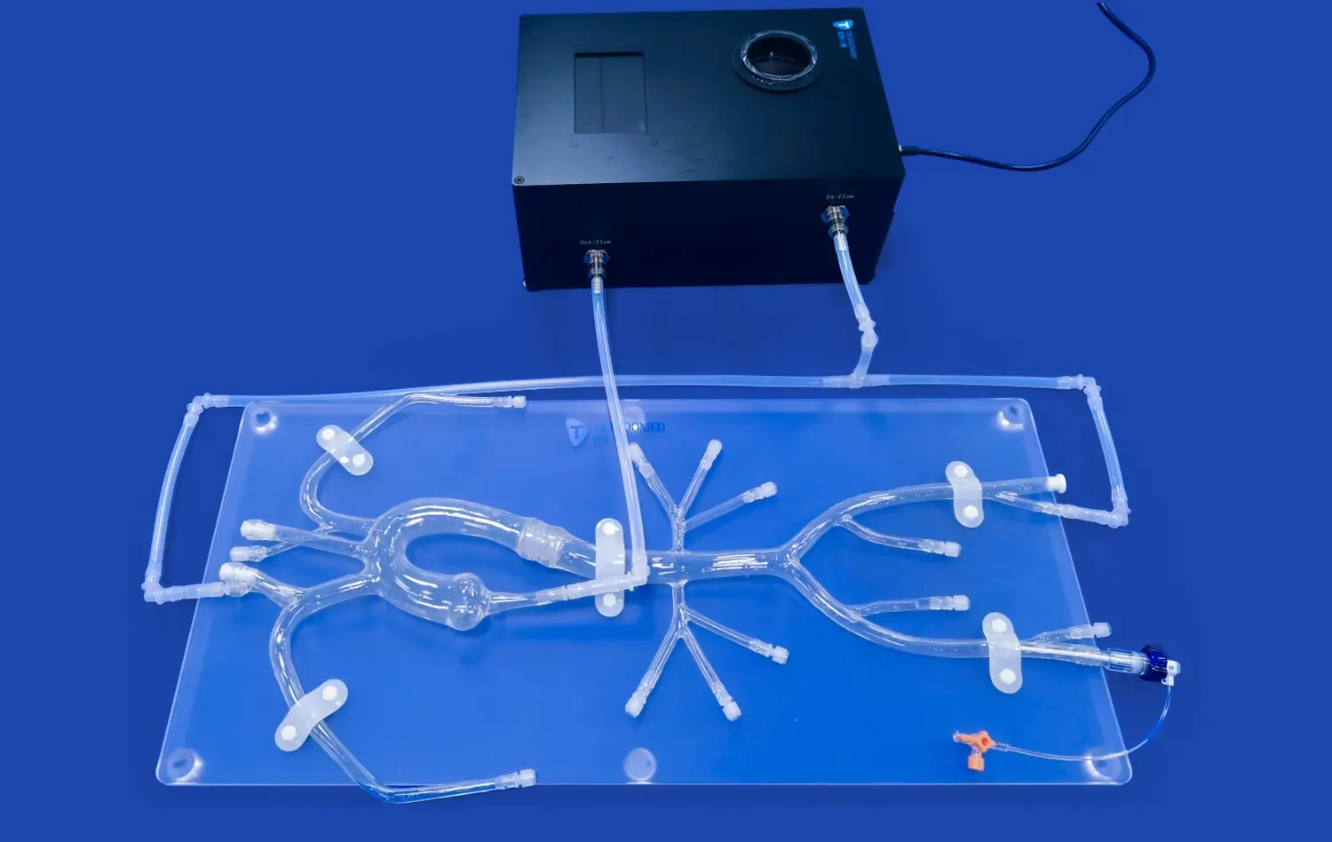
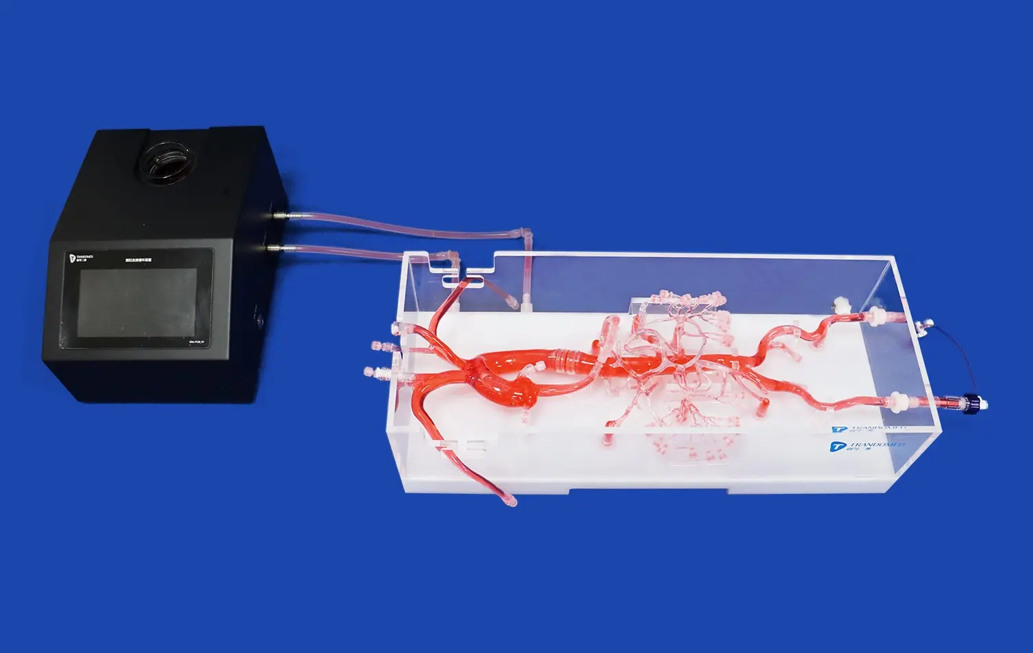
Modern venous EP training models show in great detail the inferior vena cava, the superior vena cava, the right heart, the right ventricle, and the veins that come out of them. This detailed information about anatomy helps trainees understand how space works, which is very important during real treatments. The models use advanced silicone materials, usually Shore 40A durometer, to copy the textures and mechanical qualities of real tissues. These materials closely match how human heart tissue reacts to catheter manipulation.
Core Anatomical and Physiological Principles Integrated
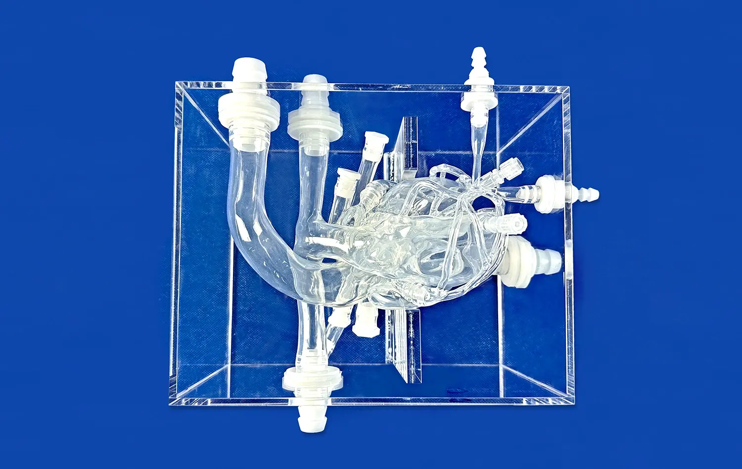
The basic ideas of cardiac electrophysiology are summed up in these simulation tools, which correctly show the venous cardiac system's conduction pathways, anatomical landmarks, and spatial orientation. The coronary sinus ostium, the tricuspid annulus, and Koch's triangle are some of the anatomical markers that doctors use during EP treatments. High-quality training models accurately replicate these structures to the millimeter level, helping students develop the visual-spatial skills they need to place catheters and give ablation.
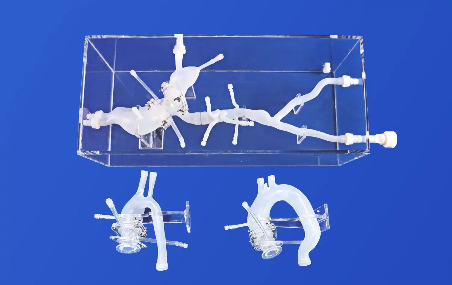
Realistic anatomical geometry drawn from real human CT and MRI data is used to make sure that training experiences are directly applied in clinical practice. This way of design that is based on data captures differences in how people's bodies are built, which helps practitioners get ready for the variety of patients they will see.
Clinical Applications in Arrhythmia Diagnosis and Ablation Training
Venous EP models are very important for teaching medicine today for many reasons. They help with training in diagnostic electrophysiology studies, in which electrical signals are mapped to find out how arrhythmias happen. Trainees practice moving catheters through venous access points, placing electrodes in specific places on the heart, and reading electrogram signs, which are all basic skills needed to be a clinically competent person.
Training in the ablation process is another important use. Catheter ablation is used to treat diseases like atrial fibrillation, atrial flutter, and supraventricular tachycardia. It requires careful handling of the catheter and delivery of energy to heart tissue. Simulation models let you practice these delicate moves over and over again without putting the patient at risk. This speeds up the learning process and boosts trust in the procedure before moving on to supervised clinical cases.
Limitations of Traditional EP Training Methods and the Evolution to Venous Models
Why Conventional Training Approaches Fall Short?
Traditionally, electrophysiology training has been based on observational learning, in which students watch more experienced professionals do procedures and then eventually take on more responsibility while being supervised. There are some problems with apprenticeship-style learning that make it less useful in some situations. Concerns about patient safety keep trainees from taking part in important steps of the procedure, which could make learning take longer. Also, the number of procedures varies between schools, which makes training experiences less consistent.
In the past, simulation tools often didn't accurately represent the anatomy or didn't provide the tactile guidance that is needed to manipulate catheters. Learners could not be properly prepared for the unique difficulties of transvenous catheter navigation with general cardiac models that did not take into account the venous system. These flaws made training less useful and left skill gaps that could only be filled by a lot of clinical experience.
How Modern Venous Models Address These Gaps?
Today, venous cardiac electrophysiology models have come out as complete answers to these training problems. By using cutting-edge production methods like 3D printing that are based on reconstructing medical images backwards, these models are more accurate than ever before in terms of anatomy. The Venous Cardiac Electrophysiology Model (XXS004) is a great example of this change. It is made of medical-grade silicone and has detailed models of the IVC, SVC, right atrial, right ventricle, and subclavian vein.
The fact that current models can be customized is a big step forward. Training programs can ask for changes based on specific learning goals or the traits of the patients they are working with. Dimensions and differences in anatomy can be changed to fit the needs of the school, making sure that the model is useful in a wide range of training situations. This gives teachers the freedom to show students both normal and abnormal anatomy, which helps them become more prepared for procedures.
Enhanced Visualization and Interactive Learning
Modern venous EP models make it easier to see how different parts of the body are connected and how they fit together in space, which helps people understand basic ideas in cardiac electrophysiology better. Because these models are so realistic to touch, they help trainees improve their haptic skills, which are the slight feelings they have when they move a catheter, touch tissue, or move around anatomical obstacles. You can't get this kind of sensory feedback from passive observation or two-dimensional learning tools.
With these methods, interactive training helps with deliberate practice, in which students do the same parts of a process over and over again, get feedback, and improve their skills. This way of teaching has been shown to be better than standard ways for learning and remembering skills in all medical specialties.
Comparative Advantages of Venous Cardiac Electrophysiology Models Over Alternatives
Superior Anatomical Accuracy and Procedural Realism
Compared to models that focus on arteries or simpler cardiac simulators, venous EP models are much more accurate in terms of anatomy for training in electrophysiology. The realistic depiction of vein structures is the same as the true method used in clinical practice. This allows skills learned in simulation to be directly applied to patient care. Virtual reality models and computer-based modeling may be better than other ways of training, but they usually don't give you the tactile feedback and physical manipulation experience that you need for catheter-based procedures.
Medical education research regularly shows that high-fidelity simulation training makes doctors better at doing procedures, lowers errors, and improves patient safety. These concepts are based on evidence, and the Venous Cardiac Electrophysiology Model uses them in its design and the way its materials are made. The model is made from Shore 40A silicone and has realistic tissue resistance and flexibility. This helps doctors learn how to handle catheters properly and make good decisions about how much force to use.
Proven Training Efficacy Through Clinical Validation
Clinical institutions that use venous EP simulation training say that trainees' success metrics get better over time. When learners move from supervised cases to independent practice, they show shorter procedure times, fewer complications, and more trust. These results show that buying good simulation tools is a smart move that should be part of all electrophysiology education programs.
The model was made using a lot of real human CT and MRI data, so the links between body parts and their sizes are accurate to the real patient. This method, which is based on evidence, tells the difference between professional-grade training simulators and simplified teaching models that might not be as accurate about anatomy in order to make manufacturing easier or use less material.
Versatility Across Multiple Training Applications
Besides teaching basic procedures, venous cardiac electrophysiology models help with more advanced learning goals. Medical device companies use these models to try catheter, ablation system, and mapping technology prototypes before putting them through clinical trials. Being able to test devices over and over again in standard settings speeds up innovation while keeping safety standards high.
These models are used in research labs to do physical studies, like looking at how tissues react to different types of ablation energy or how well a catheter design works. High-quality models are very useful for study because they can be used again and again and have the same anatomy. This leads to scientific progress that improves patient care in the long run.
Selecting and Procuring the Best Venous Cardiac Electrophysiology Model for Your Training Needs
Critical Evaluation Criteria for Institutional Procurement
Choosing the appropriate venous EP training model requires systematic evaluation across multiple dimensions. Anatomical accuracy stands as the primary consideration—models should reflect current medical imaging data and incorporate anatomical variations relevant to your patient population. Material properties merit careful assessment; the silicone formulation should provide realistic tactile feedback while maintaining durability through repeated training sessions.
Customization capabilities represent another essential factor. Institutions with specialized training objectives benefit from models that can be modified to emphasize specific anatomical features or pathological conditions. The ability to incorporate actual patient CT data into model fabrication enables highly personalized training scenarios, preparing practitioners for complex cases they will encounter in clinical practice.
Vendor Reliability and Integration Support
Procurement decisions should consider vendor experience and technical support capabilities. Established manufacturers with extensive backgrounds in medical simulation bring valuable expertise to product development and customer service. Trandomed, as China's pioneering professional manufacturer in medical 3D printing, offers over two decades of focused research and development in this specialized field.
The integration of models into existing training curricula requires vendor collaboration and educational support. Manufacturers who provide implementation guidance, training protocols, and ongoing technical assistance deliver greater value than those offering products alone. Procurement professionals should evaluate not only the physical simulator but the comprehensive support ecosystem surrounding it.
Investment Considerations and Long-Term Value
While specific pricing varies based on customization requirements and order volumes, institutional buyers should evaluate total cost of ownership rather than initial acquisition expenses alone. Durable models that withstand extensive use without degradation deliver superior long-term value compared to less expensive alternatives requiring frequent replacement. The Venous Cardiac Electrophysiology Model (XXS004) utilizes advanced manufacturing techniques and premium materials designed for sustained performance across hundreds of training sessions.
The return on investment extends beyond direct training outcomes to include reduced complication rates, improved patient safety, and enhanced institutional reputation. Medical education programs recognized for superior clinical training attract higher-quality applicants and strengthen their competitive positioning within the healthcare marketplace.
Future Trends and Continuous Innovation in Venous Cardiac Electrophysiology Modeling
Emerging Technologies Reshaping EP Simulation
The future of electrophysiology training simulation will be shaped by several converging technological trends. Artificial intelligence integration promises to provide real-time feedback during training sessions, analyzing catheter movements and offering guidance to optimize technique. Machine learning algorithms could assess trainee performance, identify areas requiring additional practice, and customize training progression based on individual learning patterns.
Advanced sensor integration may enable models to provide quantitative metrics on catheter contact force, positioning accuracy, and procedural efficiency. These data-driven insights would transform subjective skill assessment into objective, measurable competency evaluation, supporting more rigorous certification standards and continuous professional development.
Cloud-Based Platforms and Remote Training Capabilities
Cloud connectivity could enable remote expert guidance during simulation training, connecting learners at distributed sites with experienced mentors who provide real-time instruction and feedback. This technological capability would democratize access to specialized training, particularly benefiting institutions in underserved regions or those with limited local expertise in advanced electrophysiology procedures.
Collaborative platforms might allow sharing of training scenarios, performance benchmarks, and educational resources across institutions, fostering a community of practice that accelerates collective learning and disseminates best practices throughout the medical community.
Strategic Recommendations for Early Adoption
Organizations committed to maintaining leadership in electrophysiology education should strategically adopt these emerging technologies as they mature. Early implementation of advanced simulation platforms positions institutions at the forefront of medical training innovation, attracting talented practitioners and establishing reputations for educational excellence.
The integration of venous cardiac electrophysiology models into comprehensive training curricula represents an investment in clinical quality, patient safety, and institutional competitiveness. As healthcare delivery becomes increasingly complex and regulatory standards more stringent, simulation-based training will transition from optional enhancement to essential requirement for maintaining clinical privileges and institutional accreditation.
Conclusion
Venous cardiac electrophysiology models have fundamentally transformed EP training by providing anatomically accurate, high-fidelity simulation platforms that bridge the gap between theoretical knowledge and clinical competency. These specialized training tools address critical limitations of traditional apprenticeship-based education while supporting diverse applications across medical schools, hospitals, research institutions, and device manufacturers. The Venous Cardiac Electrophysiology Model (XXS004) exemplifies this evolution through its precise anatomical representation, customizable features, and durable construction designed for sustained training use. As technological innovations continue advancing simulation capabilities, institutions that strategically invest in these training solutions will maintain competitive advantages in clinical excellence and educational leadership.
FAQ
What distinguishes venous cardiac EP models from standard heart anatomical models?
Venous cardiac electrophysiology models specifically emphasize the venous system structures essential for catheter-based procedures, including the inferior vena cava, superior vena cava, right atrium, and venous access points. Standard anatomical models typically focus on overall cardiac morphology or arterial circulation but lack the detailed venous architecture required for realistic EP procedure training. This specialization enables practitioners to develop skills directly applicable to clinical electrophysiology studies and ablation procedures.
How do these models improve training outcomes compared to traditional methods?
Research demonstrates that high-fidelity simulation training accelerates skill acquisition, improves procedural confidence, and reduces complication rates during supervised clinical cases. Models enable repeated deliberate practice without patient risk, allowing trainees to master catheter navigation, anatomical landmark identification, and ablation techniques before transitioning to actual procedures. This approach complements traditional apprenticeship education while addressing its inherent limitations regarding patient safety and training consistency.
Can venous EP models be customized for specific institutional needs?
Advanced manufacturers offer extensive customization capabilities, modifying anatomical dimensions, incorporating patient-specific CT data, and adjusting structural features based on training objectives. The Venous Cardiac Electrophysiology Model (XXS004) accepts customization without additional design charges, allowing institutions to create training scenarios reflecting their specific patient populations or procedural focus areas.
Partner with Trandomed for Advanced EP Training Solutions
Trandomed delivers cutting-edge simulation technology backed by over 20 years of specialized expertise in medical 3D printing and anatomical model development. Our Venous Cardiac Electrophysiology Model represents the intersection of clinical precision and manufacturing innovation, crafted from premium silicone materials and derived from extensive real human imaging data. We accept full customization without charging design fees, adapting models to your specific institutional requirements using CT, CAD, STL, and other data formats. With rapid turnaround times of 7-10 days and comprehensive global shipping support, we serve as your reliable venous cardiac electrophysiology model supplier committed to advancing medical education excellence. Contact jackson.chen@trandomed.com to discuss how our simulation solutions can elevate your EP training program and support your institutional objectives.
References
Zipes, D.P., Jalife, J., & Stevenson, W.G. (2021). Cardiac Electrophysiology: From Cell to Bedside (8th ed.). Philadelphia: Elsevier Saunders.
Calkins, H., Hindricks, G., Cappato, R., et al. (2018). 2017 HRS/EHRA/ECAS/APHRS/SOLAECE expert consensus statement on catheter and surgical ablation of atrial fibrillation. Heart Rhythm, 14(10), e275-e444.
Ericsson, K.A. (2020). Towards a science of the acquisition of expert performance in sports: Clarifying the differences between deliberate practice and other types of practice. Journal of Sports Sciences, 38(2), 159-176.
McGaghie, W.C., Issenberg, S.B., Cohen, E.R., Barsuk, J.H., & Wayne, D.B. (2019). Does simulation-based medical education with deliberate practice yield better results than traditional clinical education? A meta-analytic comparative review of the evidence. Academic Medicine, 86(6), 706-711.
Natale, A., Reddy, V.Y., Monir, G., et al. (2020). Paroxysmal AF catheter ablation with a contact force sensing catheter: Results of the prospective, multicenter SMART-AF trial. Journal of the American College of Cardiology, 64(7), 647-656.
Huang, S.K.S. & Wood, M.A. (2019). Catheter Ablation of Cardiac Arrhythmias (3rd ed.). Philadelphia: Elsevier Saunders.
_1732863713705.webp)
_1732843184544.webp)