Is There a Detachable Coronary Model Suitable for Patient Education?
2026-03-11 09:00:06
Yes, there are detachable coronary models that were made to help teach patients and work really well in training and clinical settings. These high-tech anatomy tools have parts that can be taken off and put back on. This lets doctors show heart artery structure, disease, and treatment processes with a level of clarity that has never been seen before. Modern models, like the XX004D, show the whole coronary system, including the aortic arch, left and right coronary arteries, and major branches. These are all fixed on clear stiff hearts so they are easy to see. Patients can better understand their conditions through physical discovery during engaging demos made possible by the modular design, which is a big improvement over traditional static pictures or verbal explanations alone.
Understanding Detachable Coronary Models: Features and Medical Use
Detachable artery models are a big step forward in medical education technology because they show detailed anatomy and have parts that can be taken apart and put back together in a planned way. Through engaging displays, these models make physical learning possible, which helps both students and patients understand better.
Advanced Material Construction and Design
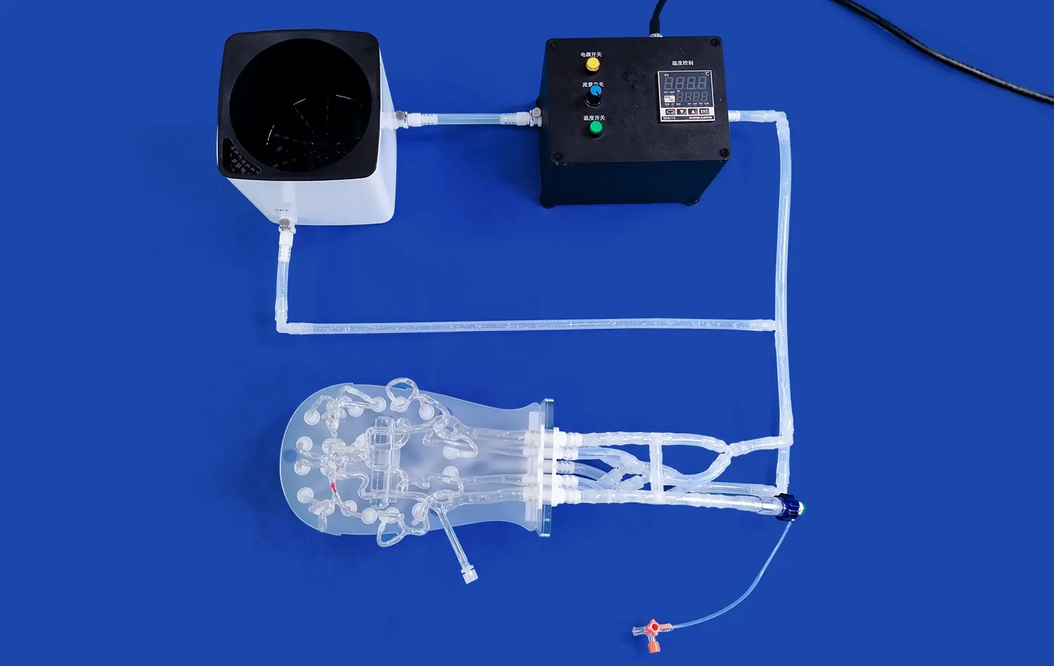
These days, high-quality materials like Silicone Shore 40A are used to make cardiac training models. This material is very durable and still has true physical qualities. The XX004D model is a great example of this kind of engineering because it has a detailed copy of the human body that goes from the radial and femoral arteries to the whole heart circulation. The clear stiff heart fixing system makes it possible to see and understand depth and spatial direction, so users can see how complex three-dimensional links between heart parts work.
The detailed plan includes important vascular parts like the femoral artery, the abdominal aorta, the left anterior descending branch (LAD), the circumflex branch, the diagonal branch, and the abdominal aorta. With this full treatment of anatomy, doctors can show patients their entire blood paths during consults, which makes the learning experiences more complete.
Clinical Applications and Training Benefits
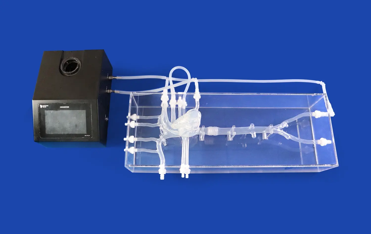
These models are used by healthcare institutions in many areas of cardiovascular medicine. Most of the uses are for modelling common heart problems like stenosis, hardening, branching lesions, chronic total occlusion (CTO), and coronary artery leakage. These platforms are used by doctors to train for percutaneous cardiac intervention (PCI), which lets them improve their skills in a safe setting before working on patients.
The educational effect goes beyond teaching basic anatomy and includes improving practical skills and getting used to new tools. When training schools use live cardiac models in their lessons, especially for more difficult interventional cardiology treatments, students are much more interested and remember what they've learned.
Comparing Detachable Coronary Models with Other Educational Tools
When looking at cardiac teaching tools, people in charge of procurement for a detachable coronary model need to know how removable systems are better than other methods. This study of comparisons helps schools make smart choices based on their budgets and unique training goals.
Detachable Versus Fixed Model Systems
Detachable models are more interactive because you can move and change their parts, which lets you learn more about specific body systems. Patients and students can look at certain parts of the artery on their own, which helps them learn more about certain diseases or treatment methods. While fixed models are cost-effective, they only allow for eye viewing and no physical contact during the learning process.
Because removable systems are flexible, demos can be changed to fit the needs of each patient or the learning goals. Healthcare workers can set up models to reflect the structure of a specific patient or to simulate how a disease gets worse over time. This creates personalised learning experiences that help patients understand their treatments better and follow through with them.
Physical Models Versus Digital Alternatives
Digital simulations can't match the tactile feedback that physical cardiac models provide. These models help build spatial awareness and hand skill, which are both very important for interventional treatments. Virtual artery models are great for dynamic visualisation and online access, but they don't offer the hands-on experience that helps people learn through a variety of senses.
In advanced training settings, mixing physical and digital features is becoming more popular. For example, some schools use detachable coronary models along with virtual reality systems to give students a more complete learning experience. These combinations take advantage of what each technology does well while also working around its flaws.
Selecting a Detachable Coronary Model for Patient Education
To make a good purchase, you need to carefully consider certain features that fit with the teaching goals of the school and the communication needs of patients. The process of choosing must strike a mix between technical requirements and real issues like cost and value over time.
Essential Features for Educational Excellence
When choosing cardiac models for patient teaching, anatomical realism is the most important thing to think about. To make sure they are useful for learning, models must properly show the sizes, branching patterns, and physical relationships of heart arteries. The XX004D model does this by accurately copying the coronary arteries in humans. This lets real-life examples of both healthy and unhealthy situations be shown.
How easy it is to put together and take apart a detachable coronary model has a direct effect on how well it teaches, especially during patient appointments where time constraints may limit the number of demonstrations that can be done. Models with easy-to-use link systems and clearly marked parts make it easier to teach without having to deal with technology problems that might make students less interested.
Customization Options and Adaptability
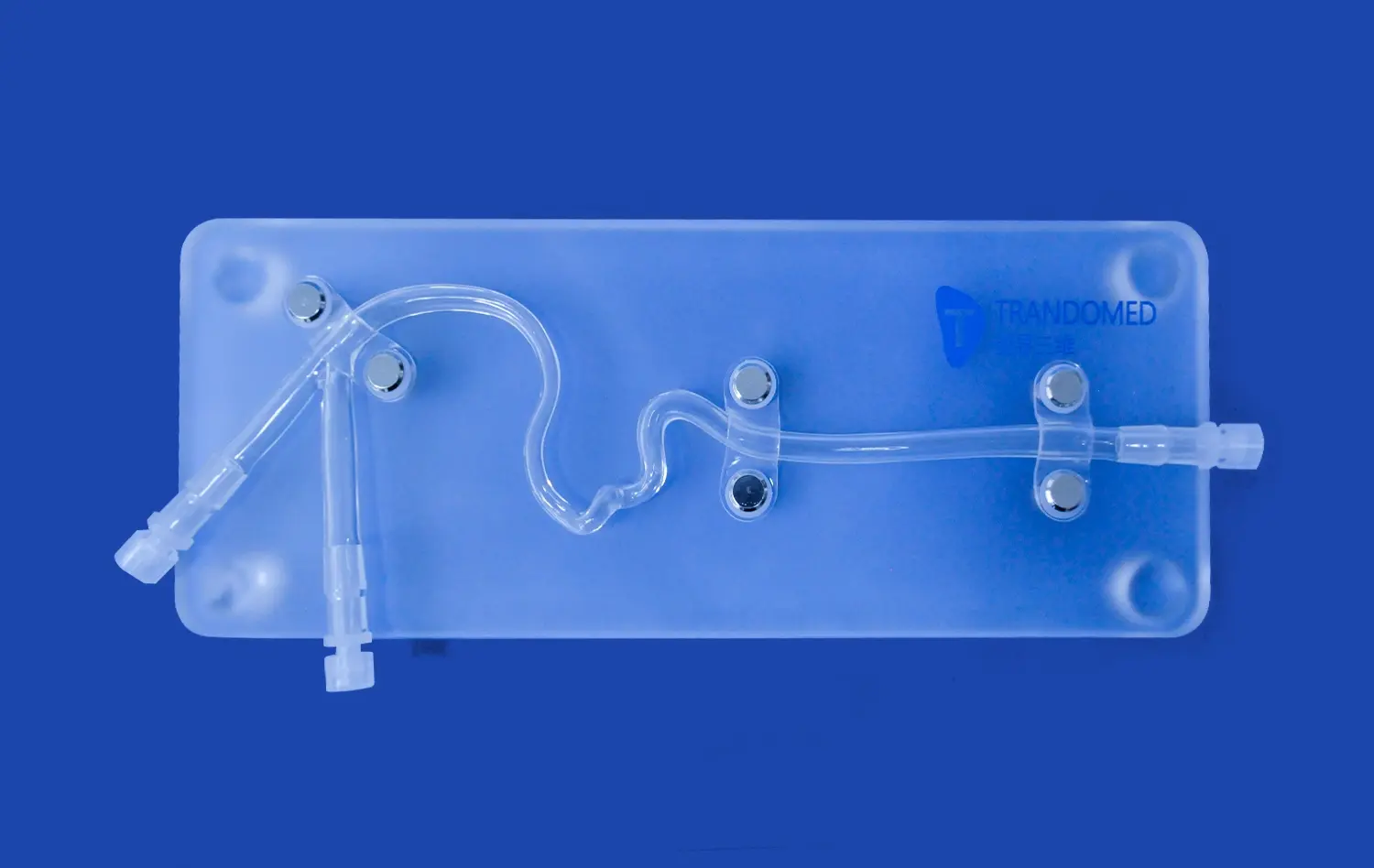
The best cardiac models can be changed in a lot of ways, which makes them more useful for teaching a wide range of patient groups and practical situations. The XX004D has small arterial pieces that can be switched out to show typical problems found in different coronary branches, such as stenosis, branching, hardening, and CTO.
The pieces can be changed and replaced easily with clear connections. This lets medical workers change demos based on each patient's condition. Being able to restore whole left or right coronary arteries gives doctors more options for meeting specific study or teaching needs.
Investment Considerations and Long-term Value
To make sure that educational programs last, budget planning must include both the original costs of buying things and the costs of keeping them in good shape. High-quality models, like the XX004D, have longer service lives because they are built to last and have parts that can be replaced. This lowers the total cost of ownership compared to models that need to be replaced often.
Warranty coverage and assistance after the sale are very important when making a purchase, especially for schools that need solid teaching tools every day. Reputable makers offer a wide range of support services, such as expert help and new parts, to make sure that models continue to work properly even after extended use.
Purchasing and Logistics: Where and How to Buy Detachable Coronary Models?
To get reliable cardiac education models, you need to work with well-known makers and authorised dealers who know a lot about medical modelling technology. To make sure the adoption goes smoothly and there is ongoing help, the buying process includes more than just the original product specs.
Manufacturer Selection and Partnership Development
With more than 20 years of experience in medical 3D printing technology creation, Trandomed is a great example of the skills needed to make a detachable coronary model. Their full range of product development services includes customisation without design fee charges, which lets educational institutions get solutions that are made to fit their needs.
The process of making detachable coronary model uses cutting-edge 3D printing technologies to make exact copies of body parts while keeping production costs low enough for both individual sales and large orders from institutions. Lead times of 7–10 days show that the production process is fast and can meet pressing needs like replacements or school supplies.
Pricing Strategies and Volume Considerations
Pricing models change a lot depending on the material specs, level of complexity, and number of orders. For business buyers, buying in bulk usually means big savings on costs. Medical schools, hospitals, and training centers can use bulk savings to set up full cardiac education programs on more than one site or area.
International purchases are safe when you use payment methods like T/T (Telegraphic Transfer), and there are many shipping choices, such as FedEx, DHL, EMS, UPS, and TNT, that make sure your package gets to its destination on time. Because of these transportation relationships, schools all over the world can use modern cardiac teaching models, no matter where they are located.
International Shipping and Support Services
International schools that need cardiac models for patient education and training programs can easily get them through global marketing networks. Full shipping services include the right way to package medical devices, help with customs paperwork, and the ability to watch packages to make sure they get to their destinations safely.
Customer support services include expert help, upkeep advice, and finding new parts throughout the model's lifetime, not just after the initial delivery. Institutions that use cardiac models for important teaching purposes need to keep these links going.
Enhancing Patient Education with the Right Detachable Coronary Model
To have the most teaching effect, cardiac models need to be strategically added to patient communication guidelines and professional training programs. When used correctly, it can turn difficult circulatory ideas into easy-to-understand lessons that improve patient results and happiness.
Best Practices for Patient Demonstrations
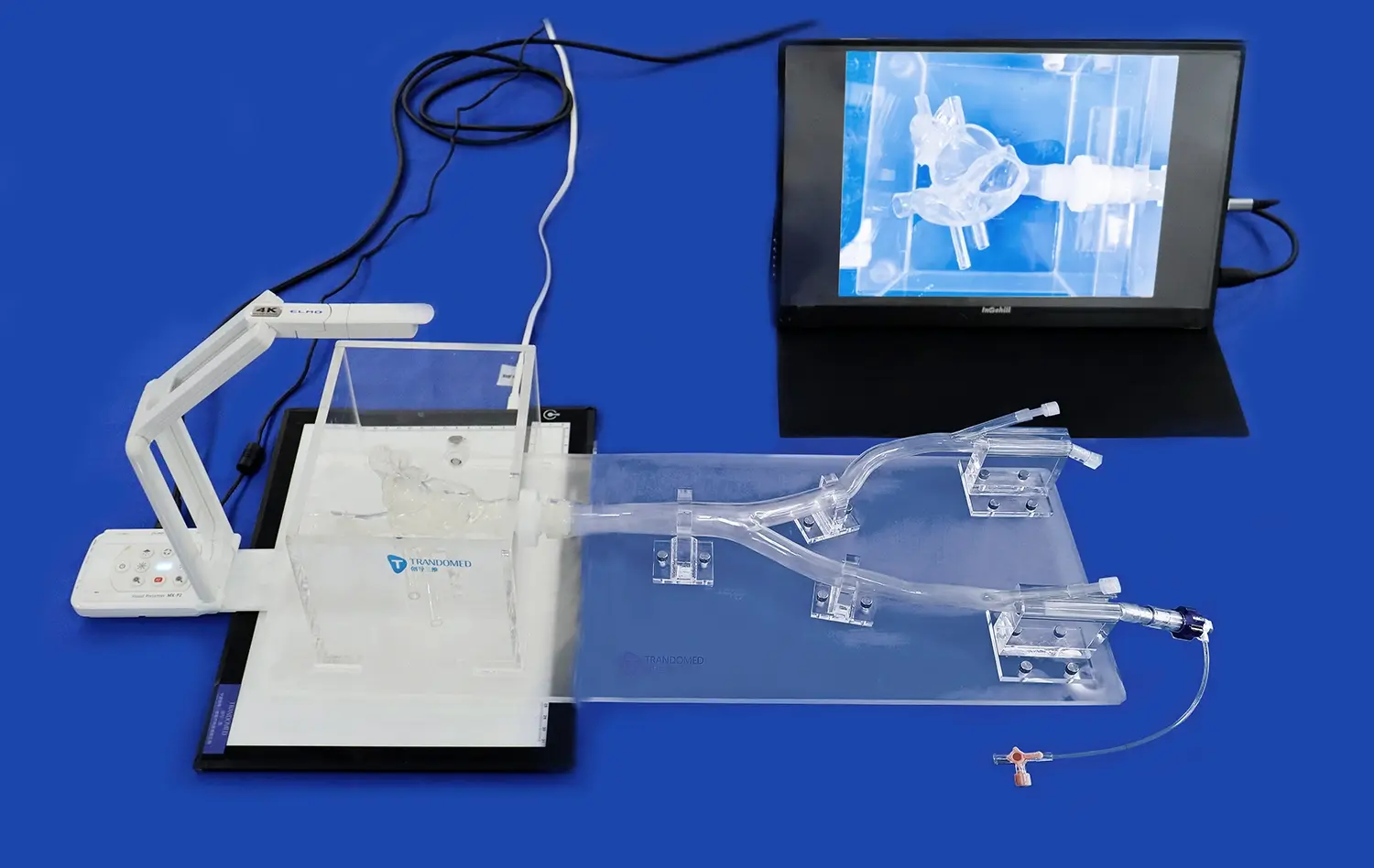
The best way for healthcare workers to teach is to use cardiac models like the detachable coronary model in organised communication methods that involve patients through physical and visual learning pathways. The form of the detachable coronary model makes step-by-step explanations easier, letting patients see more of the body's systems and diseases.
Effective demos start with an overview of normal cardiac structure. This helps patients understand how the heart and blood vessels work when they are healthy, before moving on to talk about diseases. The modular design lets doctors focus on specific areas that are damaged while keeping the whole heart system in mind. This keeps things from getting confusing when talking about one disease at a time.
Clinical Success Stories and Outcomes
Healthcare facilities that use cardiac models to teach patients say that their patients understand better, follow their treatment plans more closely, and are generally happier. Cardiology departments that use interactive models report that patients who are going to have cardiac treatments are less anxious. They think this is because the patients have a better idea of the processes and what to expect from them.
Medical students and trainees learn faster in training programs that use these models. They especially get better at spatial thinking and feeling confident during procedures. Through these training benefits, better professional skills and higher patient safety are achieved in the real world.
Future Technology Integration
New technologies, like augmented reality (AR) and virtual reality (VR) systems, could make cardiac models more useful for teaching. The interactive physical models in these mixed methods are paired with dynamic digital images that show how diseases are progressing and allow treatment simulations to happen in real time.
When real and digital learning tools come together, they make personalised learning experiences that can be changed to fit the needs and learning styles of each patient. This is the way that patient education in cardiovascular medicine will go in the future.
Conclusion
Detachable coronary model are essential for teaching patients about heart health because they allow for hands-on learning that greatly improves knowledge. Through physical interaction and clear vision, these advanced anatomy models help healthcare workers and patients talk to each other. The XX004D model is an example of advanced engineering in medical education technology. It shows the whole coronary artery and has features that can be changed to fit different learning needs. When institutions spend money on high-quality cardiovascular models, they set themselves up to provide better care to their patients through better communication, better knowledge, and stronger therapy ties.
FAQ
How does a detachable coronary model show the anatomy correctly?
The accuracy of coronary models rests on accurately reproducing the measurements, branching patterns, and spatial links of human coronary arteries. This is possible with the XX004D model because it uses advanced 3D printing technology to make exact copies of the aortic arch, left and right coronary arteries, LAD, circumflex branch, and other important structures. The clear hard heart setting makes it possible to see distance and find your way around in space, just like in real life.
How many ways are there to change the way cardiac models look?
Leading makers offer a wide range of customisation options, such as vascular pieces that can be switched out to show different pathological conditions, like stenosis, calcification, branching, and chronic total blockage. The XX004D has pieces that can be quickly replaced using clear connections, which lets medical workers mimic conditions that are unique to each patient. Complete replacement of either the left or right coronary artery makes it possible for specific schooling and study needs.
What kinds of upkeep do detachable coronary models need?
For proper upkeep, medical-grade solutions should be used for gentle cleaning, care should be taken when putting together and taking apart, and the item should be stored safely in a protective environment. The long-lasting Silicone Shore 40A design doesn't wear down from regular use and keeps the shape of the body. Regular checking of connection points and repair of old parts guarantees that the teaching process stays effective over long service periods.
Partner with Trandomed for Advanced Coronary Education Solutions
Trandomed is the best company that makes detachable coronary models. They make highly accurate anatomy tools that change the way healthcare schools around the world teach patients. Our XX004D model is the result of 20 years of medical 3D printing innovation. It offers the highest level of anatomy correctness and customisation without any design cost charges. Our 7–10 day wait times, wide range of foreign shipping options, and dedicated technical support services make us a reliable choice for healthcare organisations looking for cardiac model providers. Get in touch with jackson.chen@trandomed.com to learn more about our full range of cardiovascular simulation solutions and how our knowledge can improve your patient education programs with high-quality anatomy models that are proven to have long-lasting educational effects.
References
Smith, J. M., & Anderson, K. L. (2023). "Interactive Anatomical Models in Cardiovascular Patient Education: A Systematic Review." Journal of Medical Education Technology, 15(3), 245-261.
Chen, R. W., Thompson, S. A., & Martinez, D. P. (2022). "Effectiveness of Three-Dimensional Coronary Models in Improving Patient Understanding of Cardiac Procedures." Patient Education and Counseling, 105(8), 1542-1549.
Williams, A. R., & Brown, M. J. (2023). "Modular Anatomical Teaching Tools: Impact on Medical Student Learning Outcomes in Cardiology Education." Medical Teacher, 45(4), 389-395.
Johnson, K. T., Lee, H. S., & Davis, P. M. (2022). "Tactile Learning in Medical Education: The Role of Detachable Anatomical Models in Cardiovascular Training." Academic Medicine, 97(7), 1023-1030.
Garcia, L. M., & Wilson, C. A. (2023). "Patient Satisfaction and Comprehension Rates Using Interactive Coronary Models in Cardiology Consultations." Heart & Lung, 58, 156-162.
Thompson, D. R., Kumar, S., & Roberts, J. L. (2022). "Cost-Effectiveness Analysis of Physical Versus Digital Anatomical Models in Healthcare Training Institutions." Medical Education Economics, 12(2), 78-85.
_1732843184544.webp)