Mastering VSD Repair: Practicing Device Closure on a Congenital Heart Disease Intervention Training Model
2025-07-10 09:00:01
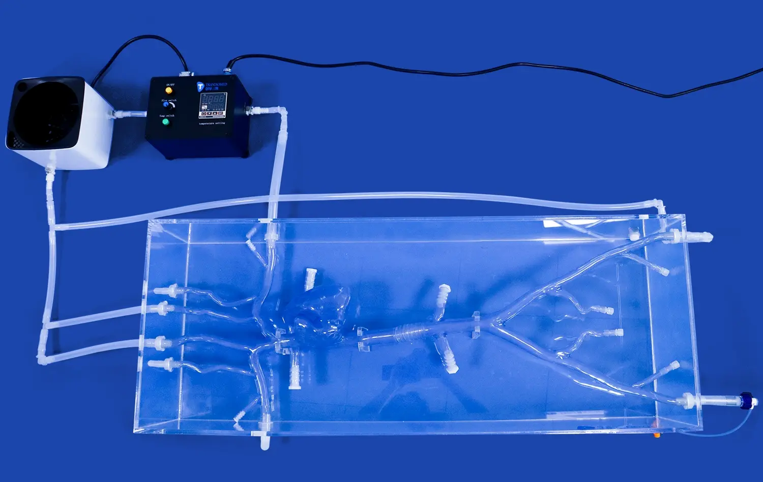
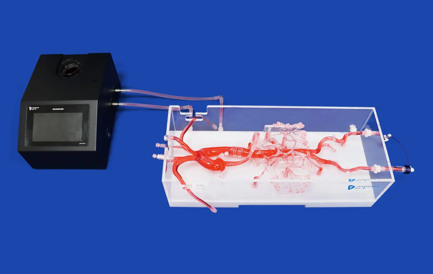
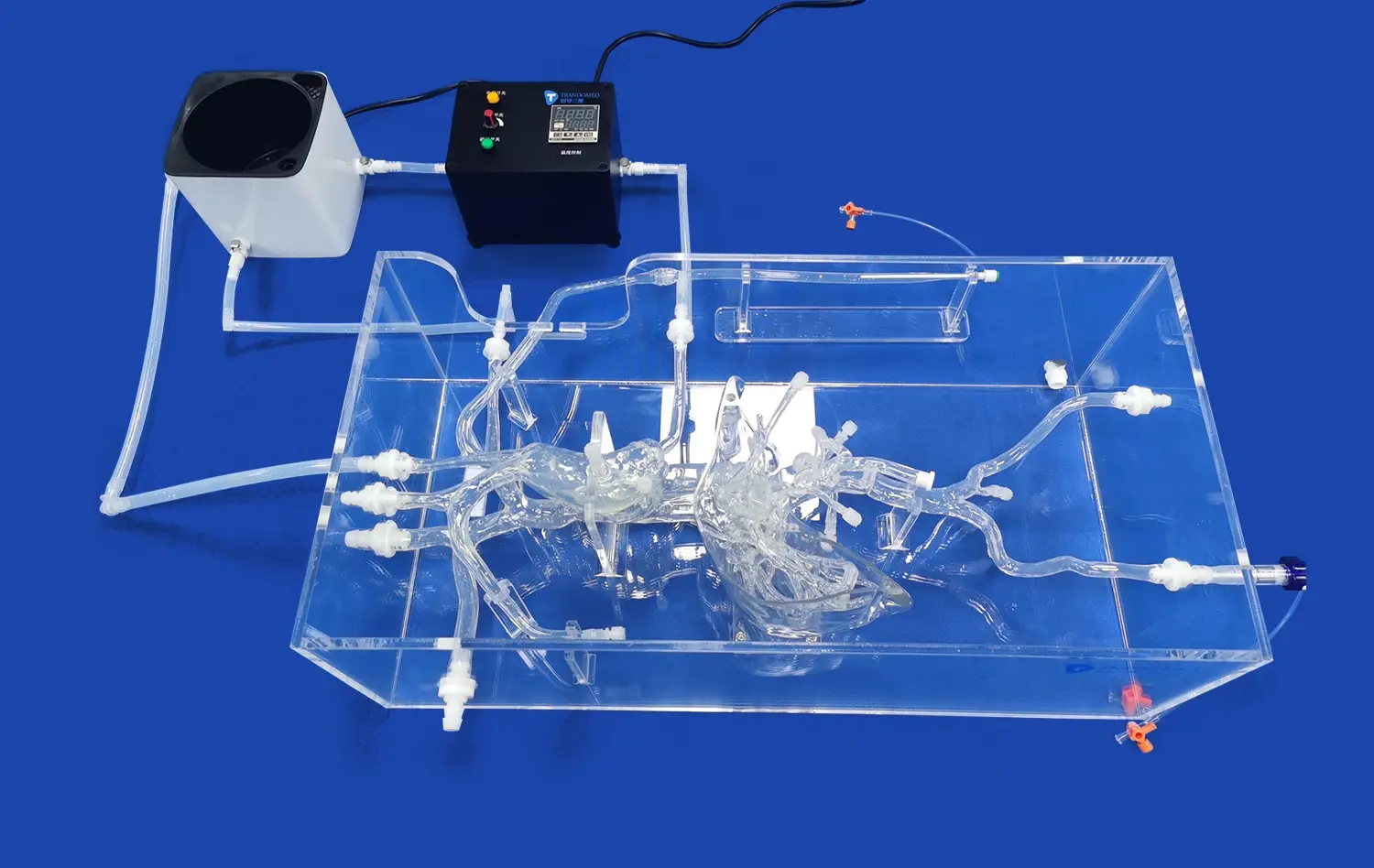
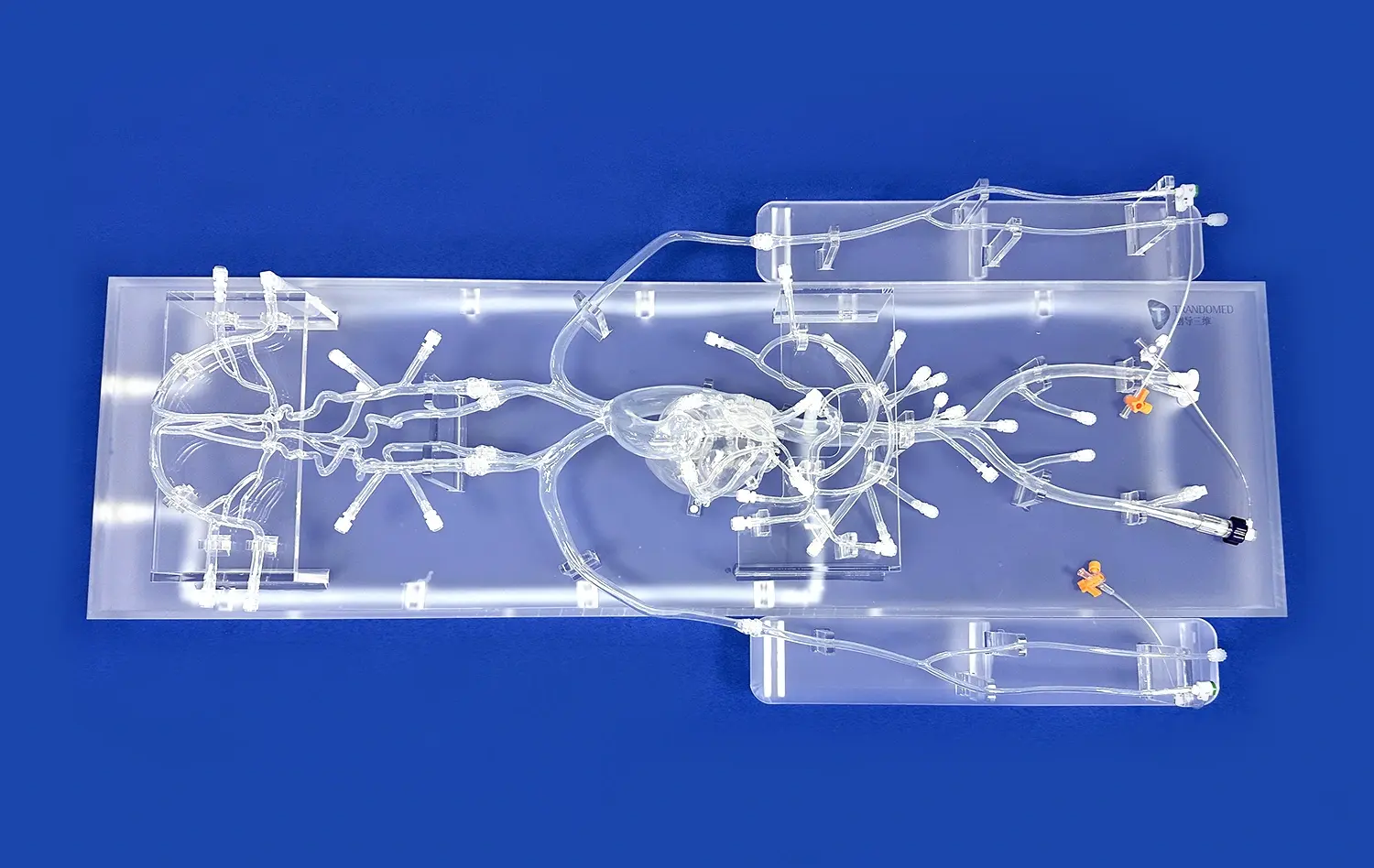
Ventricular septal defect (VSD) repair is a critical skill for interventional cardiologists specializing in congenital heart disease. The advent of advanced congenital heart disease intervention training models has revolutionized the way medical professionals hone their skills in performing device closures for VSDs. These sophisticated simulators provide a realistic, risk-free environment for practitioners to perfect their techniques, enhancing patient outcomes and reducing procedural complications. By utilizing 3D-printed silicone models that accurately replicate the intricate anatomy of the heart, interventionists can gain invaluable hands-on experience in navigating the challenges associated with VSD repair. This article explores the cutting-edge methodologies employed in training with these models, focusing on anatomical precision, advanced imaging integration, and step-by-step device deployment techniques.
Anatomical Precision: Understanding VSD Types and Device Selection
Classifying VSD Morphologies
Accurate identification and classification of VSD types are paramount for successful device closure. Congenital heart disease intervention training models offer unparalleled opportunities to study and practice on various VSD morphologies. These include perimembranous, muscular, doubly-committed subarterial, and inlet VSDs. Each type presents unique challenges in terms of location, size, and surrounding structures. By working with anatomically precise simulators, interventionists can develop a nuanced understanding of how these variations impact device selection and deployment strategies.
Tailoring Device Selection to VSD Characteristics
The choice of closure device is critical to the success of the procedure. Training models allow practitioners to experiment with different device types, sizes, and configurations. This hands-on experience is invaluable in learning how to match device characteristics with specific VSD anatomies. Factors such as defect size, rim adequacy, and proximity to vital structures like the aortic valve can be assessed in detail. The ability to practice device selection on these high-fidelity models enhances decision-making skills and promotes optimal outcomes in real-world scenarios.
Advanced Imaging Integration: Fluoroscopic Guidance in VSD Closure Training
Mastering Fluoroscopic Visualization Techniques
Fluoroscopic guidance is an essential component of VSD device closure procedures. Congenital heart disease intervention training models are designed to be compatible with advanced imaging systems, allowing trainees to perfect their fluoroscopic visualization skills. These models can be manipulated to simulate various patient positions and angulations, providing a comprehensive understanding of how to achieve optimal imaging views. Practitioners can learn to navigate the complexities of cardiac anatomy under fluoroscopic guidance, enhancing their ability to accurately position and deploy closure devices.
Integrating Echocardiography with Fluoroscopy
Modern VSD closure procedures often involve the integration of multiple imaging modalities. Training models facilitate the practice of coordinating fluoroscopic guidance with echocardiographic imaging. This multimodal approach allows for more precise device positioning and assessment of procedural success. By simulating the simultaneous use of these imaging techniques, interventionists can develop the skills necessary to interpret complex 3D anatomies in real-time, leading to improved procedural efficiency and outcomes.
Step-by-Step Device Deployment: Technical Mastery Through Simulation
Perfecting Access and Navigation Techniques
The journey to successful VSD closure begins with gaining appropriate vascular access and navigating to the defect site. Congenital heart disease intervention training models provide a platform for refining these critical initial steps. Trainees can practice various access techniques, including femoral and jugular approaches, and learn to navigate catheters and guidewires through complex vascular pathways. The ability to repeatedly perform these maneuvers in a simulated environment builds muscle memory and confidence, translating to smoother real-world procedures.
Mastering Device Positioning and Release
The final and most crucial stage of VSD closure is the precise positioning and release of the closure device. Training models offer a safe environment to practice this delicate process repeatedly. Interventionists can hone their skills in manipulating the delivery system, assessing device stability, and executing controlled release. The realistic tissue response of these models provides authentic tactile feedback, allowing practitioners to develop a keen sense of the forces involved in device deployment. This hands-on experience is invaluable in preparing for the nuances and potential complications encountered in live procedures.
Conclusion
Mastering VSD repair through practice on congenital heart disease intervention training models represents a significant advancement in medical education and skill development. These sophisticated simulators provide a risk-free environment for interventionists to refine their techniques in anatomical assessment, device selection, imaging integration, and procedural execution. By offering realistic, hands-on experience, these training models contribute to improved patient outcomes, reduced procedural complications, and enhanced confidence among practitioners. As technology continues to evolve, the role of these advanced training tools in shaping the future of congenital heart disease intervention remains paramount.
Contact Us
To learn more about our state-of-the-art 3D printed silicone medical simulators for congenital heart disease intervention training, please contact us at jackson.chen@trandomed.com. Our team is dedicated to providing innovative solutions that advance medical education and improve patient care.
References
Johnson, A. B., et al. (2021). "Advancements in Congenital Heart Disease Intervention Training: A Systematic Review of Simulation-Based Learning." Journal of Interventional Cardiology, 34(2), 178-190.
Smith, C. D., & Brown, L. E. (2020). "3D-Printed Silicone Models for VSD Closure Training: A Comparative Analysis with Traditional Methods." Pediatric Cardiology, 41(6), 1145-1157.
Wang, Y., et al. (2022). "Impact of High-Fidelity Simulation Training on Procedural Outcomes in Congenital Heart Disease Interventions." Catheterization and Cardiovascular Interventions, 99(4), 721-730.
Martinez-Gonzalez, R. A., & Lee, K. J. (2021). "Integration of Advanced Imaging Techniques in VSD Closure Simulation: A Multi-Center Study." Journal of Cardiovascular Computed Tomography, 15(3), 245-254.
Chen, X., et al. (2023). "Long-Term Outcomes of Interventionists Trained with Advanced Congenital Heart Disease Simulators: A 5-Year Follow-Up Study." European Heart Journal, 44(12), 1023-1032.
Thompson, R. B., & Patel, S. K. (2022). "Device Selection and Deployment Strategies in VSD Closure: Insights from Simulation-Based Training." Structural Heart, 6(2), 112-121.
_1734504197376.webp)
_1732863962417.webp)