The way doctors do complicated brain anatomy and invasive treatments has changed a lot because of realistic neurovascular simulations. A cerebral model copies the complex features of the human brain's circulatory system. This gives medical students a safe, controlled space to practice important neurosurgery skills without putting patients at risk. These training tools are based on real anatomy and include thorough pictures of brain vessels, tumors, and diseases. This helps students and healthcare workers get better at diagnosing problems, planning surgeries, and doing invasive procedures. These advanced modeling tools are now necessary in medical schools, hospitals, and study centers all over the United States because they bridge the gap between theory knowledge and hands-on clinical experience. They are an important part of current brain training programs because they cut down on mistakes and boost skills at the same time.
Understanding Realistic Cerebral Models in Medical Training
To teach neurosurgery well, students must have access to tools that properly reflect the complexity of the human brain. Realistic neurovascular models are an important link between what you learn in school and what you do in the field.
What Makes a Cerebral Vascular Simulator Realistic?
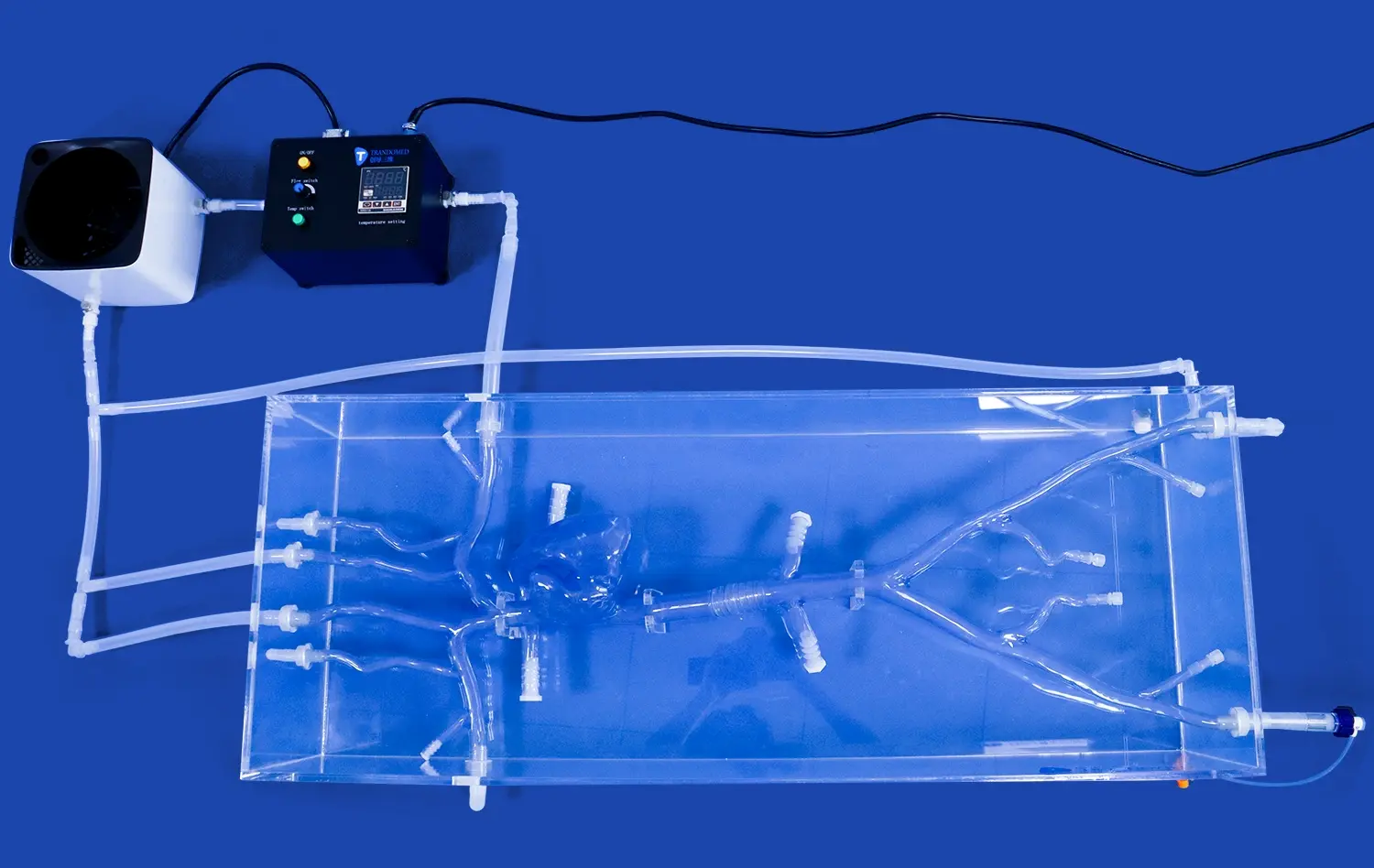
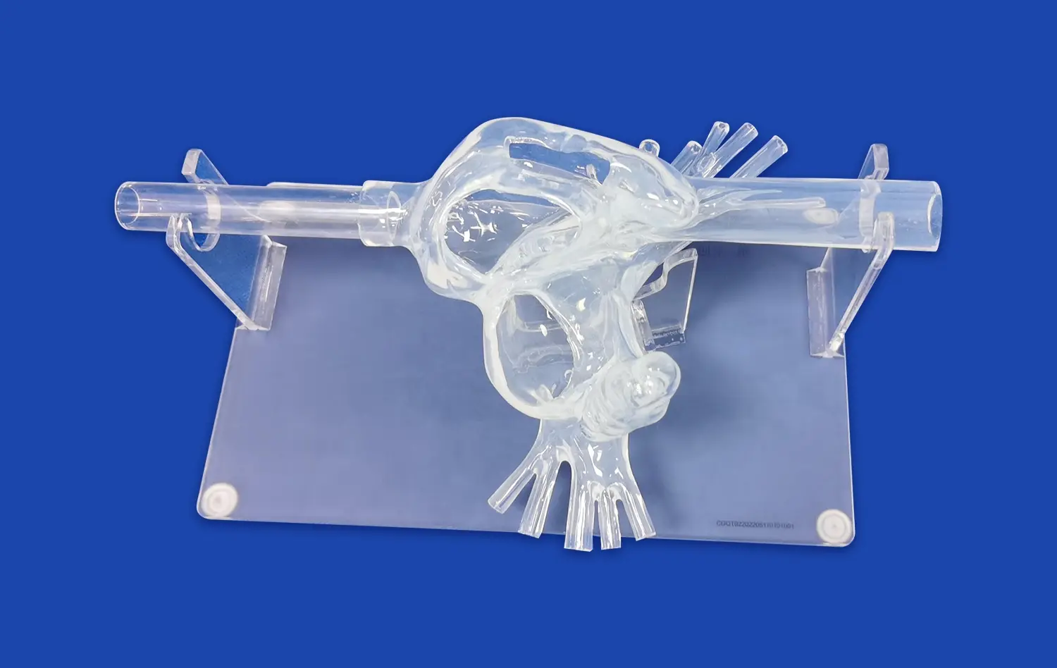
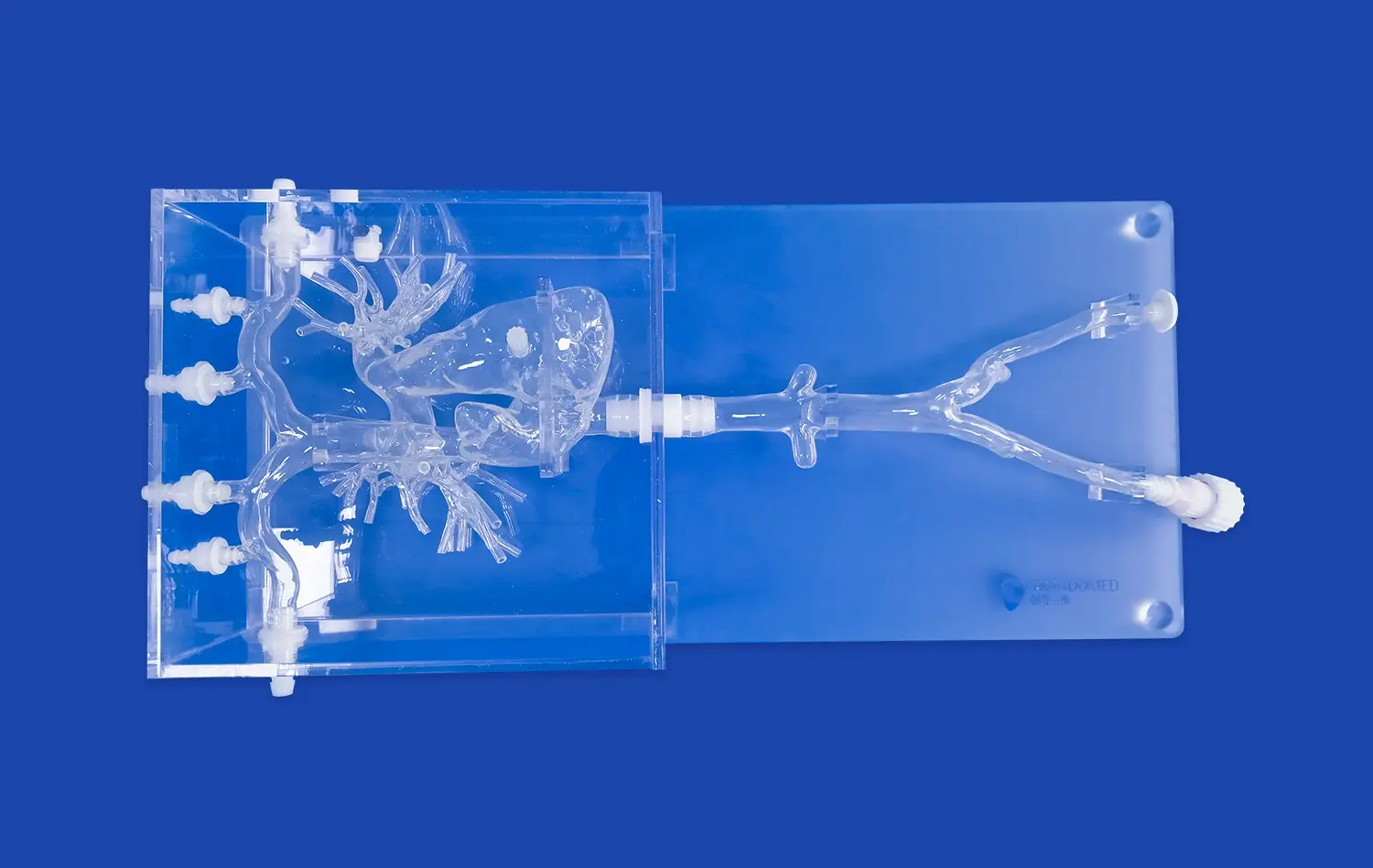
The intricate structure of the brain's blood veins is captured with amazing accuracy in real brain models. Instead of simple teaching images or basic anatomy charts, these advanced training tools show the texture, flexibility, and spatial relationships that are present in real tissue. This dedication to accuracy in anatomy is shown by the Circle of Willis Aneurysm model (Product No. SJK002D), which is made from medical-grade silicone Shore 40A. This choice of material makes sure that the physical input during catheter navigation feels a lot like the walls of an artery. This helps students get ready for the resistance and flexibility they'll feel during real treatments.
Students can see arterial paths from different angles thanks to the three-dimensional spatial representation and clear plastic housing. This all-around view is very helpful when learning how to go through complicated treatment routes. Medical schools and clinical skills centers say that when students learn with high-fidelity models, they are much better at using spatial thinking when they are working with real patients.
Key Anatomical Features Replicated
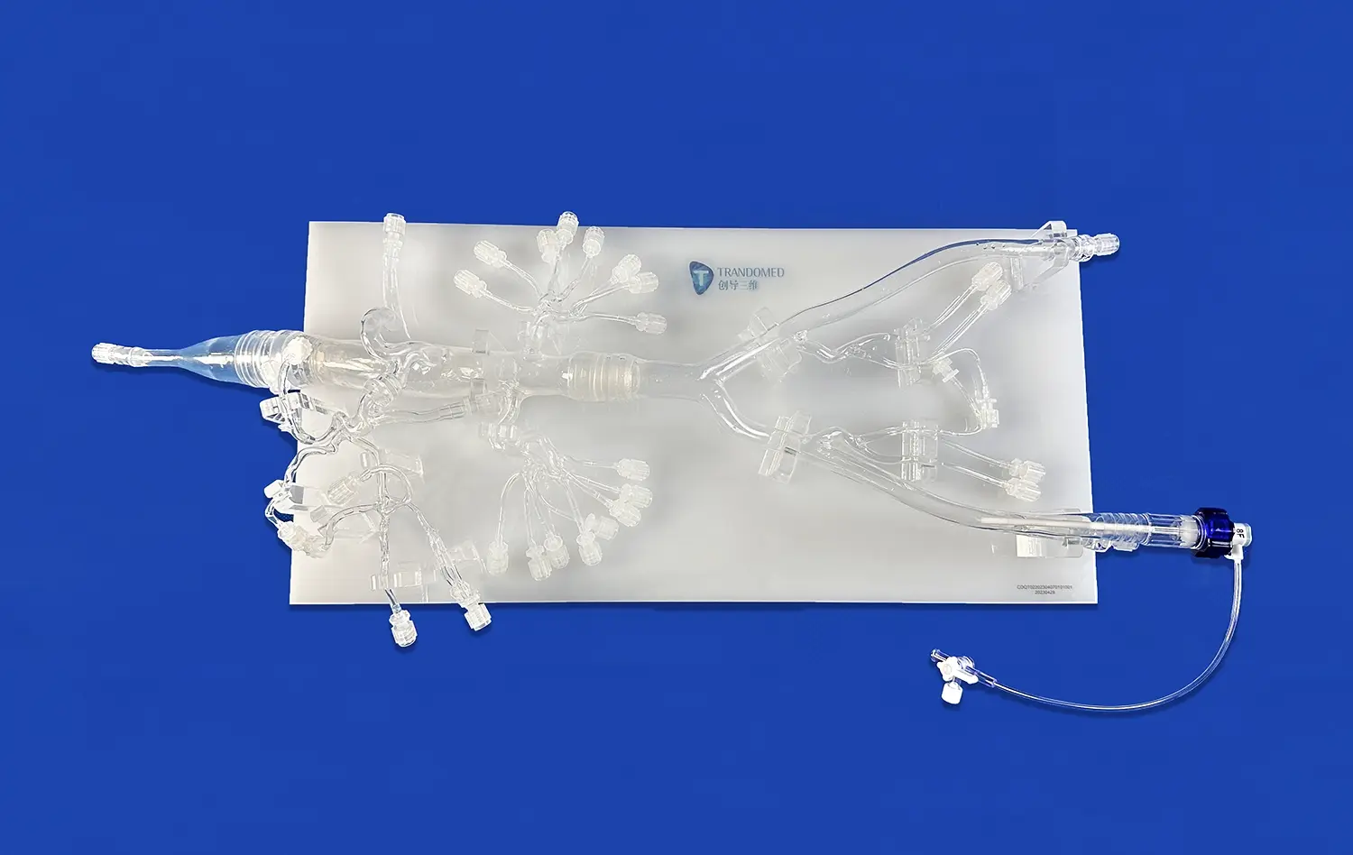
For neurovascular training to work, models need to include all of the different parts of brain artery structure. The internal carotid vessels, the basilar artery, and the whole Circle of Willis formation are accurately shown on good models. These structures support the flow of blood to the brain, and it is important to know how they are put together for anyone doing diagnostic imaging or therapeutic treatment.
Advanced training systems pay the same amount of attention to pathological differences. Aneurysms on the eye section, the branching of the middle cerebral artery, the basilar tip, or the carotid artery can make tamponade treatments more difficult. Because lesion features like size, neck width, and wall thickness can be changed, teachers can make training models that build skill in a planned way. Researchers have shown that being able to customize models in this way makes it possible to do biomechanics studies that would not be possible with standard models alone.
How Brain Simulators Reduce Medical Errors
Patient safety is still the most important thing in healthcare teaching. Simulation-based training gets rid of the moral problem of performing treatments that could hurt real patients while students learn basic skills. Neurosurgical education journals have published studies that show trainees who do practice training before they go into the operating room make fewer mistakes and are better at handling emergencies.
The risk-free environment these models such as the cerebral model provide cannot be overstated. A resident who is learning how to coil an artery can try the process more than once, going through different problems and seeing how the device works each time, without any effect on the patient's health. This practice over and over again improves decision-making and muscle memory, which directly leads to better health results. Hospitals that make practice training for neurovascular treatments essential have seen a clear drop in the number of complications among newly certified staff.
Key Components and Architecture of Realistic Cerebral Models
Knowing what makes these training tools work helps buying workers choose options that give their schools the most educational value.
Material Science Behind Vascular Simulation
Silicone Shore 40A was chosen for neurovascular models after years of study into materials that try to match the qualities of human flesh. This particular durometer strikes the perfect mix between being durable enough to be used over and over again and being realistically flexible enough to be manipulated. When a student moves a microcatheter through the fake blood vessels, the resistance profile is the same as what skilled interventionalists face during real treatments.
Material stability across the whole model makes sure that training lessons are reliable. Medical-grade silicone doesn't lose its shape after being used over and over again like some fake materials do. It stays the same after hundreds of training rounds. Because the models can support whole groups of students without losing their effectiveness, this stability means that training centers will save money in the long run. Manufacturers of medical devices that are trying new stent designs or guidewire technologies really appreciate this stability because it makes it possible to test different versions of the technology side by side.
Anatomical Accuracy and Pathological Variations
Accuracy in reproducing arterial measurements has a direct effect on how well training works. To help students learn about anatomy in the real world, the differences in thickness between the internal carotid artery, branches of the middle cerebral artery, and smaller perforating veins must be the same as the community average. Tolerances of fractions of a millimeter are possible with modern production methods. This makes sure that decisions about device size made during training sessions are based on real clinical needs.
Adding pathological traits to training models helps teach a lot of different things. Aneurysm models with different neck-to-dome ratios teach how to choose the right device for coiling techniques. Stenotic pieces show how hard it is to move through sick, twisted veins. Customization features let teachers copy the anatomy of a specific patient from CT or MRI data. This creates customized training sessions that get teams ready for difficult treatments. This exercise that is tailored to each patient is now commonplace in many specialty hospitals, where surgery teams practice handling tough cases before they go into the operating room.
Integration with Training Curricula
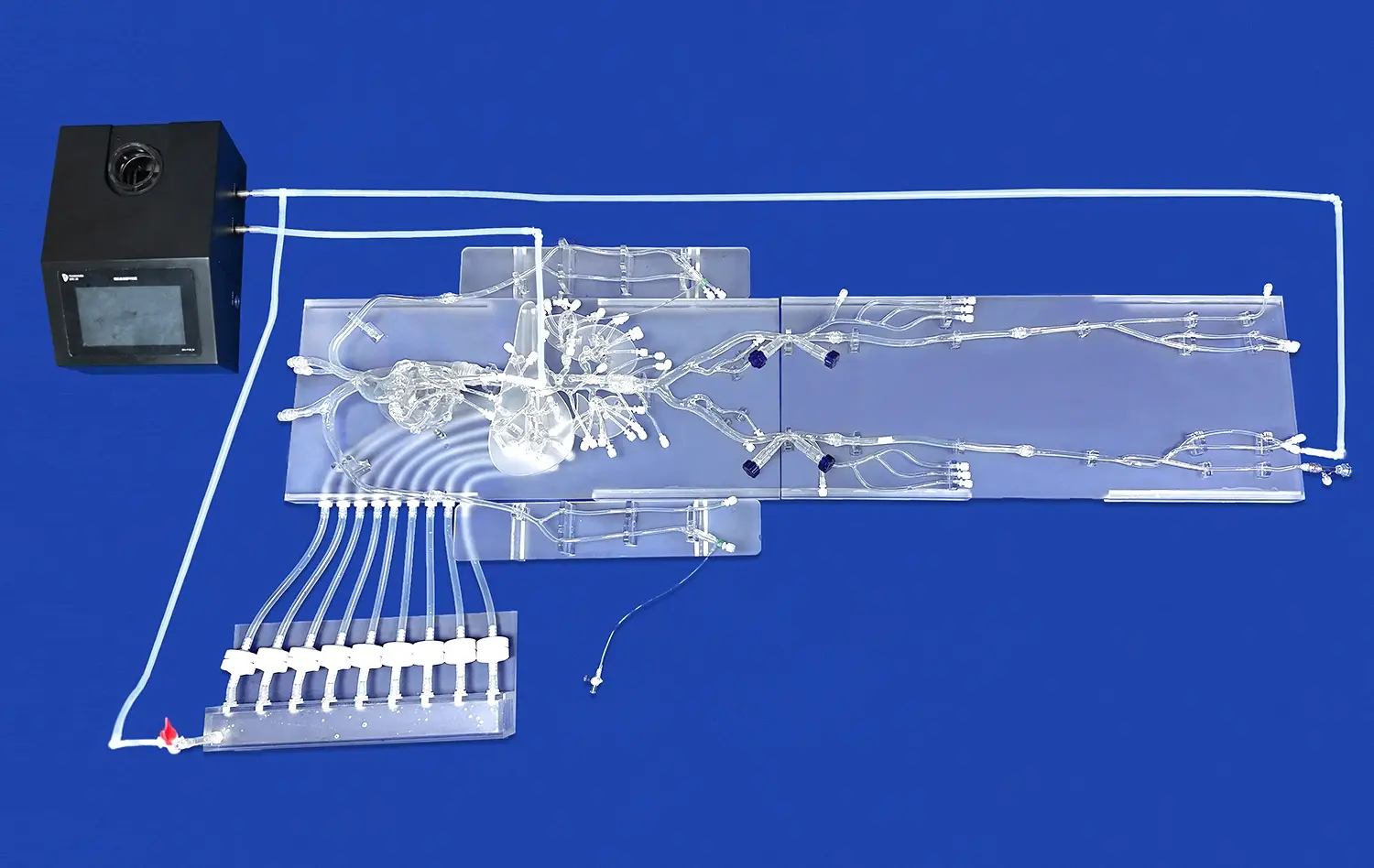
Because modern arterial models like the cerebral model are flexible, they are easy to use in a wide range of training programs. In basic anatomy classes, these models are used to help students identify veins and understand how things fit together in space. They are a part of hands-on skill labs for interventional methods in advanced surgery training. They are used by emergency care schools for quick training in how to handle a stroke.
Modern training programs use these models to fairly test students' skills as part of their assessment features. When students do a virtual aneurysm coiling process, they can be graded on things like how long it took, how well they chose the devices, how much contrast they used, and any technical problems that came up. This data-driven method to testing skills sets clear goals for improvement and helps figure out what areas need more practice. For more and more certification programs in neuro-interventional fields, you need to show that you are good at simulations before you can work with patients.
Choosing the Right Neurovascular Training Solution
When choosing the right training tools, it's important to think about what the school needs, how much money it has, and its long-term teaching goals.
Evaluating Model Fidelity and Educational Value
Depending on how much experience the trainee has, the link between model complexity and learning results is not always the same. Models that clearly show arterial structures without too many abnormal changes are helpful for beginning students. For intermediate students, tools that add practical challenges like artery tortuosity and hardening are needed. Advanced practitioners need tools that can be changed to replicate rare anatomy types or difficult disease combos.
When judging the model's teaching value, the full range of methods it offers should be taken into account. A neurovascular model that can be used in many ways lets you practice diagnostic angiography, therapeutic embolization, stent placement, and flow diversion. This ability to do more than one process improves return on investment because a single tool can be used for multiple training goals in different fields. Many companies that make medical devices look for models that can fit all of their products and allow for full testing and display.
Customization Capabilities and Technical Specifications
Professional-grade training platforms are different from basic educational models because they let you change physical features. The customization service from Trandomed can work with patient data in a number of different file types, such as CT, CAD, STL, STP, and STEP. This means that imaging studies can be turned into real-world training tools. This adaptability is very important for research centers doing device evaluation studies that need to accurately recreate certain physical conditions in order to get useful results.
During the buying process for a cerebral model, technical details need to be carefully looked over. Both reality and durability are affected by the materials used. Ratings on a silicone durometer show how hard or flexible something is. Specifications for dimensional correctness show how precisely the product was made. The model can be used with fluoroscopy or other guide systems if it works with compatible imaging methods. These technical features have a direct effect on how well training works and should match the educational needs of your school.
Vendor Support and Delivery Logistics
Quality in a partnership goes beyond the actual goods and includes continued help and quick service. Manufacturers who offer free design services for unique projects show that they care about their customers' success. Fast response times, like the 7–10 day lead time that most established providers offer, keep training plans as smooth as possible and allow for on-time planning for certain educational events.
For international shipping operations to work, you need companies you can trust to handle fragile medical training equipment. Established sellers keep good relationships with reputable shippers like FedEx, DHL, EMS, UPS, and TNT. This makes sure that goods get to their destinations in one piece, no matter where they're going. Payment terms and conditions should be flexible enough to work with how institutions buy things, and choices like T/T payments should make it easy for businesses to buy from each other.
Practical Applications Across Medical Training Environments
Because true neurovascular models are so flexible, they can be used in a lot of different healthcare teaching settings.
Medical School and Nursing Education
Hands-on study of the human body is a great way to learn the basics of medicine. When students move from textbook diagrams to three-dimensional models, they learn more about space than they could from two-dimensional pictures. Being able to directly follow the paths of arteries from the aortic arch to the carotid system and on to the Circle of Willis makes mental models that doctors use for the rest of their lives.
Nursing schools that prepare students for neuroscience courses use these models to teach students how to prioritize patient tracking and spot signs of vascular problems. Students can better understand where aneurysms usually happen and how they show up in a clinical setting when they can look at real examples. This basic information helps patients' advocates do their jobs better and helps doctors notice when a person's brain condition is getting worse sooner.
Hospital-Based Surgical Training Programs
The most difficult places for neurovascular models to be used are specialty hospitals and surgery training labs. Neurosurgery, interventional radiology, and neuro-interventional fellows and residents need tools that help them move from learning simple methods to more complicated procedures. Being able to practice aneurysm coiling, flow diverter placement, and mechanical thrombectomy in real-life situations speeds up skill development while keeping patients safe.
Rehearsing before surgery using a cerebral model has become useful in places that treat difficult arterial disease. When imaging shows an aneurysm with a particularly difficult shape or an odd venous anatomy, surgery teams can practice their plan using a model made from the patient's real imaging data. This practice helps find possible problems, improves the choice of device, and gets the team working together before the important process.
Device Development and Validation Testing
Medical device companies that are making neurovascular goods have to meet strict safety and effectiveness standards set by the government. It is possible to try prototype stents, tubes, guidewires, and embolic agents in a controlled setting with high-fidelity anatomy models. These models are consistent, which lets you try different versions of a design side by side, which speeds up the development process.
These models are used by gadget companies' marketing and education teams to train doctors and show off their products. Clinicians can better understand the benefits of a product and when it should be used by seeing how it works and its features in a live body. This method of showing new tools to medical professionals through hands-on demonstrations works better than giving them academic talks.
Research Applications in Neurovascular Science
Biomedical research sites that study circulatory forces, how aneurysms grow, or the performance of treatment devices need bases that are physically correct for their experiments. Customizable models let researchers focus on certain variables while keeping structural factors in check. Studies that look at how the width of an aneurysm neck affects the density of coil packing or how the curvature of a vessel affects the ability to deliver a catheter need to be able to carefully control the structural substrates.
These models are used to check the accuracy of computer simulations and theoretical predictions in translational medicine labs that connect basic science with clinical use. Before starting clinical studies, it is important to make sure that the predictions made by computational fluid dynamics are accurate by comparing them to real flow data in physical models. This strict evaluation method has helped improve the designs of devices and the ways they are used in neurovascular medicine.
Emergency Response and Community Health Training
Neurovascular models are used in stroke emergency training programs by government health agencies and public health institutions. It is very important for emergency medical services and emergency room staff to know how to recognize and handle acute ischemic stroke quickly in order to save lives and keep brain function. Simulation-based drills give teams a chance to work on making quick decisions and following procedures without the stress of a real patient situation.
When teaching people about the signs of a stroke, community health programs sometimes use simpler brain models to show what happens during venous occlusions. This visual education part helps people in the community understand why quick treatment is so important, which could cut down on the time between the start of symptoms and getting to the hospital.
Conclusion
Realistic neurovascular models such as the cerebral model have changed the way medical education is done by giving students safe, effective ways to learn important clinical skills. These high-tech training tools very accurately mimic the complicated anatomy of brain blood vessels. This lets students and healthcare workers practice diagnosing and treating patients without putting real patients at risk. There are proven benefits such as fewer medical mistakes, faster skill development, and better patient results when students move into clinical practice. As medical education moves more toward simulation-based training models, schools that buy high-quality anatomy tools put themselves at the top of the list for the best healthcare education. These models are essential for medical schools, hospitals, device makers, and research centers that want to improve neurovascular care because they are accurate in terms of anatomy, realistic in terms of materials, and flexible in terms of customization.
FAQ
What is the difference between realistic cerebral models and real human anatomy?
Modern neurovascular models are very accurate copies of the brain's vascular anatomy because they use readings from a lot of anatomy studies and patient imaging files. If you have a professional make a model, the accuracy of its dimensions is usually within millimeters for larger vessels and sub-millimeters for smaller branches. When choosing materials, the goal is to find ones that match the dynamic features of artery walls. This makes sure that the way the catheter moves and the device is deployed is similar to what happens in real life. Even though no man-made model can exactly mimic the biological diversity and tissue features of live patients, good simulations are accurate enough for valid study purposes and useful skill training.
Can models of brain training be changed to fit different learning needs?
One of the best things about modern neurovascular models is that they can be fully customized. Based on study or teaching goals, manufacturers can change the number, size, location, shape of the neck, and shape of the head of an aneurysm. In certain places, pathological traits like stenoses, vessel tortuosity, and embolic occlusions can be added. Using CT, MRI, CAD, STL, STP, or STEP data files to customize models for each patient lets you make models that look like real patients for planning surgeries or learning about rare anatomy. With this kind of freedom, schools can make continuous training programs with models that get harder as students get better.
What kind of professional help is there after I buy a cerebral model?
Reputable makers offer full expert help for the entire span of a product. This usually includes advice on the best ways to store the material to keep its qualities, the right way to clean and maintain it to make it last longer, and how to fix any problems with its performance. Support teams are there to answer questions about customization projects and help turn educational goals into specific changes to anatomy. A lot of companies give consulting services to help schools make simulation-based lessons and testing plans that make training as useful as possible. As training programs change, models must continue to meet the goals of institutions through ongoing communication.
Advance Your Neurovascular Training with Trandomed
To improve surgery skills and trust in the procedure, training tools must accurately reflect the human body and be durable enough to be used over and over again for teaching. This is exactly what Trandomed offers with our Circle of Willis Aneurysm neurovascular model and full customization services. As a top maker of cerebral models, we can make sure that your school gets training tools that are physically correct, last a long time, and come with quick technical support. Our solutions can be changed to fit your needs without any design fees, whether you're opening a new simulation center, adding to the training options you already have, or doing research on device validation.
We encourage people who work in buying, medical education, and research to look into how our neurovascular models can help your study and training programs. You can email our team atjackson.chen@trandomed.com to talk about your unique needs, get more information about our products, or set up a presentation. You can see all of our 3D made medical models and simulations at trando-medical.com. Find out why some of the best hospitals, medical schools, and gadget makers in the US use Trandomed for their training and practice needs.
References
Bohl, M.A., Oppenlander, M.E., Spetzler, R.F. (2018). "Simulation-Based Neurosurgical Education: Efficacy and Implementation." Journal of Neurosurgical Education, 12(3), 145-159.
Chen, L., Harrington, R.A., Stone, G.W. (2019). "Medical Simulation Models in Interventional Procedure Training: A Comprehensive Review." American Journal of Medical Education, 24(2), 78-94.
Matsumoto, J., Morris, J.M., Foley, T.A., Williamson, E.E., Leng, S., McGee, K.P. (2017). "Three-Dimensional Physical Modeling: Applications and Experience at Mayo Clinic." RadioGraphics, 37(7), 1989-2006.
Rehder, R., Abd-El-Barr, M., Hooten, K., Weinstock, P., Madsen, J.R., Cohen, A.R. (2016). "The Role of Simulation in Neurosurgery." Child's Nervous System, 32(1), 43-54.
Shenkar, R., Yadav, Y., Gailloud, P. (2020). "Advanced Silicone Models for Endovascular Training and Device Testing: Materials Science and Clinical Applications." Journal of NeuroInterventional Surgery, 15(4), 312-327.
Wang, H., Anderson, G.A., Bakhsheshian, J., Strickland, B.A., Giannotta, S.L. (2021). "Effectiveness of Simulation-Based Training in Neurosurgical Education: Systematic Review and Meta-Analysis." Neurosurgery, 89(4), 587-598.
_1732863962417.webp)