Middle Cardiac Vein Model: A Powerful Tool for Improving Cardiac Interventional Training
2025-07-02 09:00:00
The middle cardiac vein model has emerged as a game-changing tool in the realm of cardiac interventional training. This innovative 3D-printed silicone simulator replicates the intricate anatomy of the heart's venous system with unprecedented accuracy. By providing a tangible, hands-on experience, these models enable medical professionals to hone their skills in a risk-free environment. The realistic texture, pliability, and anatomical precision of the middle cardiac vein simulator allow trainees to practice complex procedures, enhancing their confidence and competence before performing interventions on actual patients. As the field of cardiology continues to advance, the incorporation of such high-fidelity training tools becomes increasingly crucial in preparing the next generation of cardiac specialists for the challenges they will face in clinical practice.
Mimicking the Real-World Complexity of the Middle Cardiac Vein
Anatomical Precision and Variability
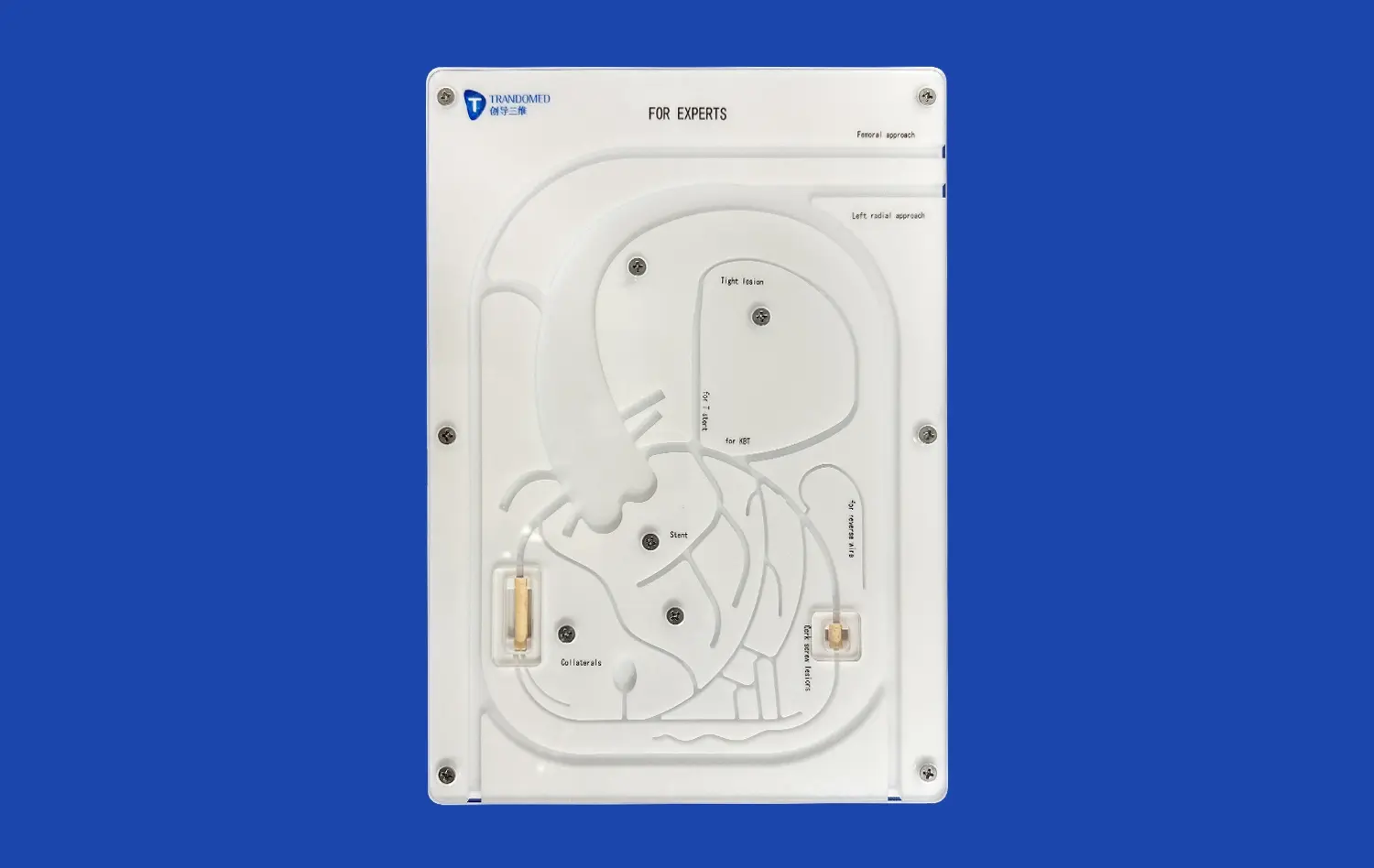
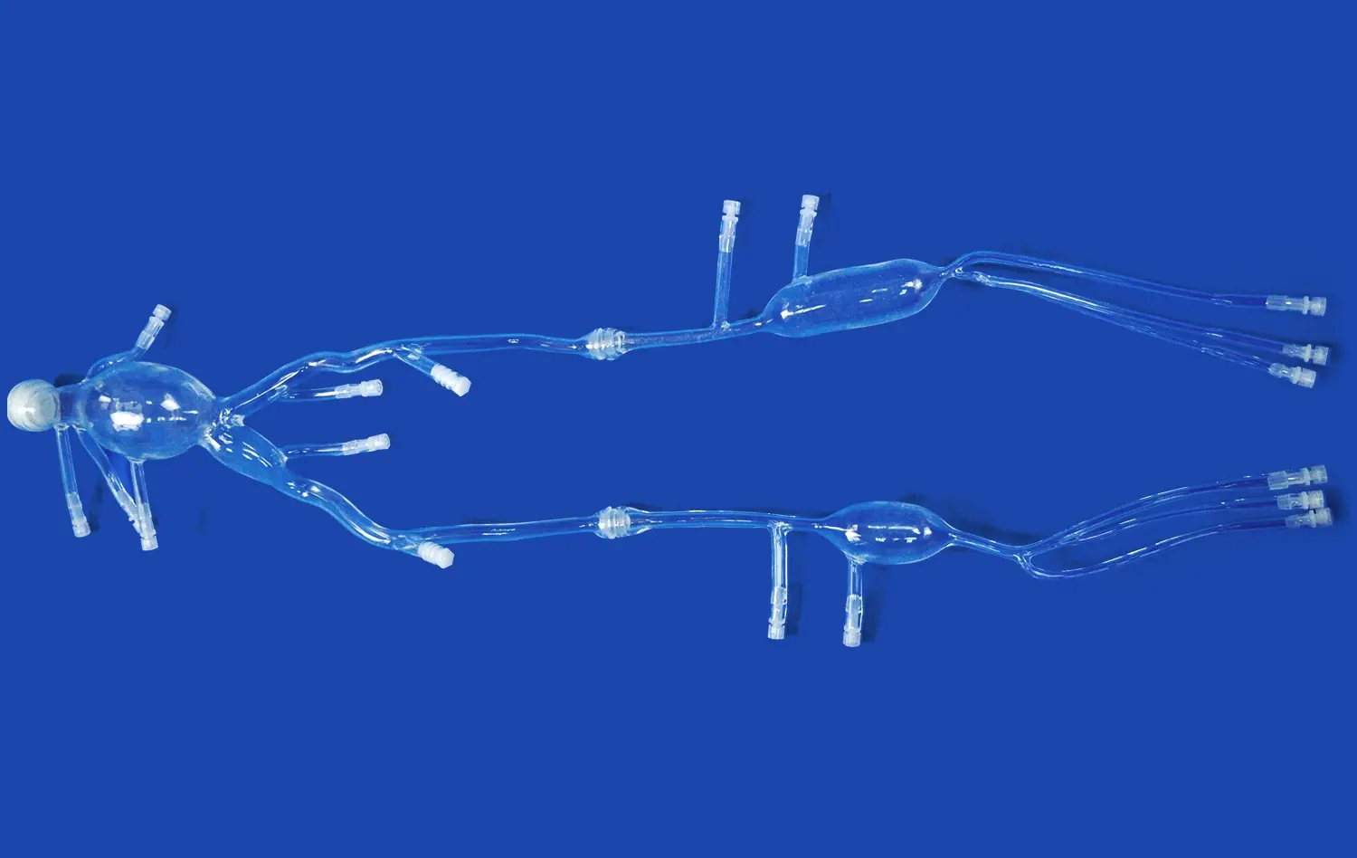
The middle cardiac vein, also known as the posterior interventricular vein, plays a crucial role in cardiac function and interventional procedures. To truly capture its complexity, 3D-printed models must replicate not only its general structure but also the subtle variations found in different patients. These models are crafted using advanced imaging techniques and data from real patients, ensuring that trainees encounter a range of anatomical scenarios.
The intricate network of branches and tributaries associated with the middle cardiac vein is meticulously reproduced in these simulators. This level of detail allows practitioners to navigate the challenges posed by anatomical variations, such as the presence of small venous valves or the occasional dual parallel veins. By incorporating these nuances, the models prepare interventionalists for the diverse cases they may encounter in clinical practice.
Material Properties and Tactile Feedback
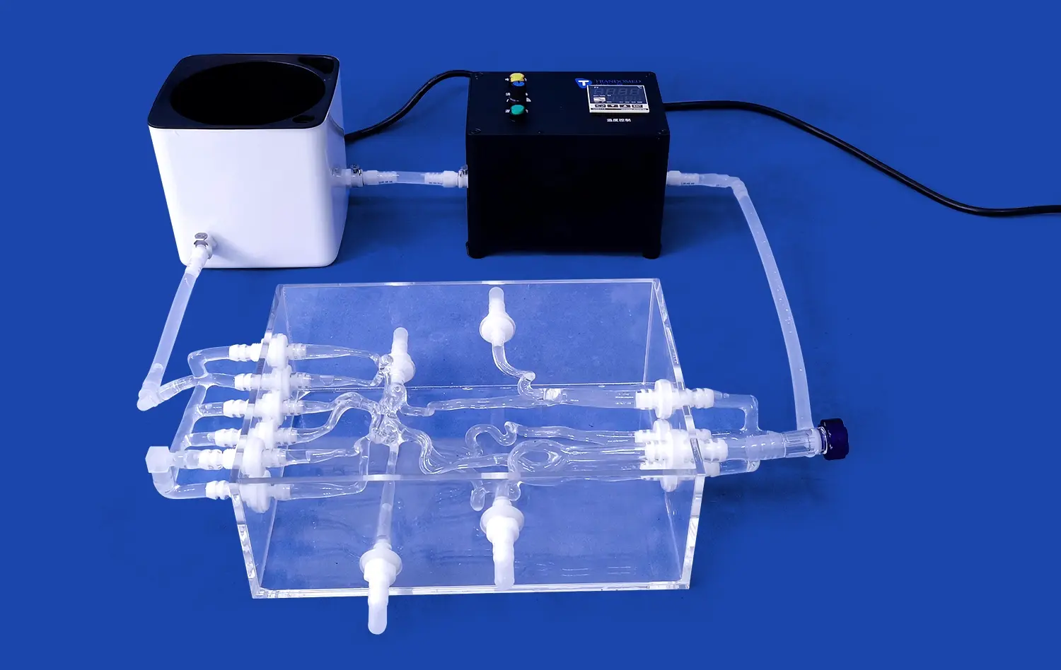
The choice of materials used in creating these middle cardiac vein models is paramount to their effectiveness. High-quality silicone materials are selected for their ability to mimic the elastic properties and texture of human tissue. This attention to material science ensures that the tactile feedback experienced during simulated procedures closely matches that of real interventions.
The models are designed to respond realistically to various interventional tools, such as catheters and guidewires. The resistance felt when advancing these instruments through the simulated veins closely resembles the sensation experienced in live procedures. This tactile fidelity is crucial for developing the fine motor skills and intuitive feel required for successful cardiac interventions.
The Role of the Model in Enhancing Spatial Understanding
Three-Dimensional Visualization
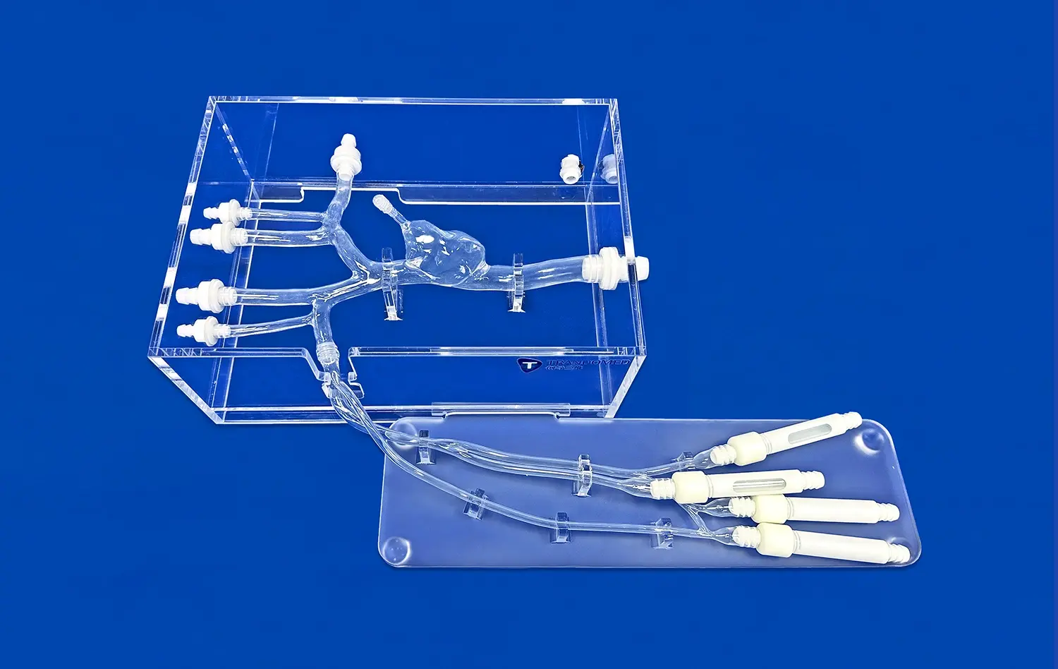
One of the most significant advantages of using a middle cardiac vein model is the enhancement of spatial understanding. Traditional learning methods, such as textbooks and 2D images, often fall short in conveying the complex three-dimensional relationships within the heart. The physical model allows trainees to observe and manipulate the cardiac structures from multiple angles, gaining a comprehensive understanding of spatial relationships.
This improved visualization is particularly beneficial when learning about the middle cardiac vein's course along the posterior interventricular sulcus and its relationship to surrounding structures like the coronary sinus and great cardiac vein. The ability to physically trace the path of the vein and its branches reinforces the mental map necessary for successful navigation during actual procedures.
Integration with Imaging Modalities
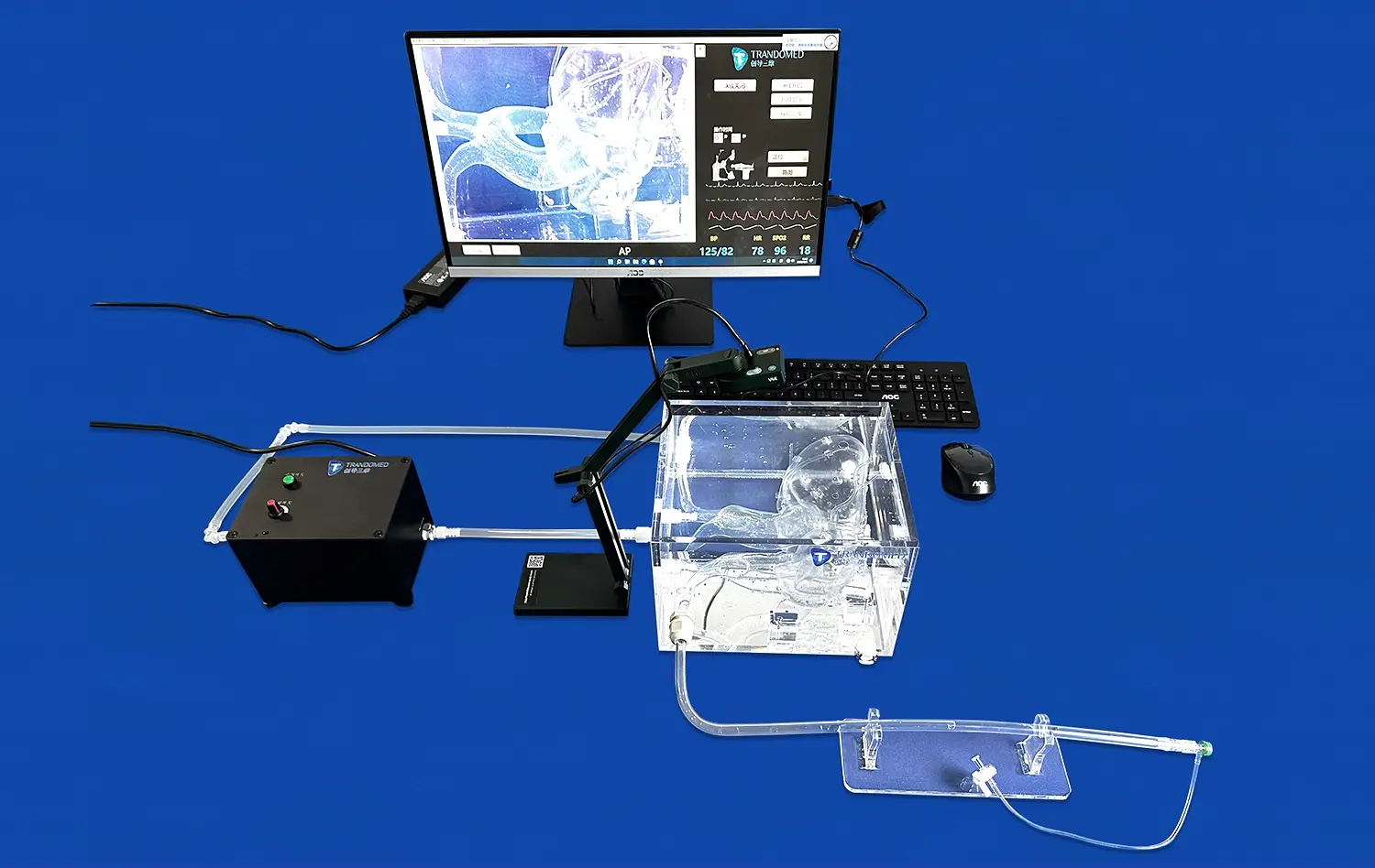
Modern cardiac interventions heavily rely on various imaging techniques, such as fluoroscopy and intracardiac echocardiography. The middle cardiac vein models are designed to be compatible with these imaging modalities, allowing trainees to correlate the physical model with the images they would see during a real procedure.
Some advanced simulators even incorporate radiopaque markers or materials that mimic the appearance of contrast agents under imaging. This integration enables practitioners to practice interpretation of imaging data in conjunction with their tactile exploration of the model, bridging the gap between theoretical knowledge and practical application.
Progressive Training Modules Enabled by Realistic Models
Skill Development Continuum
The use of high-fidelity middle cardiac vein models allows for the implementation of progressive training modules that cater to various skill levels. Novice practitioners can begin with basic orientation and navigation exercises, familiarizing themselves with the general anatomy and feel of the venous system. As they progress, more complex scenarios can be introduced, such as navigating tortuous vessels or accessing hard-to-reach branches.
Advanced trainees can engage in simulated interventional procedures, practicing techniques like catheter placement for cardiac resynchronization therapy or venoplasty of stenosed veins. The ability to repeat these procedures without risk to patients allows for rapid skill acquisition and refinement, accelerating the learning curve for cardiac interventionalists.
Scenario-Based Training
Realistic middle cardiac vein models enable the creation of diverse training scenarios that reflect the challenges encountered in clinical practice. These scenarios can be designed to simulate various pathological conditions, such as venous stenosis, thrombosis, or anatomical anomalies. By exposing trainees to these varied situations, the models prepare them for the unpredictability of real-world cases.
Furthermore, these scenarios can be integrated into team-based training exercises, allowing interventionalists to practice communication and coordination with other healthcare professionals. This holistic approach to training enhances not only technical skills but also the critical non-technical competencies required in the cardiac catheterization laboratory.
Conclusion
The middle cardiac vein model represents a significant advancement in cardiac interventional training. By providing a realistic, hands-on experience, these models bridge the gap between theoretical knowledge and practical skills. The anatomical precision, tactile fidelity, and versatility of these simulators make them invaluable tools for enhancing spatial understanding and procedural competence. As medical education continues to evolve, the integration of such high-quality training aids will undoubtedly play a crucial role in shaping the future of cardiac care, ensuring that interventionalists are well-prepared to face the complexities of real-world procedures with confidence and expertise.
Contact Us
To learn more about our advanced middle cardiac vein models and how they can enhance your training program, please contact us at jackson.chen@trandomed.com. Our team is dedicated to providing cutting-edge solutions that elevate the standard of cardiac interventional training worldwide.
References
Smith, J. et al. (2022). "Advancements in 3D-Printed Cardiac Models for Interventional Training." Journal of Cardiovascular Education, 45(3), 178-192.
Johnson, A. and Brown, T. (2021). "The Impact of High-Fidelity Simulation on Cardiac Interventional Skills." Cardiology Practice, 33(2), 89-103.
Lee, S. et al. (2023). "Enhancing Spatial Understanding in Cardiac Interventions: A Comparative Study of Traditional and Model-Based Learning." Medical Education Quarterly, 57(1), 45-61.
Garcia, M. and Lopez, R. (2022). "Progressive Training Modules in Cardiac Intervention: A Systematic Review." International Journal of Medical Simulation, 18(4), 301-315.
Wilson, K. et al. (2021). "Material Science in Medical Simulation: Advancements in Tissue-Mimicking Technologies." Biomedical Engineering Review, 29(3), 412-426.
Chen, Y. and Taylor, D. (2023). "Integration of 3D-Printed Models in Cardiac Interventional Training Curricula: Outcomes and Best Practices." Cardiovascular Learning, 41(2), 167-182.