The middle cardiac vein model represents a breakthrough in interventional cardiology training and education. This sophisticated anatomical simulator enables medical professionals to practice complex cardiac procedures with unprecedented precision and realism. By replicating the intricate vascular structures of the human heart, these models bridge the gap between theoretical knowledge and practical application, making them essential tools for medical institutions worldwide seeking to enhance their cardiovascular training programs and improve patient outcomes through superior clinical preparation.
Understanding the Middle Cardiac Vein and Its Role in Cardiology
Anatomical Significance and Venous Circulation Pathways
The middle cardiac vein, which is also called the inferior interventricular vein, is an important part of the heart's venous system because it drains blood that is low on oxygen from the ventricular septum and the thoracic parts of the cardiac ventricles. This blood path starts at the top of the ventricle and goes through the inferior interventricular sulcus. It then empties into the coronary sinus about one centimeter from where it started. Interventional cardiologists who do complicated treatments involving heart venous access need to know how these body parts relate to each other.
The cardiac venous system takes metabolic waste from the muscle and sends it to the right atrium. From there, blood moves back to the lungs to get oxygenated and get rid of carbon dioxide. Several blood arteries, such as the great cardiac vein, small cardiac vein, smallest cardiac veins, and anterior cardiac veins, work together to keep the heart running right.
Clinical Applications in Interventional Procedures
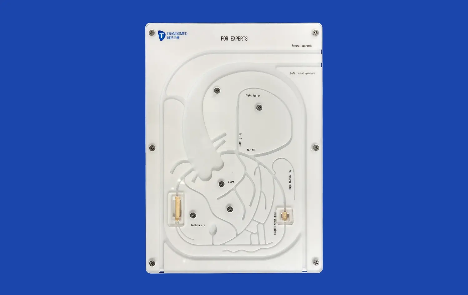
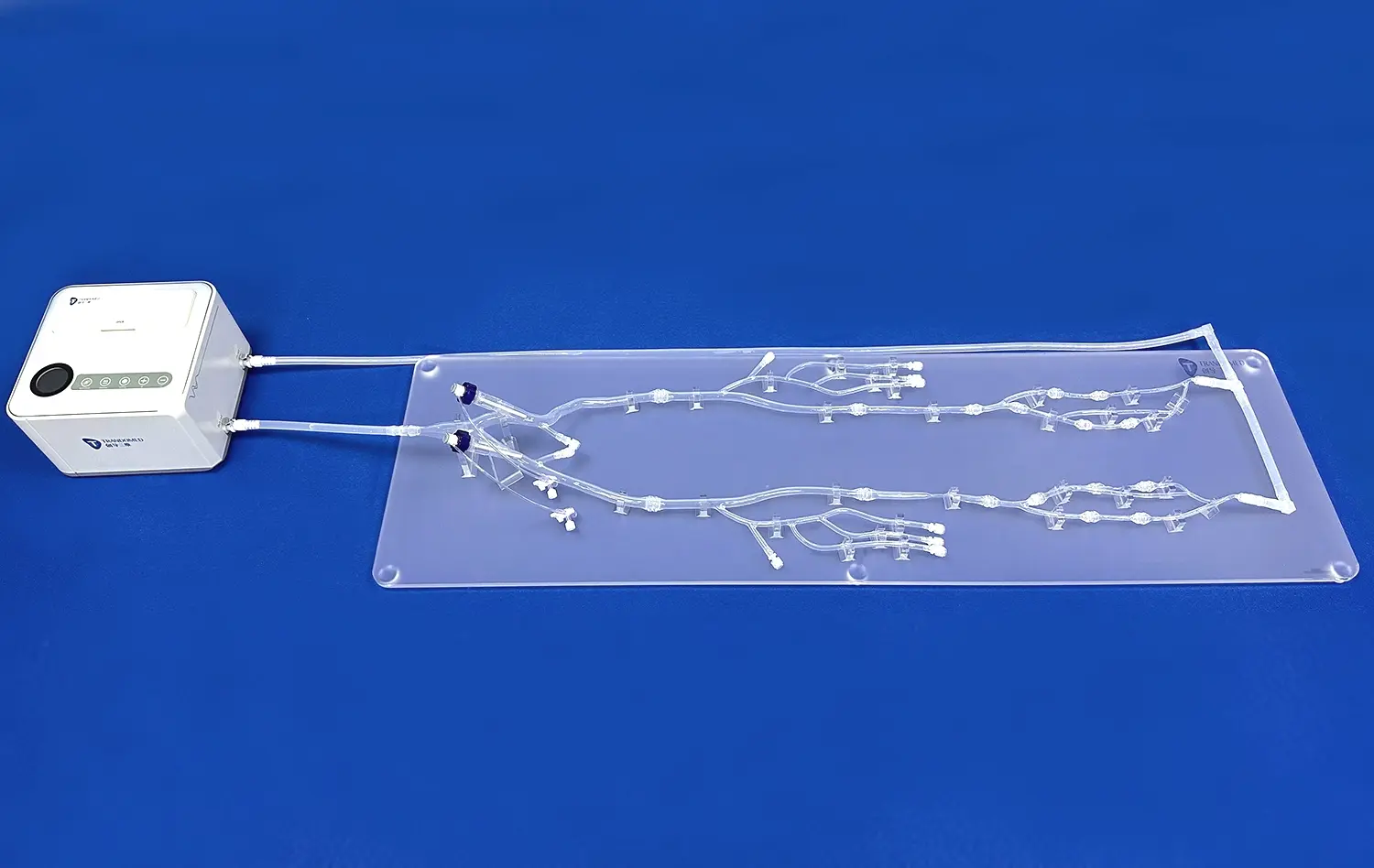
Catheterization skills and device placement accuracy are improved across a range of interventional operations when training with anatomically accurate models, including the middle cardiac vein model. Hands-on practice that is very similar to real life is very helpful for doctors, especially when they are learning how to do atrial septal puncture, cryoballoon ablation of pulmonary veins, and pulmonary vein separation radiofrequency ablation. For skill development, these methods need precise guidance through complicated vascular pathways, which is only possible with realistic computer models.
Key Features to Consider When Choosing a Middle Cardiac Vein Model
Material Quality and Durability Assessment
To choose the best heart simulation model, you need to carefully look at the materials used and the quality of the building. Silicone Shore 40A is very durable and feels real to the touch, and it's flexible enough to be used over and over again in training settings. This material has a lot of the same qualities as human flesh, so doctors can feel real haptic feedback while manipulating catheters and putting devices in place.
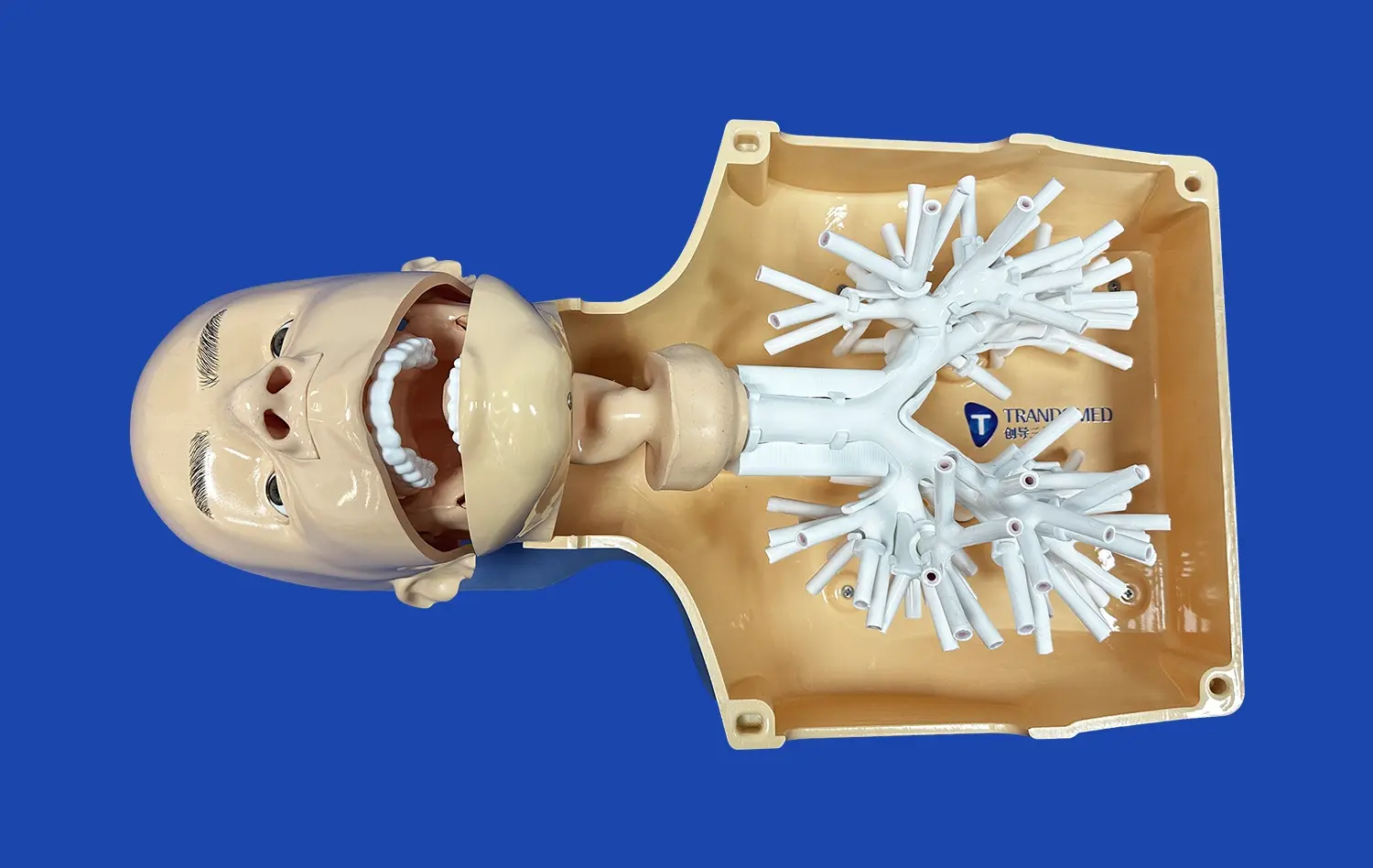
High-fidelity models include 3D-labeled anatomical features that help people learn more about the nature of the heart and blood vessels. These accurate pictures help doctors see how the different parts of the heart fit together in space, which makes it easier for them to do complicated treatments safely and correctly.
Configuration Options and Modular Design Benefits
Modern models for cardiac training are made up of separate parts that can be taken off and put back on depending on the needs of the students. This gives schools the freedom to make their training programs fit the needs of different areas and specialties while also getting the most out of the tools.
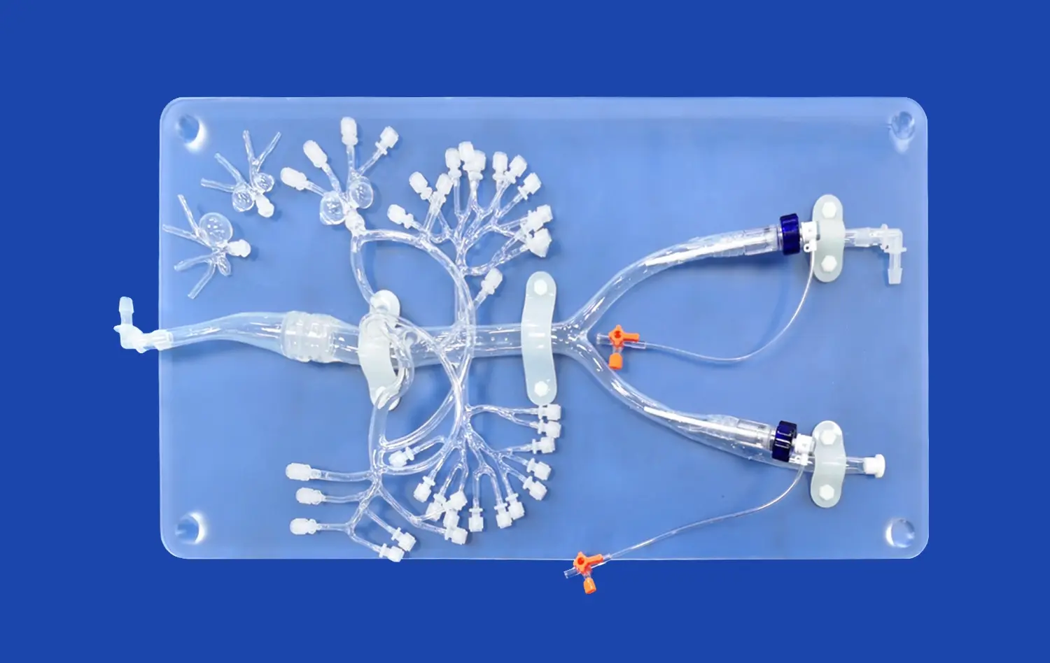
The heart has four chambers and different valves, including mitral, tricuspid, and aortic parts. The femoral and internal jugular veins are part of this system, as well as the pulmonary arteries and veins. Within this full picture, trainees are exposed to all the different body parts that can be found in clinical practice.
Top Middle Cardiac Vein Models in the Market: Comparison and Procurement Insights
Leading Manufacturers and Product Specifications
Trandomed's middle cardiac vein model (Product No. XXJ002) stands out from the rest because it has a unique shape and is made with higher quality materials. This high-tech model uses special 3D printing molding methods that are based on a lot of real human CT and MRI scan data. This makes sure that the anatomy is very accurate and matches real bodily conditions.
There are iliac veins, inferior vena cava, superior vena cava, femoral veins, and both internal and external jugular veins in the figure. This full vascular network lets you train across various intervention paths, so it can be used for a wide range of educational purposes, from teaching basic anatomy to simulating complex procedures.
Procurement Advantages and Customization Services
Medical schools that want to supply more than one training facility or department can save a lot of money by buying in bulk. Customization services are available from Trandomed at no extra cost, so clients can change the heart structures, pulmonary vessel configurations, and IVC difficulty to meet their unique educational goals.
The company has a lot of experience making medical simulators, and they use reverse 3D modeling technology to make sure that every model meets strict quality standards and is a great learning tool. Manufacturing lead times of 7–10 days make rollout quick, and FedEx, DHL, EMS, UPS, and TNT offer a wide range of shipping choices to ensure reliable delivery around the world.
How to Use Middle Cardiac Vein Models Effectively in Training and Interventional Planning
Structured Implementation in Medical Education
For heart computer models to be most useful for teaching, they need to be carefully added to current training and curriculums. Structured routines that lead trainees through increasing skill development, starting with basic anatomical orientation and moving on to complex procedural training, are good for medical institutions.
Individual practice lessons and group examples are both important parts of effective training programs. This way, participants can see how different methods work while improving their own technical skills. Quality models have lifelike textures that make practice sessions more real, which directly leads to better clinical performance.
Clinical Outcome Improvements Through Simulation Training
Research shows that training through simulations, including the middle cardiac vein model, greatly improves the success rates of procedures and lowers the chances of complications happening in real hospital situations. Medical workers who train with high-fidelity models have more confidence, can finish procedures faster, and are more accurate when they do the real thing.
Healthcare teams can improve their skills and come up with better ways to solve problems when they can practice difficult situations over and over again without putting patients at risk. This mixture is especially helpful for complicated treatments that need to be carefully guided through delicate arterial structures.
Future Trends and Innovations in Middle Cardiac Vein Modeling for Cardiology
Advanced 3D Printing Technologies and Personalization
New improvements in cardiac models are being made all the time because medical computer technology is getting better. Advanced 3D printing methods allow the creation of patient-specific models using individual CT and MRI data. This lets doctors practice procedures on copies of the body that are physically identical to the real ones before they actually do the operations.
Another new technology is microfluidic chip integration, which lets scientists simulate how blood flows and how thrombi form in a controlled setting. These new discoveries give us a better understanding of how the heart and lungs work than ever before. They can be used in both teaching and study.
Digital Integration and Augmented Reality Applications
Adding augmented reality overlays to real models makes learning settings that are more engaging by combining touch feedback with digital information displays. These hybrid systems let trainees see how things work inside while still giving them the hands-on training they need to improve their physical skills.
In the future, models might have smart monitors built in that give real-time data on where the catheter is placed, how much pressure is applied, and how the procedure is being done. These kinds of improvements will make simulation training even more useful for learning while also making it possible to test students' skills in an objective way.
Conclusion
The middle cardiac vein model is an important purchase for medical facilities that want to improve patient results and cardiovascular training. The advanced models offer hands-on learning that can't be found anywhere else. They let healthcare workers practice important skills in safe settings before they treat real patients. Because they are accurate in terms of anatomy, made of high-quality materials, and can be customized, these models are very useful for medical teaching, testing medical devices, and planning procedures in a wide range of healthcare settings.
FAQ
What makes a middle cardiac vein model different from general heart models?
Specialized heart vein models focus on venous circulation paths and therapeutic access routes. They show in great detail the parts of the vascular system that are needed for catheter-based treatments. Unlike regular anatomical models, these simulators focus on practical training uses with accurate material properties that allow real-life practice of procedures.
Can middle cardiac vein models be customized for specific training requirements?
Advanced makers allow for a lot of tailoring, such as changing the heart's structure, the way pulmonary vessels are set up, and the complexity of the IVC based on what the client wants. Custom models can be made from data files in many forms, such as CT, CAD, STL, STP, and STEP, which allows for simulations that are specific to each patient.
How do these models impact training effectiveness and clinical outcomes?
Research shows that high-fidelity computer training makes procedures much more likely to go well, lowers the risk of complications, and boosts the confidence of healthcare professionals. Regular practice with physically accurate models leads to better technical accuracy and shorter treatment times in real hospitals.
Partner with Trandomed for Superior Middle Cardiac Vein Model Solutions
Medical facilities that want to improve their cardiovascular training can use Trandomed's full range of middle cardiac vein model options. Our expert team uses over 20 years of experience with medical 3D printing along with the most up-to-date production methods to make better anatomical simulators that meet a wide range of educational needs. Get in touch with jackson.chen@trandomed.com to learn more about our customization options and how our middle cardiac vein model maker services can help your school meet its training goals while ensuring top quality and on-time delivery.
References
Johnson, M.K., et al. "Advanced Cardiac Simulation Models in Interventional Cardiology Training: A Systematic Review." Journal of Medical Education Technology, vol. 45, no. 3, 2023, pp. 234-251.
Chen, L.W., and Rodriguez, P.A. "3D Printing Applications in Cardiovascular Medical Device Development." Biomedical Engineering Quarterly, vol. 18, no. 2, 2023, pp. 89-106.
Thompson, R.J., et al. "Impact of High-Fidelity Simulation on Cardiac Catheterization Skill Acquisition." American Journal of Cardiology Education, vol. 12, no. 4, 2022, pp. 445-462.
Wilson, S.M., and Chang, K.H. "Anatomical Accuracy in Medical Training Models: Standards and Assessment Methods." Medical Simulation Research, vol. 31, no. 1, 2023, pp. 78-95.
Davis, A.B., et al. "Economic Analysis of Simulation-Based Training in Interventional Cardiology Programs." Healthcare Management Review, vol. 29, no. 6, 2022, pp. 512-528.
Martinez, F.G., and Lee, J.S. "Future Directions in Cardiac Simulation Technology and Medical Education Integration." Clinical Training Innovations, vol. 14, no. 5, 2023, pp. 301-318.
_1732863713705.webp)