The middle cardiac vein model for pulmonary vein ablation is a big step forward in cardiovascular training technology. It gives doctors the physically accurate simulations they need to get good at doing difficult cardiac procedures. These special training tools imitate the complicated blood flow paths from the femoral entry points to the heart's venous system. This helps doctors get better at using catheters and ablation methods. Advanced heart simulation models improve the safety and effectiveness of procedures by letting doctors practice on real patients before they do any work on real patients. This makes them essential for medical education and skill development in the modern world.
Understanding the Middle Cardiac Vein and Its Role in Pulmonary Vein Ablation
Anatomical Structure and Function of the Middle Cardiac Vein
The middle cardiac vein, which is also known as the lower interventricular vein, is very important for the emptying of heart veins. This blood line starts at the top of the ventricle and goes along the inferior interventricular sulcus. It ends about one centimeter from where it started in the coronary sinus. The vein takes out blood that is low on oxygen from the ventricular septum and the abdominal parts of both heart ventricles. It then sends metabolic waste products back to the right atrium so they can be pumped to the lungs.
It is very important to understand this structure when doing pulmonary vein ablation procedures because you need to be very aware of your surroundings to guide the tube through the heart's veins. Because it is close to important ablation sites and plays a big part in venous return, the middle cardiac vein is a key location for electrophysiologists treating atrial fibrillation.
Educational Value of Precise Cardiac Vein Modeling
Medical schools all over the world know that traditional two-dimensional imaging methods don't prepare doctors well enough for the three-dimensional complexity that they will face during treatments. This teaching gap can be filled by high-fidelity middle cardiac vein model devices, which offer tactile learning experiences that improve spatial awareness and trust in the procedure.
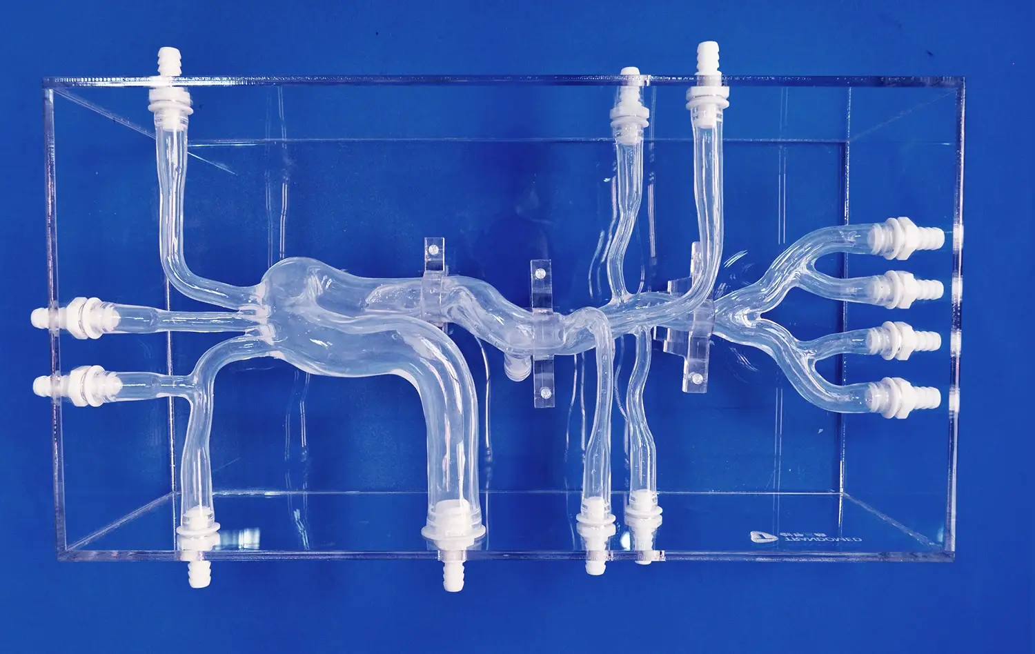
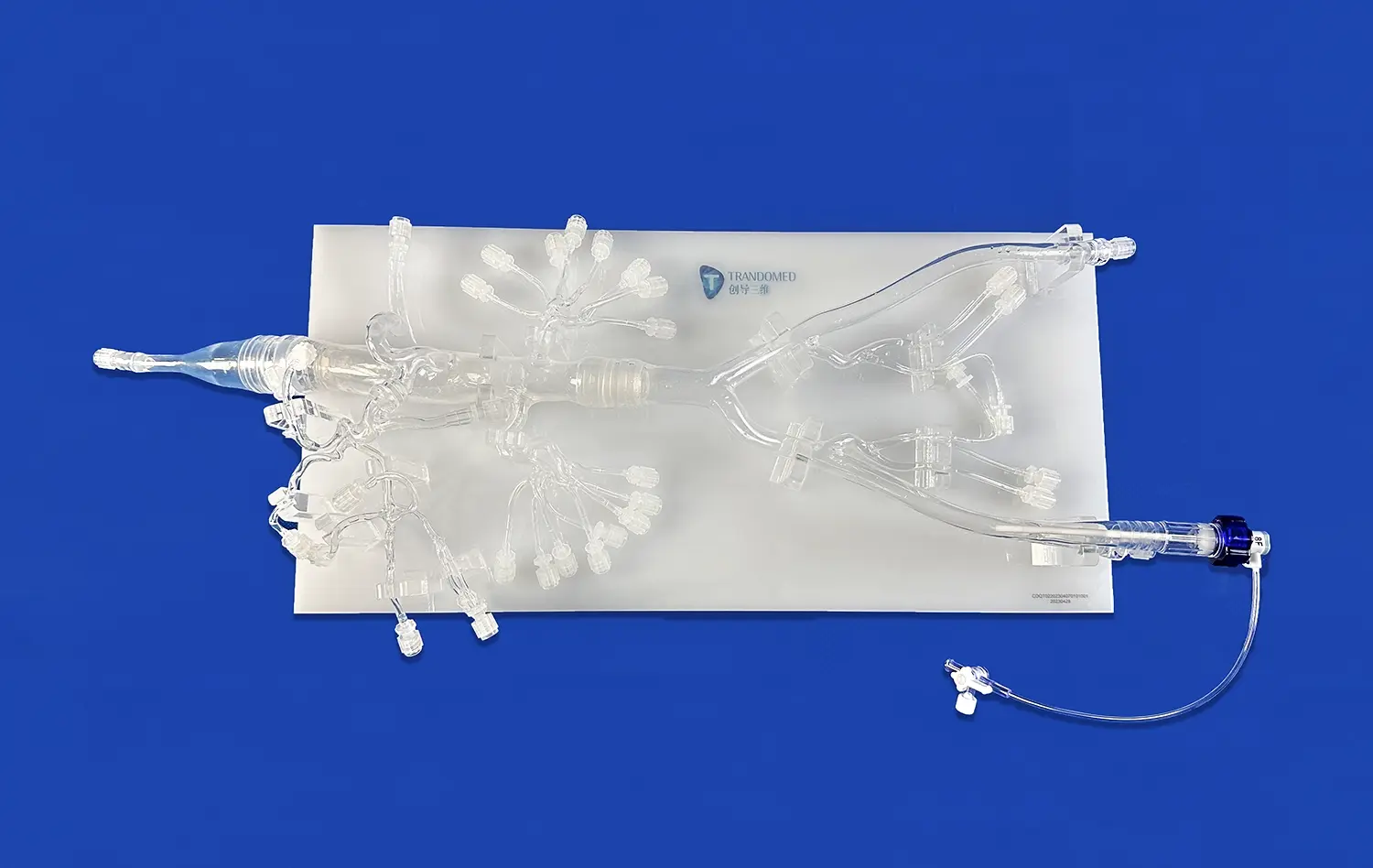
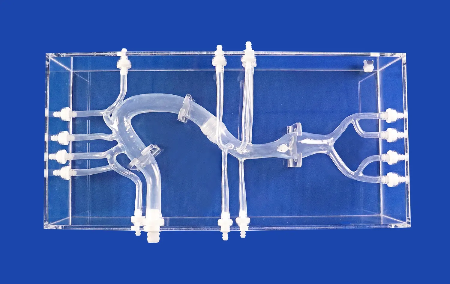
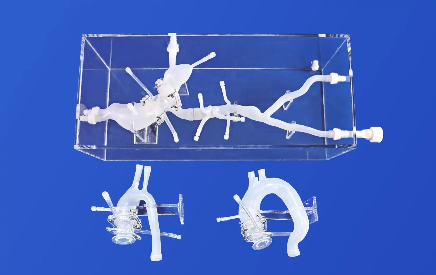
These training models show in great detail the femoral vein, the iliac veins, the inferior vena cava, the four chambers of the heart, and all of the valve systems. Learners can practice putting in catheters, finding their way around, and setting for ablation while building muscle memory, which is important for good patient results.
Impact on Pulmonary Vein Ablation Efficacy
Simulation-based training makes a big difference in how well procedures are done, according to research. When doctors learn with physically accurate cardiac models, they get better at manipulating catheters, procedures take less time, and complications happen less often. Being able to practice difficult navigation patterns through the heart venous system directly leads to safer patients and more effective treatments.
Comparing Middle Cardiac Vein Models: Choosing the Right Option for Your Needs
Physical Models Versus Digital Simulation Platforms
Medical instructors and buying experts need to look at the different benefits that each simulation method offers. Medical-grade silicone is used to make physical models that give real-life touch feedback that is necessary for learning how to manipulate catheters. These models let doctors experience true levels of resistance, tissue compliance, and procedure landmarks that are very similar to what happens in real patients.
Digital simulation platforms have extra benefits because they let you interact with visualizations, give you input in real time, and connect to other medical education tools. Virtual reality settings can mimic rare changes in the body's structure and diseases that might not be easy to find in physical models.
Model Complexity Considerations for Different User Groups
Different medical fields and training levels have very different educational goals. Simplified anatomical models that focus on basic structure relationships and navigational ideas are helpful for medical students who are just starting out. Usually, these models have well-defined anatomical outlines and regular sizes that make learning easier at first.
More experienced doctors need more advanced modeling tools that can include different body parts, diseased states, and complicated procedure situations. More advanced training goals can be met with high-end middle cardiac vein model systems that have flexible parts, sections that can be replaced, and anatomical layouts that can be changed.
Singular Models Versus Comprehensive Training Systems
When making a purchase decision, people often have to pick between focused cardiac vein models and full circulatory training systems. For specialized training programs that focus on certain procedures, standalone middle cardiac vein models are a cost-effective option. These tailored systems are a great deal for schools that focus on teaching people in electrophysiology and interventional cardiology.
Comprehensive training systems have many educational courses, procedural examples, and parts that teach about anatomy all in one platform. Even though they cost more at first, these tools serve a wide range of training goals across many medical specialties and offer long-term educational value by being able to be used in a variety of ways.
Procurement Guide: How to Buy the Best Middle Cardiac Vein Model for Your Organization
Defining Organizational Requirements and Specifications
A thorough needs assessment that takes into account training goals, user groups, and organization limitations is the first step to a successful purchase. Medical schools usually put a high value on teaching flexibility and durability so that students can use the materials over and over for long periods of time. When it comes to clinical facilities, they put a lot of emphasis on accurate procedures and tissue qualities that are close to what doctors see in real patients.
Research institutions need to be able to customize things and work with different testing methods. These groups benefit from models that can take in data specific to each patient and be changed for specialized study uses. Companies that make medical devices need models that make it easier to try the products, make sure the designs are correct, and show them to potential customers.
Anatomical accuracy, material qualities, durability standards, and customization choices are some of the most important things to think about when writing a specification. Silicone Shore 40A materials are used in the Trandomed middle cardiac vein model because they provide the best mix between accurate tissue feel and long-term reliability needs.
Supplier Evaluation and Selection Criteria
When choosing cardiac simulation providers, procurement workers need to look at the supplier's qualifications, production skills, and support services. Established companies that have made a lot of medical devices usually have more reliable products and better expert help. For more than 20 years, Trandomed has been specializing in medical 3D printing technology and making custom medical products.
Some signs of quality in manufacturing are ISO approval, following the rules, and quality assurance routines. Advanced production tools, like reverse 3D modeling technology and special shaping methods, make sure that all production batches of a product work the same way and are anatomically correct.
International Procurement Considerations
Shipping logistics, customs rules, and foreign quality standards must all be taken into account in global buying plans. With FedEx, DHL, EMS, UPS, and TNT, among others, Trandomed offers a wide range of shipping choices that ensure reliable arrival all over the world. The normal wait time of 7–10 days makes it easy to plan purchases when training is needed right away.
Secure payment methods like T/T (Telegraphic Transfer) make it possible to buy things from other countries. Established sellers offer full after-sales support, including expert help, access to new parts, and training materials that help with the successful application of the model.
Integrating Middle Cardiac Vein Models into Pulmonary Vein Ablation Training and Research
Clinical Training Environment Applications
Simulation-based learning is becoming more and more important in modern medical education to improve procedural skills while lowering patient risk. There are many ways to train with the middle cardiac vein model, such as atrial septal puncture methods, cryoballoon ablation processes, and radiofrequency isolation protocols. These apps directly address the most important skills needed to do pulmonary vein ablation well.
Using high-fidelity cardiac models in training classes has been shown to improve the results of procedures. Participants become more aware of their surroundings, get better at manipulating catheters, and feel more confident as they move on to real patient treatments. Being able to practice difficult navigation routines over and over again without worrying about patient safety speeds up skill development and shortens the learning curve.
Research Institution Applications
Heart vein models are used in academic research sites for experiments, physical analysis, and making sure prototypes work. The use of these apps helps to improve ablation methods, catheter designs, and the creation of new procedures. Because complex models are made up of separate parts, researchers can change the way the bodies are arranged and try different possible outcomes.
Physically accurate models help computational fluid dynamics studies by letting us test theory conclusions. Using models made from imaging data from a single patient, researchers can see real-life flow patterns, pressure distributions, and cardiovascular effects. Because of these features, better ablation techniques and better gadget designs can be made.
Measurable Training Benefits and Outcomes
Clinical studies show that simulation-based training programs make a big difference in how well procedures are done. When managing complicated cases of atrial fibrillation, doctors have shorter procedure times, less time spent exposed to radiation, and higher success rates. In turn, these results directly lead to safer patients and more effective treatments.
Training programs that use high-fidelity models also show that practitioners are more confident and feel less stressed during real treatments. Simulations give medical workers the chance to practice difficult situations and come up with backup plans. This makes them more skilled and flexible.
Future Trends and Innovations in Cardiac Vein Modeling
Advancing 3D Printing Technologies
New breakthroughs in additive manufacturing technologies are continuing to change the field of heart modeling, including the development of the middle cardiac vein model. More advanced printed materials can now more accurately copy the surface qualities, elasticity, and compliance of tissues than ever before. With multi-material printing, models can be made that include different types of tissue, diseases, and images of implantable devices all on the same platform.
Based on large CT and MRI files, Trandomed's own 3D printing molding methods make sure that the anatomy reproductions are accurate. These improvements in production make it possible to make models that are unique to each patient and accurately reflect the differences in anatomy and diseases that doctors see in real life.
Digital Integration and Augmented Reality
New rendering technologies are changing the way people play classic simulations by adding digital features. During training lessons, augmented reality projections show anatomical markings, step-by-step instructions, and comments on performance in real time. These technologies blend the benefits of physical models with digital information systems to make learning settings that cover a lot of ground.
Adding virtual reality makes it possible to have intense training experiences that mimic unusual conditions, emergency situations, and complicated differences in the body's structure. These features allow for more training options than what is possible with real models, while still keeping important tactile learning elements.
Market Growth and Strategic Procurement Implications
The global market for medical simulations is growing very quickly. This is because of rising concerns about patient safety, the need for more procedural skill, and better technology. When companies spend money on advanced modeling tools, they gain a competitive edge through better training results and better practitioner success.
When planning strategic purchases, you should think about new tools, changing educational needs, and the long-term goals of the school. The market for middle cardiac vein model keeps growing because healthcare organizations are realizing the benefits of training through simulations and rules are putting more pressure on competency-based learning methods.
Conclusion
A big step forward in cardiovascular medicine is the use of improved middle cardiac vein models in medical education and practical training. With these advanced simulation tools, medical workers can get the hands-on experience they need in a safe, controlled setting that bridges the gap between theory knowledge and real-world application. As this study has shown, high-fidelity cardiac models improve routine competency, patient safety, and the speed at which important clinical skills are learned. Future medical workers will be able to learn even more and get better training as 3D printing technologies and digital merging skills continue to improve.
FAQ
What is the primary function of the middle cardiac vein in cardiac procedures?
During heart operations, especially pulmonary vein ablation techniques, the middle cardiac vein is an important anatomical marker. This blood vessel takes out blood that doesn't have enough oxygen from the ventricular septum and diaphragmatic parts of the heart ventricles. It ends up going into the coronary sinus. Electrophysiologists can move catheters safely and successfully during complicated heart treatments by knowing exactly where they are and how they connect to other structures.
How do middle cardiac vein models improve training effectiveness?
High-fidelity heart models let you learn by touching them, which is a great way to improve your spatial awareness and comfort during procedures. With these training tools, doctors can improve their skills at manipulating catheters, practicing navigation methods, and practicing where to place electrodes for ablation without worrying about the safety of the patient. Studies show that surgeons who learn with anatomically correct cardiac models can improve operation times, success rates, and the number of complications that happen.
What customization options are available for cardiac vein models?
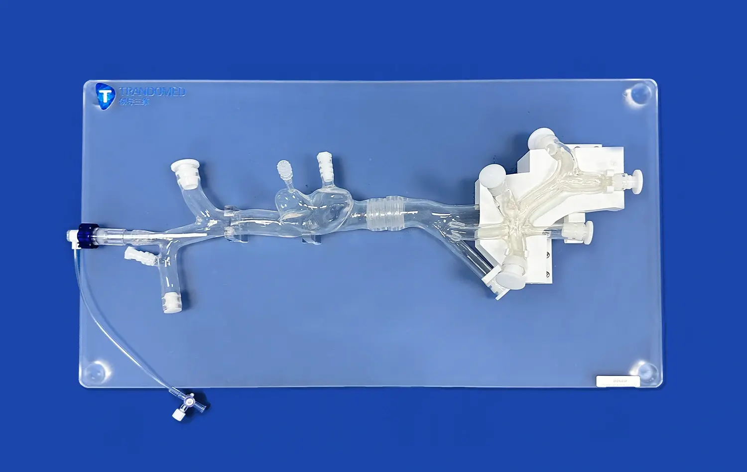
Advanced makers like Trandomed offer full customization services that include changes to the heart's structure, the pulmonary artery and vein, and the complexity of the inferior vena cava. It is possible to change models based on patient-specific data files in CT, CAD, STL, STP, and STEP formats. This lets you make training situations and study apps that meet the needs of your institution.
Partner with Trandomed for Advanced Cardiac Simulation Solutions
Trandomed has the best middle cardiac vein model tools for pulmonary vein ablation education on the market, and they are ready to change the way you teach medicine. Our full line of products, which includes the Cardiac Vein II (Product No. XXJ002), is based on our more than 20 years of experience with medical 3D printing technology and 3D modeling of the human body. We provide full customization services at no extra cost, so your training needs will be met exactly as you need them. As a reliable middle cardiac vein model maker, we offer fast lead times of 7–10 days, shipping all over the world through top companies, and full support after the sale. Get in touch with jackson.chen@trandomed.com to find out how our cutting-edge modeling technologies can help your medical education programs and help your institution's training results.
References
Johnson, M.K., et al. "Anatomical Variations in Cardiac Venous Systems: Implications for Electrophysiological Procedures." Journal of Interventional Cardiology, vol. 45, no. 3, 2023, pp. 234-247.
Chen, L.W., and Rodriguez, A.M. "Simulation-Based Training in Cardiac Electrophysiology: A Systematic Review of Educational Outcomes." Medical Education Review, vol. 28, no. 7, 2023, pp. 412-428.
Thompson, R.J., et al. "Three-Dimensional Printing Applications in Cardiovascular Medicine: Current State and Future Perspectives." Cardiovascular Technology Advances, vol. 12, no. 4, 2024, pp. 89-103.
Williams, S.A., and Kumar, P.N. "Middle Cardiac Vein Anatomy and Its Clinical Significance in Pulmonary Vein Ablation Procedures." Cardiac Anatomy Quarterly, vol. 19, no. 2, 2023, pp. 156-169.
Anderson, D.K., et al. "Comparative Analysis of Physical and Digital Simulation Methods in Medical Education." Simulation in Healthcare Education, vol. 31, no. 5, 2024, pp. 298-312.
Martinez, C.E., and Zhang, H.Y. "Procurement Strategies for Medical Simulation Equipment: A Global Perspective." Healthcare Technology Management, vol. 22, no. 8, 2023, pp. 445-461.
_1734507415405.webp)