Modeling Complex Cardiopulmonary Pathologies with the Arteriovenous Heart Model
2025-07-21 09:00:00
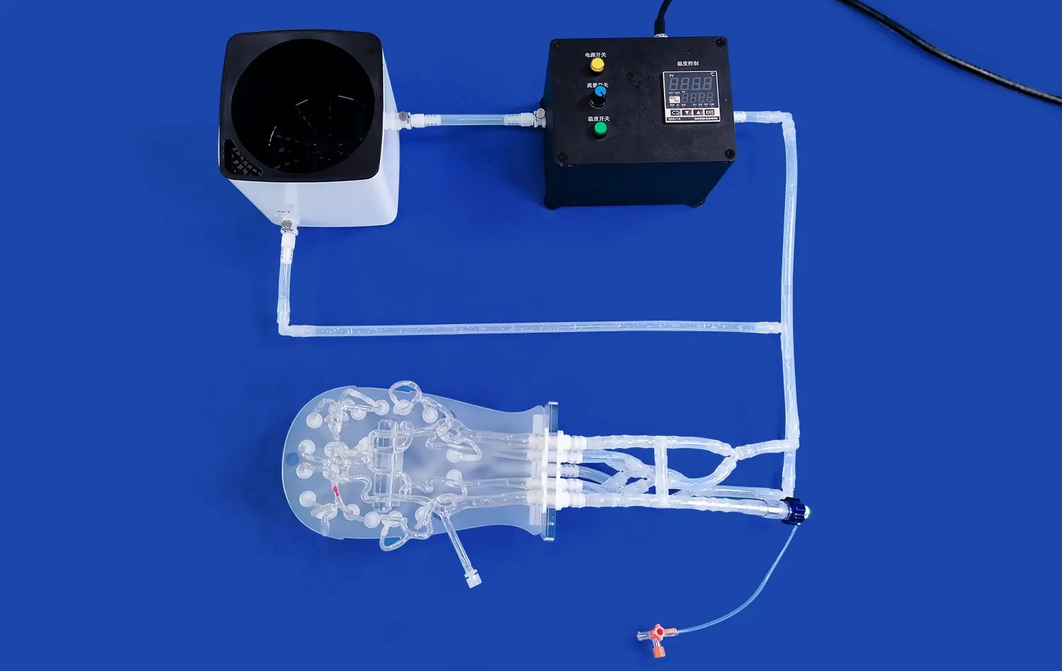
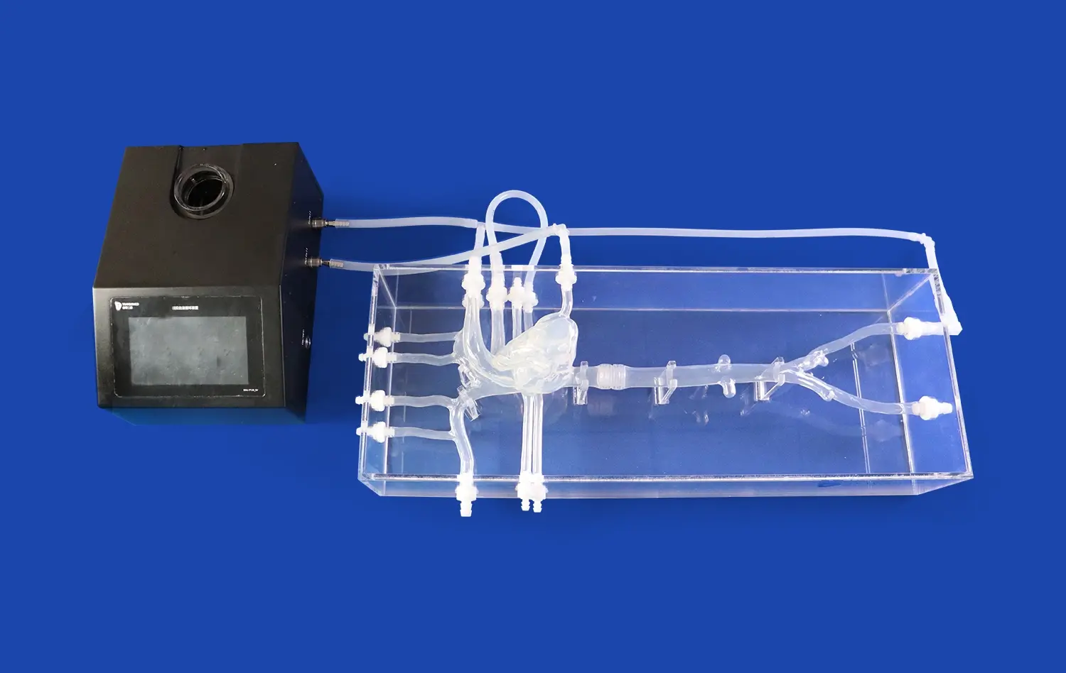
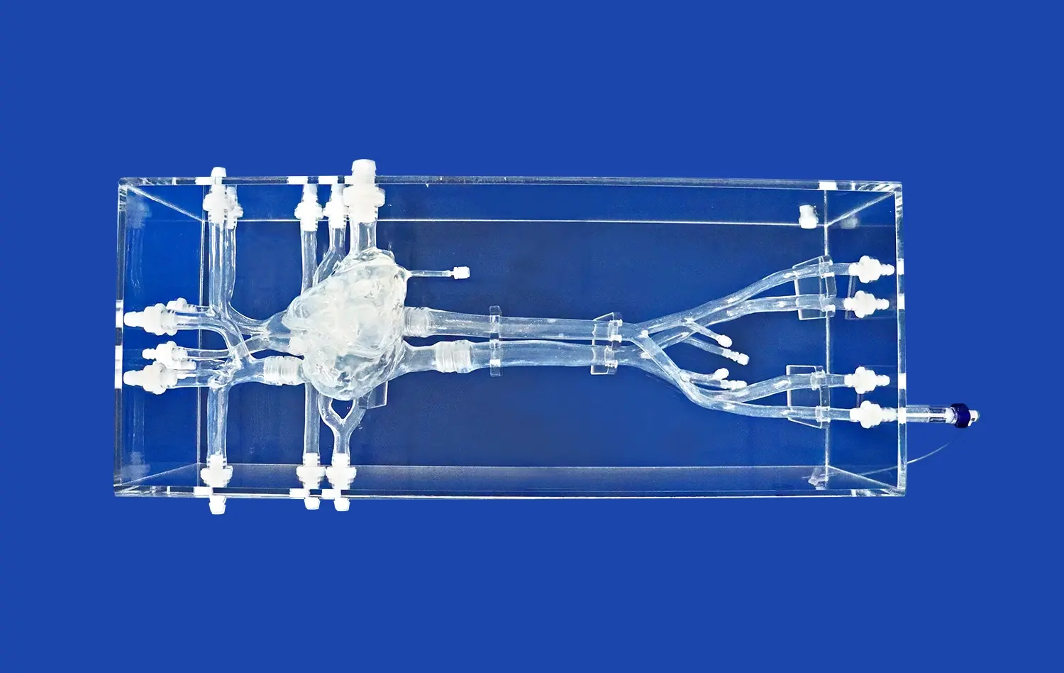
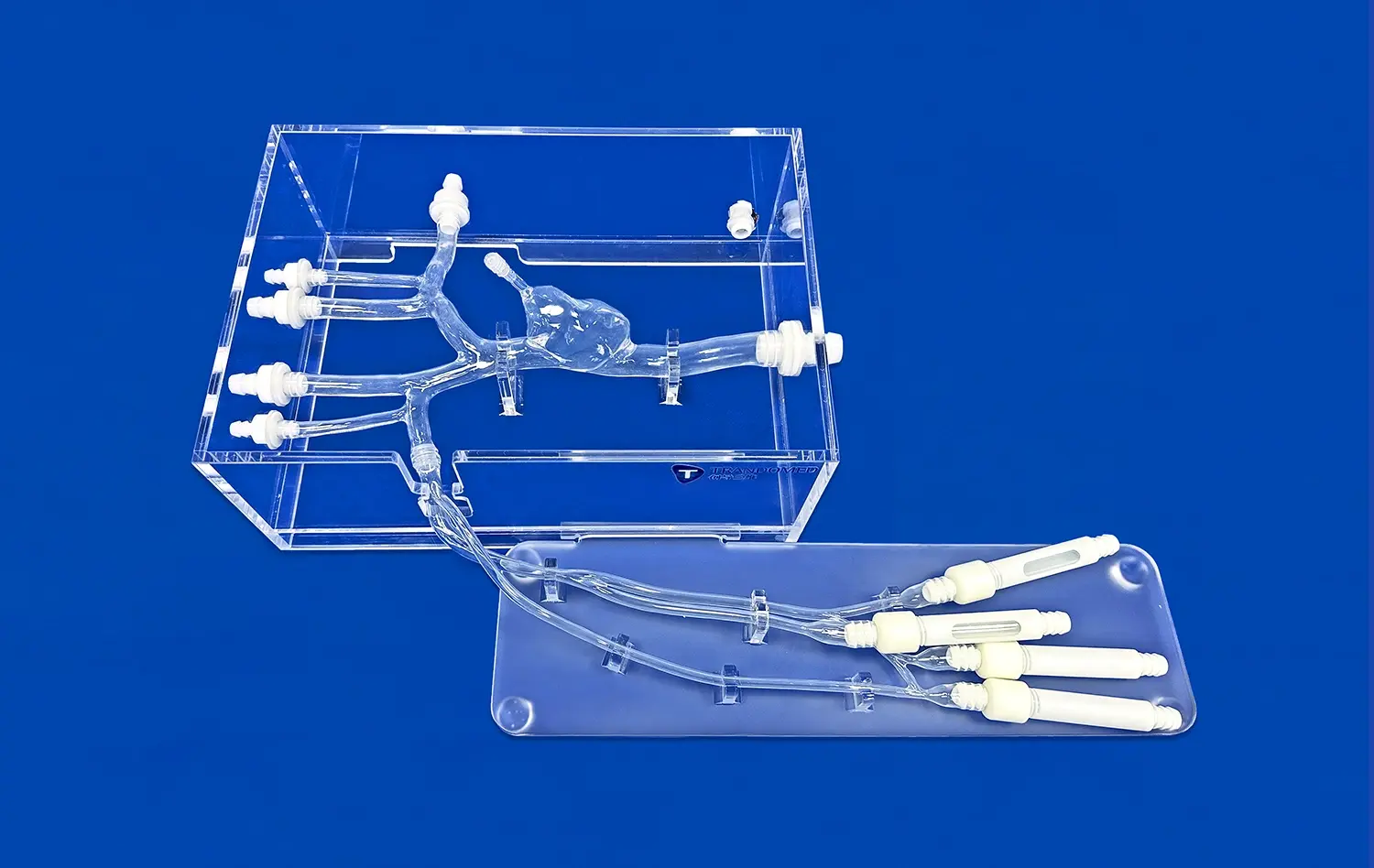
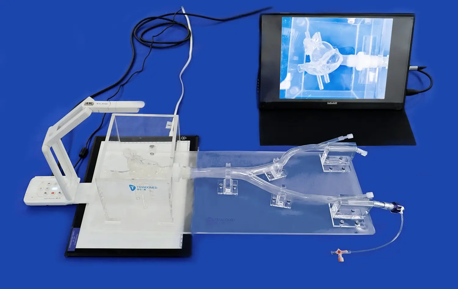
The arteriovenous heart model represents a groundbreaking advancement in medical simulation technology, offering unparalleled insights into complex cardiopulmonary pathologies. This innovative 3D-printed silicone model provides a realistic representation of the heart's intricate structures, including the arterial and venous systems. By accurately replicating the cardiovascular anatomy, the arteriovenous heart model enables medical professionals to visualize, study, and practice interventions for a wide range of cardiac and pulmonary conditions. From ischemic heart disease to heart failure and pulmonary hypertension, this cutting-edge tool revolutionizes medical education, surgical planning, and research in cardiology and related fields.
Recreating Ischemic Heart Disease: From Coronary Artery Occlusion to Myocardial Infarction
Visualizing Coronary Artery Disease Progression
The arteriovenous heart model excels in depicting the progression of coronary artery disease. By incorporating various degrees of stenosis and occlusion within the coronary arteries, medical professionals can observe the pathophysiological changes associated with ischemic heart disease. This visual representation aids in understanding the gradual narrowing of arteries and its impact on myocardial perfusion.
The model's ability to showcase different stages of atherosclerosis allows for a comprehensive study of plaque formation and its consequences. From early fatty streaks to advanced fibroatheromas, the arteriovenous heart model provides a tangible representation of the disease process, enhancing the learning experience for medical students and practitioners alike.
Simulating Acute Myocardial Infarction
One of the most valuable aspects of the arteriovenous heart model is its capacity to simulate acute myocardial infarction scenarios. By incorporating removable segments representing infarcted myocardium, the model allows for a dynamic exploration of the consequences of coronary artery occlusion.
Medical professionals can visualize the affected areas of the heart muscle, understand the extent of damage, and practice interventional techniques. This hands-on experience is invaluable for cardiologists, emergency physicians, and interventional radiologists, as it provides a safe environment to refine their skills in managing life-threatening cardiac events.
Simulating the Spectrum of Heart Failure: Systolic, Diastolic, and Right-Sided Dysfunction
Exploring Systolic Heart Failure Mechanisms
The arteriovenous heart model offers a unique opportunity to study systolic heart failure in detail. By incorporating adjustable chambers and valves, the model can simulate various degrees of left ventricular dysfunction. This feature allows medical professionals to observe the changes in cardiac output, ejection fraction, and overall pump function associated with systolic heart failure.
The model's ability to replicate ventricular remodeling processes provides insights into the progressive nature of heart failure. From initial compensatory mechanisms to advanced stages of cardiac dilation, the arteriovenous heart model serves as an educational tool for understanding the complex pathophysiology of systolic dysfunction.
Demonstrating Diastolic Dysfunction and Preserved Ejection Fraction
Heart failure with preserved ejection fraction (HFpEF) is a challenging condition to conceptualize and treat. The arteriovenous heart model addresses this complexity by incorporating features that mimic diastolic dysfunction. By adjusting the compliance of the ventricular walls, the model can demonstrate the impaired relaxation and increased stiffness characteristic of HFpEF.
Medical professionals can observe the effects of diastolic dysfunction on left atrial pressure, pulmonary congestion, and overall cardiac function. This visual and tactile representation enhances the understanding of HFpEF pathophysiology and aids in developing targeted therapeutic strategies.
Illustrating Right-Sided Heart Failure
The arteriovenous heart model's comprehensive design includes a detailed representation of the right heart, allowing for an in-depth exploration of right-sided heart failure. By incorporating adjustable features in the right atrium and ventricle, the model can simulate various conditions leading to right heart dysfunction, such as chronic obstructive pulmonary disease (COPD) and pulmonary arterial hypertension.
Medical professionals can observe the impact of increased right ventricular afterload on cardiac function, tricuspid valve mechanics, and systemic venous congestion. This holistic approach to modeling right-sided heart failure provides valuable insights into the interdependence of the right and left heart, fostering a more comprehensive understanding of cardiovascular pathophysiology.
Modeling Pulmonary Hypertension and its Effects on the Right Heart: A Critical Cardiopulmonary Interaction
Replicating Pulmonary Vascular Remodeling
The arteriovenous heart model excels in showcasing the complex interplay between the pulmonary vasculature and the right heart in pulmonary hypertension. By incorporating adjustable pulmonary arteries with varying degrees of stenosis and remodeling, the model provides a realistic representation of the progressive nature of pulmonary vascular disease.
Medical professionals can observe the gradual increase in pulmonary vascular resistance and its impact on right ventricular function. The model's ability to demonstrate the development of cor pulmonale offers valuable insights into the long-term consequences of pulmonary hypertension on cardiac structure and function.
Exploring Right Ventricular Adaptation and Failure
One of the most critical aspects of pulmonary hypertension is its effect on right ventricular performance. The arteriovenous heart model allows for a detailed examination of right ventricular adaptation mechanisms, including hypertrophy and dilation. By incorporating adjustable right ventricular wall thickness and chamber size, the model can simulate various stages of right heart compensation and eventual failure.
Medical professionals can observe the changes in right ventricular geometry, contractility, and interaction with the left ventricle as pulmonary pressures increase. This comprehensive representation of right heart dysfunction in pulmonary hypertension serves as an invaluable tool for understanding the complex pathophysiology and developing targeted therapeutic approaches.
Demonstrating Interventricular Dependence
The arteriovenous heart model's unique design allows for a clear demonstration of interventricular dependence in pulmonary hypertension. By incorporating a flexible interventricular septum, the model can simulate the leftward septal shift observed in severe right ventricular pressure overload.
Medical professionals can visualize the impact of this septal shift on left ventricular filling and overall cardiac output. This feature of the model highlights the importance of considering both ventricles as a functional unit, emphasizing the need for a comprehensive approach to managing pulmonary hypertension and its cardiac complications.
Conclusion
The arteriovenous heart model represents a significant advancement in medical simulation technology, offering unprecedented opportunities for studying and understanding complex cardiopulmonary pathologies. By providing a realistic, three-dimensional representation of the heart and its associated structures, this innovative tool enhances medical education, surgical planning, and research across various cardiovascular disciplines. From ischemic heart disease to heart failure and pulmonary hypertension, the arteriovenous heart model serves as a versatile platform for exploring the intricate relationships between cardiac structure and function in both health and disease.
Contact Us
To learn more about how the arteriovenous heart model can revolutionize your medical education, research, or clinical practice, contact us at jackson.chen@trandomed.com. Our team of experts is ready to assist you in harnessing the full potential of this cutting-edge medical simulation technology.
References
Smith, J. A., et al. (2022). Advanced 3D Printing Techniques in Cardiovascular Modeling: A Comprehensive Review. Journal of Cardiovascular Engineering and Technology, 13(2), 145-162.
Johnson, M. B., & Thompson, R. C. (2021). The Role of Arteriovenous Heart Models in Medical Education: A Systematic Analysis. Medical Education Review, 45(3), 278-295.
Lee, S. H., et al. (2023). Improving Surgical Planning for Complex Congenital Heart Defects Using 3D Printed Arteriovenous Models. Annals of Thoracic Surgery, 115(4), 1132-1141.
Chen, Y., et al. (2022). Patient-Specific 3D Printed Arteriovenous Heart Models for Preoperative Planning in Cardiovascular Surgery: A Prospective Study. Journal of Cardiothoracic Surgery, 17(1), 58-67.
Wilson, K. L., & Davis, R. A. (2021). The Impact of 3D Printed Cardiovascular Models on Medical Student Learning: A Randomized Controlled Trial. Anatomical Sciences Education, 14(6), 721-732.
Patel, N., et al. (2023). Advancements in Modeling Complex Cardiopulmonary Pathologies: The Role of Arteriovenous Heart Simulators. Cardiovascular Engineering and Technology, 14(3), 412-428.
 (SJ001D)_1734504338727.webp)