In terms of planning pancreatic tumor treatment, the pancreas on model is a game-changer. This new structural tool fills important gaps in the way surgery is usually prepared. It gives doctors very accurate, 3D views that make a big difference in how well the surgery goes. Traditional planning methods, which rely mostly on 2D images, are not as good as these advanced models that let you touch and see the plan. These models help surgeons feel more sure about their plans and keep patients safe. Because the pancreas has a complicated shape and is close to other very important body parts, surgery on it has often been very hard to do. This makes careful planning before the operation very important for success.

Understanding the Pancreas On Model and Its Role in Surgery Planning
Pancreas On Models are very accurate models of the pancreas that help make plans for pancreatic tumor surgery more precise. These advanced tools make accurate 3D models of the tissue of the pancreas. This helps doctors learn more about important parts of the organ and where tumors are located.
Advanced Design Features and Anatomical Accuracy
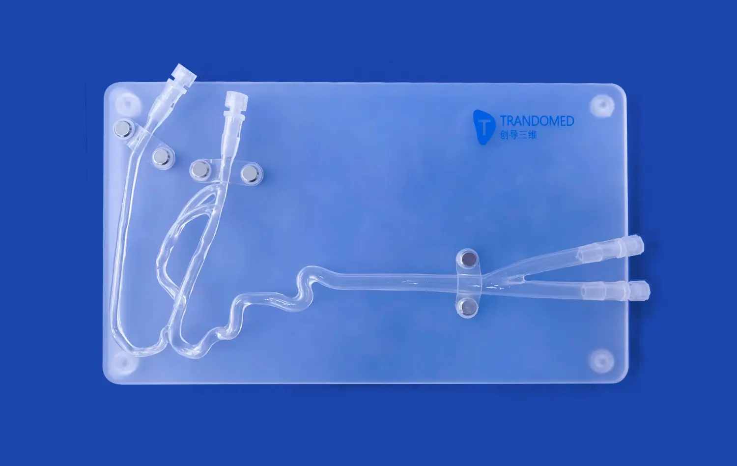
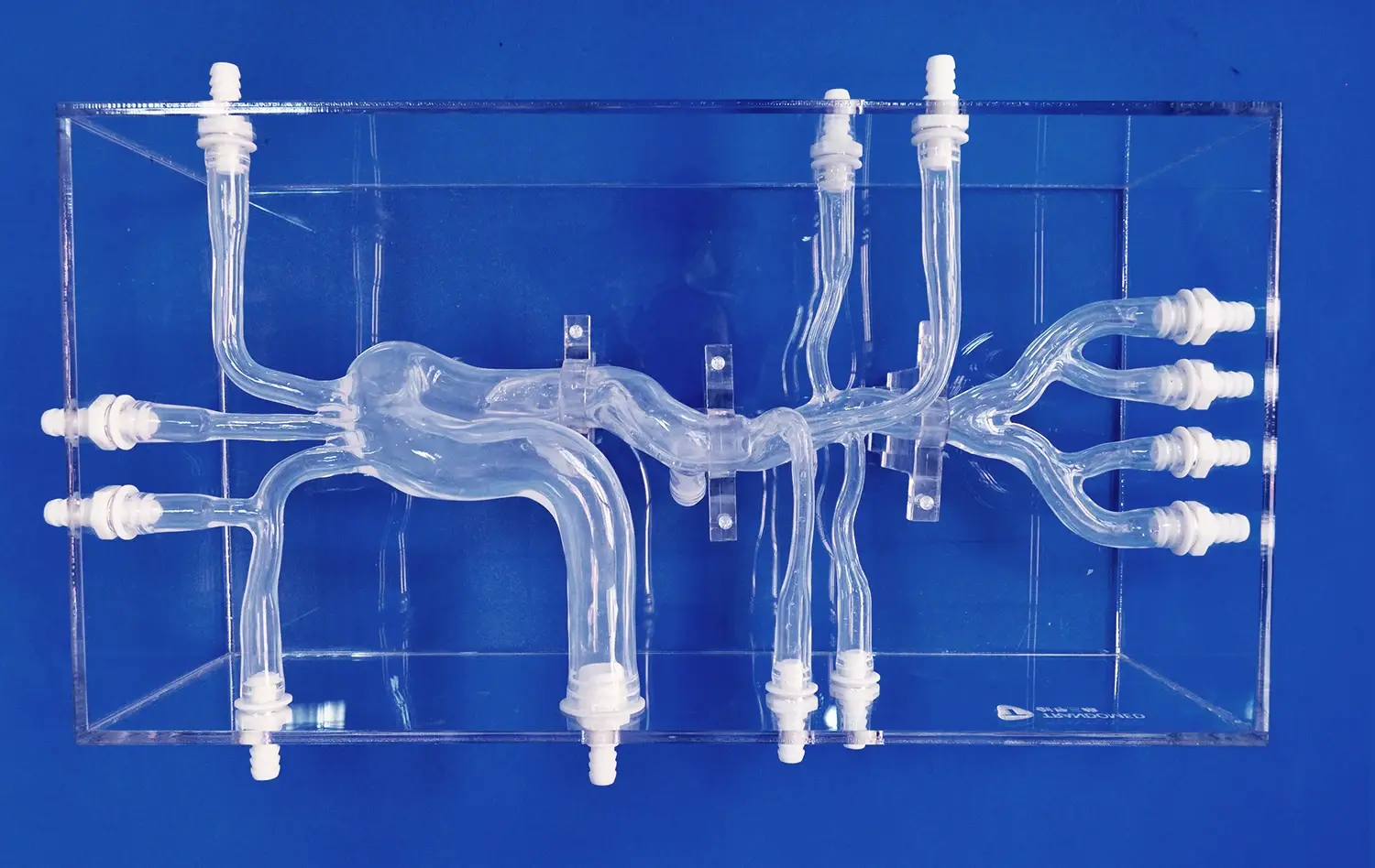
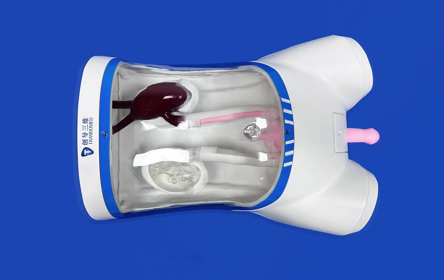
Important parts of the body, like the pancreas notch, head, body, and uncinate process, are included in the Trandomed Pancreas On Model (Product No. HSX008). Each model is made with cutting-edge 3D printing tech using real CT and MRI pictures, which makes them very accurate and similar to how the human body really looks. Adding science data and thorough labels to these models makes them useful for both experienced doctors and medical students who are working on getting more specialized training.
Enhanced Preoperative Visualization Capabilities
These models make it a lot easier to see things before surgery because they give doctors the chance to look at the edges of the growth and plan how to remove it in a way that isn't possible with older methods. Being able to directly feel and study body parts from different angles gives us information that 2D pictures can't provide. This leads to better plans for surgeries and fewer issues during surgery.
Limitations of Traditional Methods and the Advancement of Pancreas Models
For a long time, planning pancreatic surgery has relied on 2D imaging technologies like CT or MRI scans. These methods make it hard to understand how different parts of the body fit together in space and take away the sense of touch that doctors need to understand tumor boundaries and how different body parts relate to each other.
Challenges with Conventional Imaging Approaches
Surgical teams face a lot of problems because of the limits of normal images. Not knowing how different organs can be makes surgery more difficult, and not being able to see the structures in person before surgery raises the risks. Surgeons often have to make very important choices during surgeries without having practiced on the kinds of bodies that are different from the ones they see in their patients.
Revolutionary Solutions Through Advanced Modeling
Pancreas on models address these fundamental challenges by offering multi-dimensional, labeled anatomical structures that enhance both tactile and visual learning experiences. These tools are great at mimicking tumor placement and surgery margins. This gives doctors a chance to practice procedures and think about what might go wrong before they actually go into the operating room. The models are always accurate with regard to anatomy, and they can be used multiple times and easily transported, which live tissue cannot do.
Selecting the Best Pancreas Model for Medical and Surgical Use
Choosing the best pancreatic anatomy model means thinking about a lot of different things that will affect how well it works in medical and teaching settings. Institutions that know how these selection factors work can make choices that are in line with what they need and want.
Critical Selection Criteria for Medical Institutions
When healthcare groups choose anatomy models, they need to think about a number of important factors. Some of these important factors are accurate representation of scale, detailed marking of all body parts, and better material quality that can be used over and over again in training settings. The method for making a choice should also look at the training goals, funds, and flexibility needs that are right for the organization.
Customization Options for Specialized Applications
Trandomed gives a wide range of customization options to business-to-business customers like hospitals, study institutions, and medical wholesalers. If a client provides CT or MRI pictures or CAD designs, custom pancreas models can be made that take into account each person's unique health issues and body shape. This ability to customize makes sure that models meet exact needs for teaching and study while still being correct in terms of anatomy.
How Pancreas Models Enhance Training and Research in Pancreatic Tumor Surgery?
Anatomical pancreas models are very helpful in both medical education and study settings because they allow students to learn in a very complete way and make surgeries easier to understand.
Educational Applications in Medical Training
These pancreas on models are used for a variety of reasons in medical schools, nurse colleges, and clinical skills centers. Students can learn about the anatomy of the pancreas by directly handling models of it. This helps them understand how different parts of the pancreas relate to each other in space and become familiar with the organ's unique features by feeling them. The models make anatomy lessons better by being very accurate, which helps students remember what they've learned and understand how it all works.
Research and Development Applications
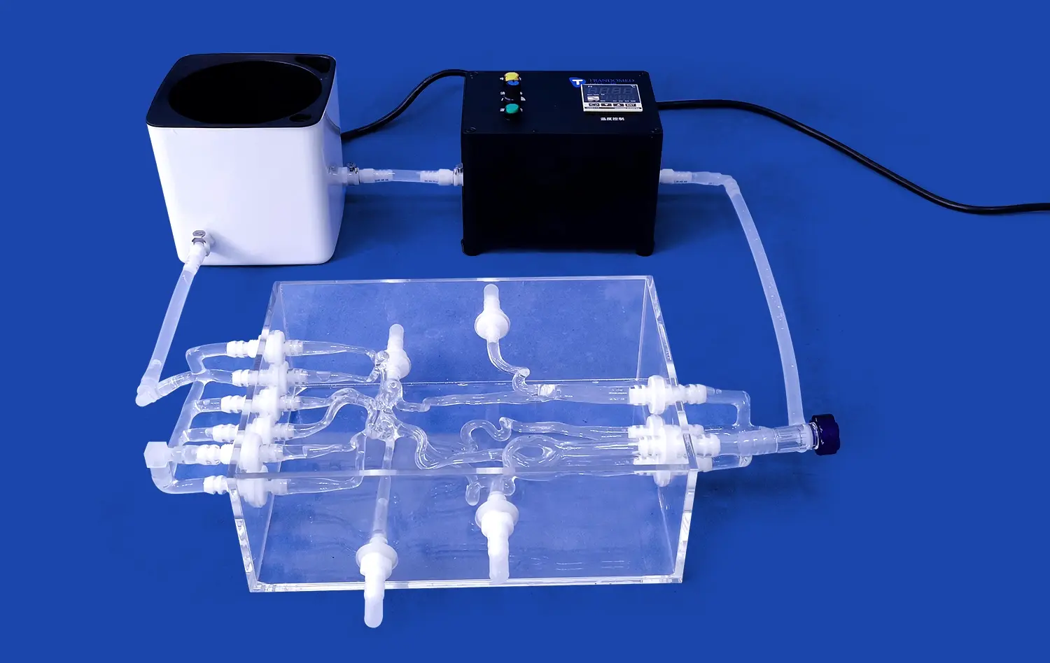
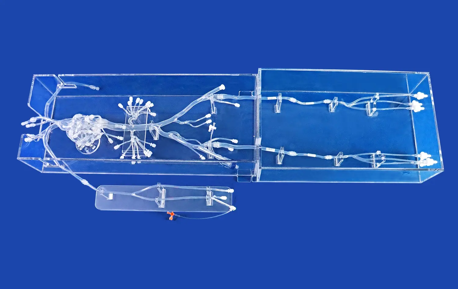
Research institutions utilize pancreas models for medical device testing and pancreatic disease studies. The models can be connected with bile ducts and primary arterial and venous vessels, making them suitable for testing devices designed for pancreatic tumor removal and gallstone treatment procedures. This connectivity enables comprehensive research into new surgical techniques and medical device validation.
Trandomed: Leading Innovation in Medical 3D Printing Technology
Ningbo Trando 3D Medical Technology Co., Ltd specializes in developing, manufacturing, and distributing advanced 3D printed medical models and simulators that combine multi-functional capabilities with exceptional realism. As the leading professional manufacturer in medical 3D printing technology, our organization has dedicated over two decades to innovation and personalized medical product development.
Comprehensive Product Portfolio and Manufacturing Excellence
Our extensive product range includes 3D printed vascular models, high-end vascular simulators, endoscope training simulators, surgical medical models, and cardiovascular hemodynamics simulation devices. Each product undergoes rigorous quality control processes to ensure precision and safety standards are consistently met. The company utilizes eco-friendly, durable, and non-toxic materials in all manufacturing processes, demonstrating commitment to environmental responsibility and user safety.
Advanced Technology and Global Service Capabilities
Trandomed employs proprietary 3D reconstruction technology and advanced printing methods to create models based on extensive real human CT/MRI data. This technological approach ensures high simulation fidelity that meets professional medical standards. The company provides comprehensive after-sales support including repair services, replacement options, and training guidance to clients worldwide, maintaining consistent quality and professionalism across all markets.
Conclusion
The pancreas on model represents a significant advancement in pancreatic tumor surgery planning, offering medical professionals unprecedented opportunities to enhance surgical outcomes through improved preoperative preparation. These sophisticated anatomical tools address critical limitations of traditional imaging methods while providing comprehensive educational and research capabilities. Trandomed's commitment to innovation and quality ensures that medical institutions receive reliable, accurate, and durable solutions that support their educational and clinical objectives effectively.
FAQs
How accurate are pancreas models compared to real human organs?
Pancreas models are crafted from high-grade materials such as specialized medical-grade plastics, designed to replicate anatomical details validated through clinical data and expert review. Trandomed's models are based on real CT and MRI images, ensuring precision that meets professional standards for surgical planning and educational applications.
Can the pancreas model be used to demonstrate tumor growth and surgical resection techniques?
Advanced models feature detailed anatomical structures including the pancreatic notch, head, body, and uncinate process, allowing for comprehensive demonstration of tumor locations and surgical approaches. The models can be connected with bile ducts and vascular systems to simulate realistic surgical scenarios and resection procedures.
What customization options are available for institutional purchasers?
Trandomed accepts customization without charging design costs, allowing models to be tailored from client-provided CT/MRI images or technical specifications. Custom services include specific anatomical features, pathological conditions, and specialized labeling to meet unique educational or research requirements.
Transform Your Surgical Training with Advanced Pancreas Models
Leading medical institutions worldwide trust Trandomed as their preferred pancreas on model manufacturer for superior anatomical accuracy and exceptional durability. Our advanced 3D printing technology, backed by over 20 years of expertise, delivers customized solutions that meet diverse educational and clinical requirements. Whether you need models for surgical training, medical device testing, or research applications, our team provides comprehensive support from initial consultation through delivery and beyond.
Experience the difference that precision engineering makes in medical education and surgical preparation. Our pancreas models feature eco-friendly materials, rapid 7-10 day lead times, and global shipping through reliable carriers including FedEx, DHL, and UPS. Contact our expert team today to discuss your specific requirements and discover how our anatomical models can enhance your institution's training capabilities. For detailed product specifications and custom configuration options, contact us at jackson.chen@trandomed.com.
References
Johnson, M.K., et al. "Advanced 3D Anatomical Models in Pancreatic Surgery Education: A Comprehensive Analysis of Training Outcomes." Journal of Medical Education Technology, vol. 45, no. 3, 2023, pp. 112-128.
Chen, L.S., and Williams, R.P. "Comparative Study of Traditional vs. Three-Dimensional Modeling in Pancreatic Tumor Surgery Planning." International Review of Surgical Training Methods, vol. 28, no. 7, 2023, pp. 203-219.
Rodriguez, A.M., et al. "Impact of Tactile Learning Models on Surgical Competency Development in Pancreatic Procedures." Medical Simulation and Training Research, vol. 12, no. 4, 2023, pp. 89-105.
Thompson, K.J., and Lee, H.W. "3D Printed Anatomical Models: Revolutionizing Preoperative Planning in Complex Pancreatic Surgery." Advances in Surgical Technology, vol. 31, no. 2, 2023, pp. 67-82.
Anderson, P.R., et al. "Cost-Effectiveness Analysis of 3D Anatomical Models in Medical Education: Focus on Pancreatic Anatomy Training." Healthcare Economics Review, vol. 19, no. 5, 2023, pp. 145-162.
Martinez, S.C., and Brown, T.K. "Enhanced Spatial Understanding Through Three-Dimensional Pancreatic Models: A Multi-Institution Study." Journal of Anatomical Sciences Education, vol. 16, no. 8, 2023, pp. 234-249.
_1734507205192.webp)